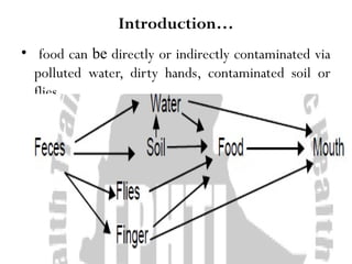
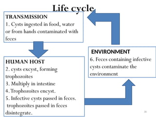
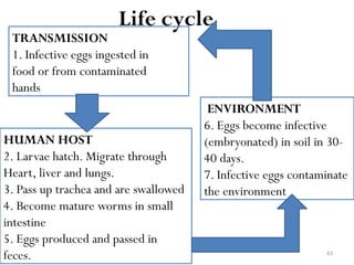
1
CHAPTER THREE
Oral-fecal transmitted diseases
2
Oral-fecal transmitted diseases
Learning Objectives
At the end of this chapter, students will be able to:
• Identify the five important “Fs” in oral-fecal disease
transmission.
• State diseases transmitted mainly in water and in soil.
• List diseases commonly transmitted by having direct
contact with feces.
• Participate in the diagnosis and treatment of cases.
• Implement preventive and control methods of oral-
fecal transmitted diseases.
3
Introduction
• The causative organisms are excreted in the stools
of infected persons (rarely animals)
• The portal of entry for these diseases is the mouth.
• This is known as the fece-oral transmission route
• As indicated in the schematic diagram below, food
takes a central position
4
Introduction…
• food can be directly or indirectly contaminated via
polluted water, dirty hands, contaminated soil or
flies.
5
Introduction…
• The five “Fs” which play an important role in fecal oral
diseases transmission are:
–Finger
–Flies
–Food
–Fomites and
–Fluid
6
Typhoid fever
7
Typhoid fever
Definition
• A systemic infectious disease characterized by
high continuous fever, malaise and
involvement of lymphoid tissues.
Infectious agent
• Salmonella typhi
• Salmonella enteritidis (rare cause)
8
Typhoid…
Epidemiology
• It occurs worldwide, particularly in poor socioeconomic
areas
• In endemic areas the disease is most common in
preschool and school aged children
Reservoir- Humans
Mode of transmission- water and food contaminated
by feces and urine of patients and carriers.
• Flies may infect foods.
Incubation period –1-3 weeks
9
Typhoid…
Period of communicability
• As long as the bacilli appears in excreta
• Usually from the first week through out convalescence
• 10% of untreated patients will discharge bacilli for 3 months
after onset of symptoms
• 2-5% becomes chronic carriers
• Susceptibility increased in individuals with gastric
achlorhydria or in HIV positive individuals
10
Typhoid…
Clinical manifestations
First week
• Mild illness characterized by: Fever rising stepwise
(ladder type)
• Anorexia
• Lethargy, Malaise
• General aches
11
Typhoid…
• Dull and continuous frontal head ache (prominent )
• Nose bleeding
• Vague abdominal pain
• Constipation in 10 % pts
• Cough ( due to bronchitis)
12
Typhoid…
Second week
• Sustained temperature (fever)
• Sever illness with weakness
• Mental dullness or delirium
• Abdominal discomfort and distension
• Diarrhea ( feces may contain blood )
13
Typhoid…
Third week
• Fever and exhaustion will increase
• Improvement and reduction of temperature if no
complication
14
Typhoid…
Suggestive clinical manifestations
• Fever- Sustained fever (ladder fashion)
• Rose spots- Small pallor, blanching, slightly raised macules
usually seen on chest and abdomen in the first week in 25% of
white people.
• Relative bradycardia- Slower than would be expected from
the level of temperature.
• Leucopoenia- White cell count is less than 4000/mm3 of
blood.
15
Typhoid…
Diagnosis
• Based on clinical grounds but this is confused with
wide variety of diseases.
• Widal reaction against somatic and flagellar antigens.
• Blood, feces or urine culture.
Treatment
1. Ampicillin or co-trimoxazole for carriers and mild
cases.
2. Chloramphenicol or ciprofloxacin or ceftriaxone for
seriously ill patients.
16
Typhoid…
Prevention and control
1.Treatment of patients and carriers
2. Education on hand washing, particularly food
handlers, patients and childcare givers
3. Sanitary disposal of feces and control of flies
4. Provision of safe and adequate water
5. Safe handling of food
17
Typhoid…
6. Exclusion of typhoid carriers and patients from
handling of food and patients
7. Immunization for people at special risk (e.g.Travelers
to endemic areas)
8. Regular check-up of food handlers in food and
drinking establishments
18
Bacillary Dysentery (Shigellosis)
19
Bacillary Dysentery (Shigellosis)
Definition
• An acute bacterial disease involving the large and distal
small intestine, caused by the bacteria of the genus
shigella.
Infectious agent
Shigella is comprised of four species or serotypes.
• GroupA= Shigella dysentraie (most common cause)
• Group B= Shigella flexneri
• Group C= Shigella boydii
• Group D= Shigella sonnei
20
Bacillary Dysentery…
Occurrence
• World wide
• Endemic in both tropics & temperate climates
• Out breaks commonly occur in:
– over crowding and poor personal hygiene
– refuge camps
– mental hospitals and
–day care centers
21
Bacillary Dysentery…
Reservoir : Humans (children are the main reservoir)
Mode of transmission:
• Mainly by direct or indirect fecal- oral transmission
from a patient or carrier
• Through contaminated water and milk
• Flies can transfer organisms from latrines
Incubation period -usually 1-3 days
22
Bacillary Dysentery…
Period of communicability -During acute infection
and until the infectious agent is no longer present in
feces
Susceptibility and resistance
• more sever in young children, the elderly &
malnourished
• Breast feeding is protective for infants and young
children
23
Bacillary Dysentery…
Clinical manifestation
• Fever, rapid pulse, vomiting & abd.cramp are prominent
• Diarrhea usually appears after 48 hrs with dysentery
• Generalized abdominal tenderness
• Tenasmus is present and feces are bloody, mucoid & of small
quantity.
• Dehydration is common & dangerous = cause muscular cramp,
oliguria and shock
24
Bacillary Dysentery…
Diagnosis
• Clinical grounds
• Stool microscopy (presence of pus cells and
erythrocytes)
• Stool culture confirms the diagnosis
25
Bacillary Dysentery…
Treatment
• Antibiotics are only indicated in case of sever systemic
symptoms
• Fluid and electrolyte replacement ( rehydration is the
first priority)
• Co-trimaxazole in sever cases or Nalidixic acid in the
case of resistance
• Gentamycin 80mg im 2x/d for 10 days
26
Bacillary Dysentery…
Prevention and control
• Detection of carriers & treatment of cases
• Hand washing after toilet and before handling or
eating food
• Proper excreta disposal
• Adequate and safe water supply
• Control of flies
• Cleanliness in food handling and preparation.
27
Amoebiasis (Amoebic Dysentery)
28
Amoebiasis (Amoebic Dysentery)
Definition
• An infection due to a protozoan parasite that causes
intestinal or extra-intestinal disease.
Infectious agent
• Entamoeba histolytica
Occurrence- worldwide but most common in the
tropics and sub-tropics.
• Prevalent in areas with poor sanitation, in mental
institutions and homosexuals
29
Amoebiasis …
Reservoir- human (lumen of large intestine)
Mode of transmission
• Feco- oral transmission
• Ingestion of food or water contaminated by feces
containing the cyst
Incubation period
• Few days to several months or years commonly 2-4
weeks.
Period of communicability
• During the period of passing cysts of E.histolytica which
may continue for years
30
TRANSMISSION
1. Cysts ingested in food, water
or from hands contaminated with
feces
HUMAN HOST
2. cysts excyst, forming
trophozoites
3. Multiply in intestine
4.Trophozoites encyst.
5. Infective cysts passed in feces.
trophozoites passed in feces
disintegrate.
ENVIRONMENT
6. Feces containing infective
cysts contaminate the
environment
Life cycle
31
Amoebiasis …
Clinical Manifestation
• Starts with a prodormal episode of diarrhea,
abdominal cramps, nausea, vomiting and tenesmus.
• With dysentery, feces are generally watery, containing
mucus and blood.
Diagnosis
• Demonstration of Etamoeba histolytica cyst or
trophozoite in stool
Treatment
• Metronidazole orTinidazole
32
Amoebiasis …
Prevention and control
1.Adequate treatment of cases
2. Provision of safe drinking water
3. Proper disposal of human excreta (feces)
4. Hand washing following defecation.
5.Cleaning and cooking of local foods to avoid eating food
contaminated with feces.
(e.g. raw vegetables)
33
Giardiasis
34
Giardiasis
Definition
• A protozoan infection principally of the upper small
intestine associated with symptoms of:
– chronic diarrhea
– steatorrhea
–abdominal cramps
–bloating
–frequent loose and pale greasy stools
– fatigue and weight loss.
35
Giardiasis…
Reservoir – Humans
Period of communicability – Entire period of
infection, often months
Susceptibility and resistance
• Asymptomatic carrier rate is high
• Infection is frequently self-limited
• Persons withAIDS may have more serious & prolonged
infection
36
Giardiasis…
Infectious agent
• Giardia lamblia
Occurrence
• World wide
• Children are more affected than adults
• Highly prevalent in areas of poor sanitation
37
TRANSMISSION
1. Cysts ingested in food, water
or from hands contaminated with
feces
HUMAN HOST
2. cysts excyst, forming
trophozoites
3. Multiply in intestine
4.Trophozoites encyst
5. Infective cysts passed in feces.
trophozoites passed in feces
disintegrate
ENVIRONMENT
6. Feces containing infective
cysts contaminate the
environment
Life cycle
38
Giardiasis…
Clinical manifestation
• Ranges from asymptomatic infection to sever failure to
thrive and malabsorption
• Young children usually have diarrhea but abdominal
distension and bloating are frequent
• Adults have abdominal cramps, diarrhea, anorexia, nausea,
malaise, bloating,
• many patients complain of sulphur testing (belching)
39
Giardiasis…
Diagnosis -Demonstration of Giardia lamblia cyst or
trophozoite in feces
Treatment -Metronidazole orTinidazole
Prevention and control
• Good personal hygiene and hand washing
• Sanitary disposal of feces
• Protection of public water supply from contamination
of feces
• Case treatment
• Safe water supply
40
Cholera
41
Cholera
Definition
• Cholera is an infectious
gastroenteritis caused by
enterotoxin-producing strains of the
bacterium Vibrio cholerae
Infectious agent
• Gram-negative Vibrio cholerae
• That produces cholera toxin, an
enterotoxin
42
Cholera…
Occurrence- has made periodic outbreaks in
different parts of the world and given rise to
pandemics.
• Endemic predominantly in children.
Reservoir- Humans
Mode of transmission- by ingestion of food or
water directly or indirectly contaminated with feces
or vomitus of infected person.
43
Cholera…
Incubation period- from a few hours to 5 days,
usually 2-3 days.
Period of communicability- for the duration of
the stool positive stage, usually only a few days after
recovery.
• Antibiotics shorten the period of communicability.
Susceptibility and resistance
• gastric achlorhydria increases risk of illness
• very sensitive to gastric acid
• breast feeding infants are protected.
44
Cholera…
Clinical Manifestation
• Abrupt painless watery diarrhea; the diarrhea looks like
rice water.
• In severe cases, several liters of liquid may be lost in few
hours leading to shock.
• Severely ill patients are cyanotic, have sunken eyes and
cheeks, scaphoid abdomen, poor skin turgor, and slow
pulse.
• Loss of fluid continues for 1-7 days.
45
Cholera…
 Diagnosis
• Based on clinical grounds
• Culture (stool) confirmation
 Treatment
1. Prompt replacement of fluids and electrolytes
• Rapid IV infusions of large amounts
• Isotonic saline solutions alternating with isotonic sodium
bicarbonate or sodium lactate.
2. Antibiotics like tetracycline dramatically reduce the duration
and volume of diarrhea resulting in early eradication of vibrio
cholerae.
46
Cholera…
Nursing care
• wear gown and glove
• wash your hands
• monitor out put including stool out put
• patients are treated on “ cholera beds”- beds with a
central hole through which the continuous stools can
pass in to a bucket & be measured
• Protect the patient family by administering tetracycline
• Health education
47
Cholera…
• Prevention and control
1. Case treatment
2. Safe disposal of human excreta and control of flies
3. Safe public water supply
4. Hand washing and sanitary handling of food
5. Control and management of contact case
48
Class activity
Assume that there are patients who are suffered
with sever watery diarrhoea and they are far
from health institution. If you see those patients,
how you are going to help them at that place?
49
Infectious hepatitis
50
Infectious hepatitis
(Viral hepatitis A, Epidemic hepatitis, type A
hepatitis)
Definition
• An acute viral disease characterized by abrupt onset of:
– fever
– malaise
– anorexia
– nausea and abdominal discomfort followed within a
few days by jaundice.
Infectious agent
• HepatitisA virus
51
Infectious hepatitis…
Occurrence
• World wide distribution in sporadic & epidemic forms
• Infection is common where environmental sanitation is
poor
• Adults are usually immune (long-lasting)
Reservoir- Humans
Mode of transmission
• person to person by feco-oral route
• water and food contaminated by infected food handlers
52
Infectious hepatitis…
Incubation period- 15-55 days, average 28-30 days.
Period of communicability- High during the later
half of the incubation period and continuing for few
days following onset of jaundice.
• Most cases are non-infectious following first week of
jaundice.
Susceptibility and resistance- Susceptibility is
general.
• Immunity following infection probably lasts for life.
53
Infectious hepatitis…
Clinical manifestation
• Abrupt onset of fever
• Malaise
• Anorexia
• Nausea and
• Abdominal discomfort, followed in few days by
jaundice.
• Complete recovery without sequel or recurrence as a
rule.
54
Diagnosis
• Based on clinical and epidemiological grounds
• Demonstration of IgM (IgM anti-HAV) in the serum
of acutely or recently ill patients
Treatment
• Symptomatic: Rest, high carbohydrate diet with low
fat and protein.
Infectious hepatitis…
55
Preicteric phase (before onset of Jaundice): anorexia,
nausea, vomiting, tenderness of Liver
Icteric phase: Jaundice, Hepatomegaly
Recovery slow.
56
Milder disease than Hepatitis B;
No chronic form of the disease.
No carrier status.
Immunity – long lasting
Asymptomatic infections are very common, especially in children.
Adults, especially pregnant women, may develop more severe
disease.
57
Prevention and control
1. Public education about good sanitation and personal
hygiene, with special emphasis on careful hand washing
and sanitary disposal of feces.
2. Proper water treatment and distribution systems and
sewage disposal.
3. Proper management of day care centers to minimize
possibility of fecal-oral transmission.
4. HA vaccine for all travelers to intermediate or highly
endemic areas.
5. Protection of day care centers’ employees by vaccine.
Infectious hepatitis…
58
Feces Mainly in Soil
• The diseases in this category are mainly transmitted
through fecal contamination of soil.
• These infections are acquired through man’s exposure
to fecally contaminated soil.
59
Ascariasis
60
Ascariasis
Definition
• A helminthic infection of the small intestine generally
associated with few or no symptoms.
Occurrence
• The most common parasite of humans where sanitation
is poor
• School children (5-10 yrs) are most affected
• Highly prevalent in moist tropical countries (not too
dry & loose soil)
A female may produce up to 200,000 eggs daily
61
Ascariasis…
Reservoir –Humans, eggs in soil
Mode of transmission
• Ingestion of infective eggs /embryonated eggs/ from
the soil contaminated with human feces.
• Uncooked vegetable (e.g. salad) contaminated with soil
containing infective eggs ( especially when the field has
been manured with human feces)
• No transmission from fresh feces
• No direct transmission from person to person
62
Ascariasis…
Incubation period - 4-8 wks.
Period of communicability
• As long as mature fertilized female worms live in
the intestine
• Usual life span of the adult worm is 12 months
63
TRANSMISSION
1. Infective eggs ingested in
food or from contaminated
hands
HUMAN HOST
2. Larvae hatch. Migrate through
Heart, liver and lungs.
3. Pass up trachea and are swallowed
4. Become mature worms in small
intestine
5. Eggs produced and passed in
feces.
ENVIRONMENT
6. Eggs become infective
(embryonated) in soil in 30-
40 days.
7. Infective eggs contaminate
the environment
Life cycle
64
65
Ascariasis…
Clinical Manifestation
• Most infections go unnoticed until large worm is passed
in feces and occasionally the mouth and nose.
• Migrant larvae may cause itching, wheezing and
dyspnea, fever, cough productive of bloody sputum may
occur.
• Abdominal pain may arise from intestinal or duct
(biliary, pancreatic) obstruction.
• Serious complications include bowel obstruction due to
knotted/intertwined worms.
66
Ascariasis…
Diagnosis
• Microscopic identification of eggs in a stool sample
• Adult worms passed from anus, mouth or nose.
Treatment
1.Albendazole or
2. Mebendazole or
3. Piperazine or
4. Levamisole
67
Prevention and control
1.Treatment of cases
2. Sanitary disposal of feces
3. Prevent soil contamination in areas where children
play
4. Promote good personal hygiene (hand washing).
Ascariasis…
68
Trichuriasis
69
Trichuriasis
Definition
• A nematode infection of the large intestine, usually
asymptomatic in nature.
Infectious agent
• Trichuris trichuria (whip worm)
Occurrence- Worldwide, especially in warm
moist regions.
• Common in children 3-11 years of age.
Reservoir- Humans
70
Trichuriasis…
Mode of transmission- Indirect, particularly
through pica or ingestion of contaminated vegetables.
• Not immediately transmissible from person to person.
Incubation period- Indefinite
Period of communicability- Several years in
untreated carriers.
Susceptibility and resistance- Susceptibility is
universal
71
TRANSMISSION
1. Infective eggs ingested in food
or from contaminated hands
HUMAN HOST
2. Larvae hatch.
Develop in small intestine.
Migrate to caecum.
3. Become mature worms.
4. Eggs produced and passed
in feces.
ENVIRONMENT
6. Eggs become infective
(embryonated) in soil after 3
weeks.
7. Infective eggs contaminate
the environment
Life cycle
72
Trichuriasis…
Clinical manifestation
• Severity is directly related to the number of infecting
worms.
• Most infected people are asymptomatic.
• Abdominal pain, tiredness, nausea and vomiting,
diarrhea or constipation are complaints by patients.
• Rectal prolapse may occur in heavily infected very
young children
73
Trichuriasis…
Diagnosis
• Demonstration of eggs in feces.
Treatment
1.Albendazole or
2. Mebendazole
Prevention and control
1. Sanitary disposal of feces
2. Maintaining good personal hygiene (i.e. washing hands and
vegetables and other soil contaminated foods)
3. Cutting nails especially in children
4.Treatment of cases.
74
Entrobiasis
(pinworm infection)
75
Entrobiasis
(pinworm infection)
Definition
• A common intestinal helminthic infection that is often
asymptomatic.
Infectious agent
• Entrobius vermicularis
 Occurrence
• world wide
• Prevalence is high in school aged children followed by pre-
school children
• Infection usually occurs in more than one family member
 Reservoir - human
76
Entrobiasis …
Mode of transmission
• Direct transfer of infective eggs by hand from anus
to mouth of the same person (auto infection ) or
another person
• Indirect transmission through clothing, bedding,
food or other articles contaminated with eggs.
• Retro infection- in moist conditions eggs on the peri
anal skin may hatch – larvae-enter the host via anus.
• Air born infection through inhalation of dust
containing eggs
77
Entrobiasis…
Incubation period - 2-6 weeks
Period of communicability
• As long as gravid females are discharging eggs on peri
anal skin
• Eggs remain infective in an indoor environment for
about 2 weeks
Susceptibility and resistance- Susceptibility is
universal.
78
Life cycle
Adult worms
in caecum
Gravid females migrate through
the anus to the perianal skin and
deposit eggs (usually during the
night)
Eggs become infective in a few
hours in perianal area
Ingestion of eggs by
Man
Larvae
hatch in
duodenum
Migrate down
to caecum
79
Entrobiasis…
Clinical manifestation
• Perianal itching, disturbed sleep, irritability and some
times secondary infection of the scratched skin.
Diagnosis
• Stool microscopy for eggs or female worms.
Treatment
• Mebendazole
80
Entrobiasis…
Prevention and control
1. Educate the public about hygiene (i.e. handwashing
before eating or preparing food, keeping nails short and
discourage nail biting).
2.Treatment of cases
3. Reduce overcrowding in living accommodations.
4. Provide adequate toilets.
81
Strongyloidiasis
82
Strongyloidiasis
Definition
• An often asymptomatic helminthic infection of the duodenum
and upper jejunum.
Infectious agent
• Strongyloides stercolaris
• The parasite is unique in that it has both parasitic and free-
living generations
• parasitic males are absent.
• parasitic females are present in the submucosa of the small
intestine and produces eggs parthenogenetically
83
Strongyloidiasis…
Occurrence- In tropical and temperate areas.
• More common in warm and wet regions.
Reservoir- Human
Mode of transmission- Infective (filariform) larvae
penetrate the skin and enter the venous circulation
85
TRANSMISSION
1. Infective filariform larvae
penetrate skin, e.g. feet.
Autoinfection also occurs
HUMAN HOST
2. Larvae migrate, pass up trachea and are
swallowed.
3. Become mature worms in small
intestine
4. Eggs laid. Hatch rhabditi form
larvae in intestine.
5. Rhabditiform larvae:
- Passed in feces, or
- Become filariform larvae in
intestine, causing
atutoinfection.
ENVIRONMENT
6. In soil larvae become freeliving
worms produce more
rhabditiform larvae
Free-living cycle can be
repeated several times
7. Become infective filariform
larvae in the soil
Life cycle
86
Life cycle
87
Strongyloidiasis…
Clinical Manifestation
• Pneumonia occurs during heavy larval migration.
• Mild peptic ulcer like epigastric discomfort to severe
watery diarrhea.
• Heavy infection may result in malabsorption syndrome
Diagnosis
• Identification of larvae in stool specimen
88
Strongyloidiasis…
Treatment
1.Albendazole or
2.Thiabendazole
Prevention and control
1. Proper disposal of human excreta (feces)
2. Personal hygiene including use of footwear.
3. Case treatment.
89
Hookworm disease
90
Hookworm disease
Definition
• A common chronic parasitic infection with a
variety of symptoms usually in proportion of the
degree of anemia
Infectious agent
• Ancylostoma duodenale
(predominantly old world species)
• Necator americanus
(predominantly new world species)
91
Hookworm disease…
Occurrence- Widely endemic in tropical and
subtropical countries where sanitary disposal of
human feces is not practiced and the soil moisture and
temperature conditions favor development of
infective larvae.
Reservoir- Humans
Mode of transmission- Through skin penetration
by the infective larvae.
92
Hookworm disease…
Incubation period- Symptoms may develop after a
few weeks to many months depending on intensity of
infection and iron intake of the host.
Period of communicability- Infected people can
contaminate the soil for several years in the absence of
treatment.
Susceptibility and resistance- Susceptibility is
universal.
• No evidence that immunity develops with infection.
93
TRANSMISSION
1. Infective filariform larvae
penetrate the skin, e.g. feet.
A. duodenale also transmitted by
ingestion of larvae.
HUMAN HOST
2. Larvae migrate. Pass up trachea
and are swallowed.
3. Become mature worms in small
intestine (attach to wall and suck
blood).
4. Eggs produced and passed in
feces.
ENVIRONMENT
5. Eggs develop;
Rhabditiform larvae hatch.
Feed in soil.
6. Develop into infective
filariform larvae in about 1
week.
7. Filariform larvae
contaminate soil
Life cycle
94
95
Hookworm disease…
Clinical Manifestation
• The clinical manifestation is related to:
1. Larval migration of the skin
• Produces transient, localized maculopapular rash associated
with itching called ground itch.
2. Migration of larva to the lungs.
• Produces cough, wheezing and transient pneumonitis.
3. Blood sucking
• Light infection-no symptoms
• Heavy infection-result in symptoms of peptic ulcer disease
like epigastric pain and tenderness.
96
Hookworm disease…
• Further loss of blood leads to anemia manifested by exertional
dyspenea, weakness and light-headedness.
Diagnosis
• Demonstration of eggs in stool specimen.
Treatment
1. Mebendazole or
2.Albendazole or
3. Levamisole
Prevention and control
1. Sanitary disposal of feces
2.Wearing of shoes
3. Case treatment.
97
Direct Contact with Feces
• These are diseases transmitted mainly through
direct contact with feces of the infected
person.
98
Poliomyelitis (Infantile paralysis)
99
Poliomyelitis (Infantile paralysis)
• A viral infection most often recognized by the acute onset
of flaccid paralysis
Infectious agent
• Poliovirus- types 1,2 and 3
Pathogenesis :All types cause paralysis, but typ1 is the
most paralytogenic
• Virus multiplies in the alimentary tract
• Viremia may then follow with invasion of the CNS and
selective involvement of motor cells resulting in flaccid
paralysis most commonly of the lower extremities
100
Poliomyelitis …
• Paralysis of muscles of respiration and swallowing
frequently threatens life
• The site of paralysis depends upon location of nerve cell
destruction in spinal cord or brain stem, but is
characteristically asymmetrical
• There is never any sensory deficit
101
Poliomyelitis …
Occurrence
• World wide prior to the advent of immunization
• Cases of polio occur both sporadically & in epidemic
• Primarily a disease of infants & young children
• 70-80% cases are less than three yrs of age
• More than 90% of infections are unapparent
Reservoir -humans, especially children
102
Poliomyelitis …
Mode of transmission
• Primarily person to person
• Spread principally through the fecal- oral route, mainly
via sewerage- contaminated water
• In rare conditions, milk food stuffs and other materials
contaminated with feces have been incriminated as
vehicles
Incubation period -commonly 7-14 days
Period of communicability
• not precisely known
• but transmission is possible as long as the virus is excreted
103
Poliomyelitis …
Susceptibility and resistance
• Susceptibility is common in children but paralysis
rarely occurs
• Infection confers permanent immunity
Clinical manifestation
• Usually asymptomatic or non-specific fever is
manifested in 90% of cases.
• If it progresses to major illness, severe muscle pain,
stiff neck and back with or without flaccid paralysis
may occur.
104
Poliomyelitis …
• Paralysis is asymptomatic and occurs within three to four
days of illness.
• The legs are more affected than other part of the body.
• Paralysis of respiratory and swallowing muscles is life
threatening.
Diagnosis
• based on clinical and epidemiological grounds
• virus may be isolated from the stools and less often from
CSF or throat swabs
• virus excretion in the stools can persist for 6-12 weeks
after the acute illness
105
Poliomyelitis …
Treatment
• physiotherapy, bed rest
Prevention and control
• Educate public about the advantage of immunization in
early child hood
• Trivalent live attenuated vaccine (OPV) at birth
• Safe disposal of human excreta.
106
Hydatid Disease (Echinococcosis)
Definition
• The tapeworm Echinococcus granulosus is the
most common species of Echinococcus and
causes cystic hydatid disease.
Infectious agent
• Echinococcus granulosus, a small tapeworm of
dog
107
• Epidemiology
• Occurrence – occurs on all continents except
Antarctica.
• Especially common in grazing countries where
dogs consume viscera containing cysts.
• Reservoir- Domestic dogs and canids are
definitive hosts; they may harbor in their
intestines without signs of infection. Sheep act
as intermediate hosts.
108
• Mode of transmission – directly with hand to
mouth transfer of eggs after association with
infected dogs or indirectly through
contaminated food, water, soil or fomites.
• Incubation period – variable from 12 months
to many years,
109
• Period of communicability – Infected dogs
begin to pass eggs approximately 7weeks
after infection.
• Susceptibility and resistance – Children are
more likely to be exposed to infection.
110
• according to location of the cyst and number.
• Ruptured or leaking cysts can cause severe
anaphylactic reactions.
• Cysts are typically spherical, thick walled and
unilocular and are most frequently found in
the liver and lungs.
111
Diagnosis
• History of residence in an endemic area
• Sonography and CT scan
• Serologic test
Treatment
• 1. Surgical resection of isolated cysts
• 2. Albendazol (mebendazol)
• 3. If cysts rupture, praziquantel
112
• Prevention and control
• 1. Educate the public at risk to avoid exposure
to dog feces.
• 2. Interrupt transmission from intermediate to
definitive hosts
• 3. Safe disposal of infected viscera.
• 4. Periodical treatment of high-risk dogs.
113
Exercise
• What does fecal-oral transmission mean?
• State some of the common preventive and control measures
of oral-fecal transmitted diseases.

Chapter 3 (1).pptx.communicable diseases

  • 1.
  • 2.
    2 Oral-fecal transmitted diseases LearningObjectives At the end of this chapter, students will be able to: • Identify the five important “Fs” in oral-fecal disease transmission. • State diseases transmitted mainly in water and in soil. • List diseases commonly transmitted by having direct contact with feces. • Participate in the diagnosis and treatment of cases. • Implement preventive and control methods of oral- fecal transmitted diseases.
  • 3.
    3 Introduction • The causativeorganisms are excreted in the stools of infected persons (rarely animals) • The portal of entry for these diseases is the mouth. • This is known as the fece-oral transmission route • As indicated in the schematic diagram below, food takes a central position
  • 4.
    4 Introduction… • food canbe directly or indirectly contaminated via polluted water, dirty hands, contaminated soil or flies.
  • 5.
    5 Introduction… • The five“Fs” which play an important role in fecal oral diseases transmission are: –Finger –Flies –Food –Fomites and –Fluid
  • 6.
  • 7.
    7 Typhoid fever Definition • Asystemic infectious disease characterized by high continuous fever, malaise and involvement of lymphoid tissues. Infectious agent • Salmonella typhi • Salmonella enteritidis (rare cause)
  • 8.
    8 Typhoid… Epidemiology • It occursworldwide, particularly in poor socioeconomic areas • In endemic areas the disease is most common in preschool and school aged children Reservoir- Humans Mode of transmission- water and food contaminated by feces and urine of patients and carriers. • Flies may infect foods. Incubation period –1-3 weeks
  • 9.
    9 Typhoid… Period of communicability •As long as the bacilli appears in excreta • Usually from the first week through out convalescence • 10% of untreated patients will discharge bacilli for 3 months after onset of symptoms • 2-5% becomes chronic carriers • Susceptibility increased in individuals with gastric achlorhydria or in HIV positive individuals
  • 10.
    10 Typhoid… Clinical manifestations First week •Mild illness characterized by: Fever rising stepwise (ladder type) • Anorexia • Lethargy, Malaise • General aches
  • 11.
    11 Typhoid… • Dull andcontinuous frontal head ache (prominent ) • Nose bleeding • Vague abdominal pain • Constipation in 10 % pts • Cough ( due to bronchitis)
  • 12.
    12 Typhoid… Second week • Sustainedtemperature (fever) • Sever illness with weakness • Mental dullness or delirium • Abdominal discomfort and distension • Diarrhea ( feces may contain blood )
  • 13.
    13 Typhoid… Third week • Feverand exhaustion will increase • Improvement and reduction of temperature if no complication
  • 14.
    14 Typhoid… Suggestive clinical manifestations •Fever- Sustained fever (ladder fashion) • Rose spots- Small pallor, blanching, slightly raised macules usually seen on chest and abdomen in the first week in 25% of white people. • Relative bradycardia- Slower than would be expected from the level of temperature. • Leucopoenia- White cell count is less than 4000/mm3 of blood.
  • 15.
    15 Typhoid… Diagnosis • Based onclinical grounds but this is confused with wide variety of diseases. • Widal reaction against somatic and flagellar antigens. • Blood, feces or urine culture. Treatment 1. Ampicillin or co-trimoxazole for carriers and mild cases. 2. Chloramphenicol or ciprofloxacin or ceftriaxone for seriously ill patients.
  • 16.
    16 Typhoid… Prevention and control 1.Treatmentof patients and carriers 2. Education on hand washing, particularly food handlers, patients and childcare givers 3. Sanitary disposal of feces and control of flies 4. Provision of safe and adequate water 5. Safe handling of food
  • 17.
    17 Typhoid… 6. Exclusion oftyphoid carriers and patients from handling of food and patients 7. Immunization for people at special risk (e.g.Travelers to endemic areas) 8. Regular check-up of food handlers in food and drinking establishments
  • 18.
  • 19.
    19 Bacillary Dysentery (Shigellosis) Definition •An acute bacterial disease involving the large and distal small intestine, caused by the bacteria of the genus shigella. Infectious agent Shigella is comprised of four species or serotypes. • GroupA= Shigella dysentraie (most common cause) • Group B= Shigella flexneri • Group C= Shigella boydii • Group D= Shigella sonnei
  • 20.
    20 Bacillary Dysentery… Occurrence • Worldwide • Endemic in both tropics & temperate climates • Out breaks commonly occur in: – over crowding and poor personal hygiene – refuge camps – mental hospitals and –day care centers
  • 21.
    21 Bacillary Dysentery… Reservoir :Humans (children are the main reservoir) Mode of transmission: • Mainly by direct or indirect fecal- oral transmission from a patient or carrier • Through contaminated water and milk • Flies can transfer organisms from latrines Incubation period -usually 1-3 days
  • 22.
    22 Bacillary Dysentery… Period ofcommunicability -During acute infection and until the infectious agent is no longer present in feces Susceptibility and resistance • more sever in young children, the elderly & malnourished • Breast feeding is protective for infants and young children
  • 23.
    23 Bacillary Dysentery… Clinical manifestation •Fever, rapid pulse, vomiting & abd.cramp are prominent • Diarrhea usually appears after 48 hrs with dysentery • Generalized abdominal tenderness • Tenasmus is present and feces are bloody, mucoid & of small quantity. • Dehydration is common & dangerous = cause muscular cramp, oliguria and shock
  • 24.
    24 Bacillary Dysentery… Diagnosis • Clinicalgrounds • Stool microscopy (presence of pus cells and erythrocytes) • Stool culture confirms the diagnosis
  • 25.
    25 Bacillary Dysentery… Treatment • Antibioticsare only indicated in case of sever systemic symptoms • Fluid and electrolyte replacement ( rehydration is the first priority) • Co-trimaxazole in sever cases or Nalidixic acid in the case of resistance • Gentamycin 80mg im 2x/d for 10 days
  • 26.
    26 Bacillary Dysentery… Prevention andcontrol • Detection of carriers & treatment of cases • Hand washing after toilet and before handling or eating food • Proper excreta disposal • Adequate and safe water supply • Control of flies • Cleanliness in food handling and preparation.
  • 27.
  • 28.
    28 Amoebiasis (Amoebic Dysentery) Definition •An infection due to a protozoan parasite that causes intestinal or extra-intestinal disease. Infectious agent • Entamoeba histolytica Occurrence- worldwide but most common in the tropics and sub-tropics. • Prevalent in areas with poor sanitation, in mental institutions and homosexuals
  • 29.
    29 Amoebiasis … Reservoir- human(lumen of large intestine) Mode of transmission • Feco- oral transmission • Ingestion of food or water contaminated by feces containing the cyst Incubation period • Few days to several months or years commonly 2-4 weeks. Period of communicability • During the period of passing cysts of E.histolytica which may continue for years
  • 30.
    30 TRANSMISSION 1. Cysts ingestedin food, water or from hands contaminated with feces HUMAN HOST 2. cysts excyst, forming trophozoites 3. Multiply in intestine 4.Trophozoites encyst. 5. Infective cysts passed in feces. trophozoites passed in feces disintegrate. ENVIRONMENT 6. Feces containing infective cysts contaminate the environment Life cycle
  • 31.
    31 Amoebiasis … Clinical Manifestation •Starts with a prodormal episode of diarrhea, abdominal cramps, nausea, vomiting and tenesmus. • With dysentery, feces are generally watery, containing mucus and blood. Diagnosis • Demonstration of Etamoeba histolytica cyst or trophozoite in stool Treatment • Metronidazole orTinidazole
  • 32.
    32 Amoebiasis … Prevention andcontrol 1.Adequate treatment of cases 2. Provision of safe drinking water 3. Proper disposal of human excreta (feces) 4. Hand washing following defecation. 5.Cleaning and cooking of local foods to avoid eating food contaminated with feces. (e.g. raw vegetables)
  • 33.
  • 34.
    34 Giardiasis Definition • A protozoaninfection principally of the upper small intestine associated with symptoms of: – chronic diarrhea – steatorrhea –abdominal cramps –bloating –frequent loose and pale greasy stools – fatigue and weight loss.
  • 35.
    35 Giardiasis… Reservoir – Humans Periodof communicability – Entire period of infection, often months Susceptibility and resistance • Asymptomatic carrier rate is high • Infection is frequently self-limited • Persons withAIDS may have more serious & prolonged infection
  • 36.
    36 Giardiasis… Infectious agent • Giardialamblia Occurrence • World wide • Children are more affected than adults • Highly prevalent in areas of poor sanitation
  • 37.
    37 TRANSMISSION 1. Cysts ingestedin food, water or from hands contaminated with feces HUMAN HOST 2. cysts excyst, forming trophozoites 3. Multiply in intestine 4.Trophozoites encyst 5. Infective cysts passed in feces. trophozoites passed in feces disintegrate ENVIRONMENT 6. Feces containing infective cysts contaminate the environment Life cycle
  • 38.
    38 Giardiasis… Clinical manifestation • Rangesfrom asymptomatic infection to sever failure to thrive and malabsorption • Young children usually have diarrhea but abdominal distension and bloating are frequent • Adults have abdominal cramps, diarrhea, anorexia, nausea, malaise, bloating, • many patients complain of sulphur testing (belching)
  • 39.
    39 Giardiasis… Diagnosis -Demonstration ofGiardia lamblia cyst or trophozoite in feces Treatment -Metronidazole orTinidazole Prevention and control • Good personal hygiene and hand washing • Sanitary disposal of feces • Protection of public water supply from contamination of feces • Case treatment • Safe water supply
  • 40.
  • 41.
    41 Cholera Definition • Cholera isan infectious gastroenteritis caused by enterotoxin-producing strains of the bacterium Vibrio cholerae Infectious agent • Gram-negative Vibrio cholerae • That produces cholera toxin, an enterotoxin
  • 42.
    42 Cholera… Occurrence- has madeperiodic outbreaks in different parts of the world and given rise to pandemics. • Endemic predominantly in children. Reservoir- Humans Mode of transmission- by ingestion of food or water directly or indirectly contaminated with feces or vomitus of infected person.
  • 43.
    43 Cholera… Incubation period- froma few hours to 5 days, usually 2-3 days. Period of communicability- for the duration of the stool positive stage, usually only a few days after recovery. • Antibiotics shorten the period of communicability. Susceptibility and resistance • gastric achlorhydria increases risk of illness • very sensitive to gastric acid • breast feeding infants are protected.
  • 44.
    44 Cholera… Clinical Manifestation • Abruptpainless watery diarrhea; the diarrhea looks like rice water. • In severe cases, several liters of liquid may be lost in few hours leading to shock. • Severely ill patients are cyanotic, have sunken eyes and cheeks, scaphoid abdomen, poor skin turgor, and slow pulse. • Loss of fluid continues for 1-7 days.
  • 45.
    45 Cholera…  Diagnosis • Basedon clinical grounds • Culture (stool) confirmation  Treatment 1. Prompt replacement of fluids and electrolytes • Rapid IV infusions of large amounts • Isotonic saline solutions alternating with isotonic sodium bicarbonate or sodium lactate. 2. Antibiotics like tetracycline dramatically reduce the duration and volume of diarrhea resulting in early eradication of vibrio cholerae.
  • 46.
    46 Cholera… Nursing care • weargown and glove • wash your hands • monitor out put including stool out put • patients are treated on “ cholera beds”- beds with a central hole through which the continuous stools can pass in to a bucket & be measured • Protect the patient family by administering tetracycline • Health education
  • 47.
    47 Cholera… • Prevention andcontrol 1. Case treatment 2. Safe disposal of human excreta and control of flies 3. Safe public water supply 4. Hand washing and sanitary handling of food 5. Control and management of contact case
  • 48.
    48 Class activity Assume thatthere are patients who are suffered with sever watery diarrhoea and they are far from health institution. If you see those patients, how you are going to help them at that place?
  • 49.
  • 50.
    50 Infectious hepatitis (Viral hepatitisA, Epidemic hepatitis, type A hepatitis) Definition • An acute viral disease characterized by abrupt onset of: – fever – malaise – anorexia – nausea and abdominal discomfort followed within a few days by jaundice. Infectious agent • HepatitisA virus
  • 51.
    51 Infectious hepatitis… Occurrence • Worldwide distribution in sporadic & epidemic forms • Infection is common where environmental sanitation is poor • Adults are usually immune (long-lasting) Reservoir- Humans Mode of transmission • person to person by feco-oral route • water and food contaminated by infected food handlers
  • 52.
    52 Infectious hepatitis… Incubation period-15-55 days, average 28-30 days. Period of communicability- High during the later half of the incubation period and continuing for few days following onset of jaundice. • Most cases are non-infectious following first week of jaundice. Susceptibility and resistance- Susceptibility is general. • Immunity following infection probably lasts for life.
  • 53.
    53 Infectious hepatitis… Clinical manifestation •Abrupt onset of fever • Malaise • Anorexia • Nausea and • Abdominal discomfort, followed in few days by jaundice. • Complete recovery without sequel or recurrence as a rule.
  • 54.
    54 Diagnosis • Based onclinical and epidemiological grounds • Demonstration of IgM (IgM anti-HAV) in the serum of acutely or recently ill patients Treatment • Symptomatic: Rest, high carbohydrate diet with low fat and protein. Infectious hepatitis…
  • 55.
    55 Preicteric phase (beforeonset of Jaundice): anorexia, nausea, vomiting, tenderness of Liver Icteric phase: Jaundice, Hepatomegaly Recovery slow.
  • 56.
    56 Milder disease thanHepatitis B; No chronic form of the disease. No carrier status. Immunity – long lasting Asymptomatic infections are very common, especially in children. Adults, especially pregnant women, may develop more severe disease.
  • 57.
    57 Prevention and control 1.Public education about good sanitation and personal hygiene, with special emphasis on careful hand washing and sanitary disposal of feces. 2. Proper water treatment and distribution systems and sewage disposal. 3. Proper management of day care centers to minimize possibility of fecal-oral transmission. 4. HA vaccine for all travelers to intermediate or highly endemic areas. 5. Protection of day care centers’ employees by vaccine. Infectious hepatitis…
  • 58.
    58 Feces Mainly inSoil • The diseases in this category are mainly transmitted through fecal contamination of soil. • These infections are acquired through man’s exposure to fecally contaminated soil.
  • 59.
  • 60.
    60 Ascariasis Definition • A helminthicinfection of the small intestine generally associated with few or no symptoms. Occurrence • The most common parasite of humans where sanitation is poor • School children (5-10 yrs) are most affected • Highly prevalent in moist tropical countries (not too dry & loose soil) A female may produce up to 200,000 eggs daily
  • 61.
    61 Ascariasis… Reservoir –Humans, eggsin soil Mode of transmission • Ingestion of infective eggs /embryonated eggs/ from the soil contaminated with human feces. • Uncooked vegetable (e.g. salad) contaminated with soil containing infective eggs ( especially when the field has been manured with human feces) • No transmission from fresh feces • No direct transmission from person to person
  • 62.
    62 Ascariasis… Incubation period -4-8 wks. Period of communicability • As long as mature fertilized female worms live in the intestine • Usual life span of the adult worm is 12 months
  • 63.
    63 TRANSMISSION 1. Infective eggsingested in food or from contaminated hands HUMAN HOST 2. Larvae hatch. Migrate through Heart, liver and lungs. 3. Pass up trachea and are swallowed 4. Become mature worms in small intestine 5. Eggs produced and passed in feces. ENVIRONMENT 6. Eggs become infective (embryonated) in soil in 30- 40 days. 7. Infective eggs contaminate the environment Life cycle
  • 64.
  • 65.
    65 Ascariasis… Clinical Manifestation • Mostinfections go unnoticed until large worm is passed in feces and occasionally the mouth and nose. • Migrant larvae may cause itching, wheezing and dyspnea, fever, cough productive of bloody sputum may occur. • Abdominal pain may arise from intestinal or duct (biliary, pancreatic) obstruction. • Serious complications include bowel obstruction due to knotted/intertwined worms.
  • 66.
    66 Ascariasis… Diagnosis • Microscopic identificationof eggs in a stool sample • Adult worms passed from anus, mouth or nose. Treatment 1.Albendazole or 2. Mebendazole or 3. Piperazine or 4. Levamisole
  • 67.
    67 Prevention and control 1.Treatmentof cases 2. Sanitary disposal of feces 3. Prevent soil contamination in areas where children play 4. Promote good personal hygiene (hand washing). Ascariasis…
  • 68.
  • 69.
    69 Trichuriasis Definition • A nematodeinfection of the large intestine, usually asymptomatic in nature. Infectious agent • Trichuris trichuria (whip worm) Occurrence- Worldwide, especially in warm moist regions. • Common in children 3-11 years of age. Reservoir- Humans
  • 70.
    70 Trichuriasis… Mode of transmission-Indirect, particularly through pica or ingestion of contaminated vegetables. • Not immediately transmissible from person to person. Incubation period- Indefinite Period of communicability- Several years in untreated carriers. Susceptibility and resistance- Susceptibility is universal
  • 71.
    71 TRANSMISSION 1. Infective eggsingested in food or from contaminated hands HUMAN HOST 2. Larvae hatch. Develop in small intestine. Migrate to caecum. 3. Become mature worms. 4. Eggs produced and passed in feces. ENVIRONMENT 6. Eggs become infective (embryonated) in soil after 3 weeks. 7. Infective eggs contaminate the environment Life cycle
  • 72.
    72 Trichuriasis… Clinical manifestation • Severityis directly related to the number of infecting worms. • Most infected people are asymptomatic. • Abdominal pain, tiredness, nausea and vomiting, diarrhea or constipation are complaints by patients. • Rectal prolapse may occur in heavily infected very young children
  • 73.
    73 Trichuriasis… Diagnosis • Demonstration ofeggs in feces. Treatment 1.Albendazole or 2. Mebendazole Prevention and control 1. Sanitary disposal of feces 2. Maintaining good personal hygiene (i.e. washing hands and vegetables and other soil contaminated foods) 3. Cutting nails especially in children 4.Treatment of cases.
  • 74.
  • 75.
    75 Entrobiasis (pinworm infection) Definition • Acommon intestinal helminthic infection that is often asymptomatic. Infectious agent • Entrobius vermicularis  Occurrence • world wide • Prevalence is high in school aged children followed by pre- school children • Infection usually occurs in more than one family member  Reservoir - human
  • 76.
    76 Entrobiasis … Mode oftransmission • Direct transfer of infective eggs by hand from anus to mouth of the same person (auto infection ) or another person • Indirect transmission through clothing, bedding, food or other articles contaminated with eggs. • Retro infection- in moist conditions eggs on the peri anal skin may hatch – larvae-enter the host via anus. • Air born infection through inhalation of dust containing eggs
  • 77.
    77 Entrobiasis… Incubation period -2-6 weeks Period of communicability • As long as gravid females are discharging eggs on peri anal skin • Eggs remain infective in an indoor environment for about 2 weeks Susceptibility and resistance- Susceptibility is universal.
  • 78.
    78 Life cycle Adult worms incaecum Gravid females migrate through the anus to the perianal skin and deposit eggs (usually during the night) Eggs become infective in a few hours in perianal area Ingestion of eggs by Man Larvae hatch in duodenum Migrate down to caecum
  • 79.
    79 Entrobiasis… Clinical manifestation • Perianalitching, disturbed sleep, irritability and some times secondary infection of the scratched skin. Diagnosis • Stool microscopy for eggs or female worms. Treatment • Mebendazole
  • 80.
    80 Entrobiasis… Prevention and control 1.Educate the public about hygiene (i.e. handwashing before eating or preparing food, keeping nails short and discourage nail biting). 2.Treatment of cases 3. Reduce overcrowding in living accommodations. 4. Provide adequate toilets.
  • 81.
  • 82.
    82 Strongyloidiasis Definition • An oftenasymptomatic helminthic infection of the duodenum and upper jejunum. Infectious agent • Strongyloides stercolaris • The parasite is unique in that it has both parasitic and free- living generations • parasitic males are absent. • parasitic females are present in the submucosa of the small intestine and produces eggs parthenogenetically
  • 83.
    83 Strongyloidiasis… Occurrence- In tropicaland temperate areas. • More common in warm and wet regions. Reservoir- Human Mode of transmission- Infective (filariform) larvae penetrate the skin and enter the venous circulation
  • 84.
    85 TRANSMISSION 1. Infective filariformlarvae penetrate skin, e.g. feet. Autoinfection also occurs HUMAN HOST 2. Larvae migrate, pass up trachea and are swallowed. 3. Become mature worms in small intestine 4. Eggs laid. Hatch rhabditi form larvae in intestine. 5. Rhabditiform larvae: - Passed in feces, or - Become filariform larvae in intestine, causing atutoinfection. ENVIRONMENT 6. In soil larvae become freeliving worms produce more rhabditiform larvae Free-living cycle can be repeated several times 7. Become infective filariform larvae in the soil Life cycle
  • 85.
  • 86.
    87 Strongyloidiasis… Clinical Manifestation • Pneumoniaoccurs during heavy larval migration. • Mild peptic ulcer like epigastric discomfort to severe watery diarrhea. • Heavy infection may result in malabsorption syndrome Diagnosis • Identification of larvae in stool specimen
  • 87.
    88 Strongyloidiasis… Treatment 1.Albendazole or 2.Thiabendazole Prevention andcontrol 1. Proper disposal of human excreta (feces) 2. Personal hygiene including use of footwear. 3. Case treatment.
  • 88.
  • 89.
    90 Hookworm disease Definition • Acommon chronic parasitic infection with a variety of symptoms usually in proportion of the degree of anemia Infectious agent • Ancylostoma duodenale (predominantly old world species) • Necator americanus (predominantly new world species)
  • 90.
    91 Hookworm disease… Occurrence- Widelyendemic in tropical and subtropical countries where sanitary disposal of human feces is not practiced and the soil moisture and temperature conditions favor development of infective larvae. Reservoir- Humans Mode of transmission- Through skin penetration by the infective larvae.
  • 91.
    92 Hookworm disease… Incubation period-Symptoms may develop after a few weeks to many months depending on intensity of infection and iron intake of the host. Period of communicability- Infected people can contaminate the soil for several years in the absence of treatment. Susceptibility and resistance- Susceptibility is universal. • No evidence that immunity develops with infection.
  • 92.
    93 TRANSMISSION 1. Infective filariformlarvae penetrate the skin, e.g. feet. A. duodenale also transmitted by ingestion of larvae. HUMAN HOST 2. Larvae migrate. Pass up trachea and are swallowed. 3. Become mature worms in small intestine (attach to wall and suck blood). 4. Eggs produced and passed in feces. ENVIRONMENT 5. Eggs develop; Rhabditiform larvae hatch. Feed in soil. 6. Develop into infective filariform larvae in about 1 week. 7. Filariform larvae contaminate soil Life cycle
  • 93.
  • 94.
    95 Hookworm disease… Clinical Manifestation •The clinical manifestation is related to: 1. Larval migration of the skin • Produces transient, localized maculopapular rash associated with itching called ground itch. 2. Migration of larva to the lungs. • Produces cough, wheezing and transient pneumonitis. 3. Blood sucking • Light infection-no symptoms • Heavy infection-result in symptoms of peptic ulcer disease like epigastric pain and tenderness.
  • 95.
    96 Hookworm disease… • Furtherloss of blood leads to anemia manifested by exertional dyspenea, weakness and light-headedness. Diagnosis • Demonstration of eggs in stool specimen. Treatment 1. Mebendazole or 2.Albendazole or 3. Levamisole Prevention and control 1. Sanitary disposal of feces 2.Wearing of shoes 3. Case treatment.
  • 96.
    97 Direct Contact withFeces • These are diseases transmitted mainly through direct contact with feces of the infected person.
  • 97.
  • 98.
    99 Poliomyelitis (Infantile paralysis) •A viral infection most often recognized by the acute onset of flaccid paralysis Infectious agent • Poliovirus- types 1,2 and 3 Pathogenesis :All types cause paralysis, but typ1 is the most paralytogenic • Virus multiplies in the alimentary tract • Viremia may then follow with invasion of the CNS and selective involvement of motor cells resulting in flaccid paralysis most commonly of the lower extremities
  • 99.
    100 Poliomyelitis … • Paralysisof muscles of respiration and swallowing frequently threatens life • The site of paralysis depends upon location of nerve cell destruction in spinal cord or brain stem, but is characteristically asymmetrical • There is never any sensory deficit
  • 100.
    101 Poliomyelitis … Occurrence • Worldwide prior to the advent of immunization • Cases of polio occur both sporadically & in epidemic • Primarily a disease of infants & young children • 70-80% cases are less than three yrs of age • More than 90% of infections are unapparent Reservoir -humans, especially children
  • 101.
    102 Poliomyelitis … Mode oftransmission • Primarily person to person • Spread principally through the fecal- oral route, mainly via sewerage- contaminated water • In rare conditions, milk food stuffs and other materials contaminated with feces have been incriminated as vehicles Incubation period -commonly 7-14 days Period of communicability • not precisely known • but transmission is possible as long as the virus is excreted
  • 102.
    103 Poliomyelitis … Susceptibility andresistance • Susceptibility is common in children but paralysis rarely occurs • Infection confers permanent immunity Clinical manifestation • Usually asymptomatic or non-specific fever is manifested in 90% of cases. • If it progresses to major illness, severe muscle pain, stiff neck and back with or without flaccid paralysis may occur.
  • 103.
    104 Poliomyelitis … • Paralysisis asymptomatic and occurs within three to four days of illness. • The legs are more affected than other part of the body. • Paralysis of respiratory and swallowing muscles is life threatening. Diagnosis • based on clinical and epidemiological grounds • virus may be isolated from the stools and less often from CSF or throat swabs • virus excretion in the stools can persist for 6-12 weeks after the acute illness
  • 104.
    105 Poliomyelitis … Treatment • physiotherapy,bed rest Prevention and control • Educate public about the advantage of immunization in early child hood • Trivalent live attenuated vaccine (OPV) at birth • Safe disposal of human excreta.
  • 105.
    106 Hydatid Disease (Echinococcosis) Definition •The tapeworm Echinococcus granulosus is the most common species of Echinococcus and causes cystic hydatid disease. Infectious agent • Echinococcus granulosus, a small tapeworm of dog
  • 106.
    107 • Epidemiology • Occurrence– occurs on all continents except Antarctica. • Especially common in grazing countries where dogs consume viscera containing cysts. • Reservoir- Domestic dogs and canids are definitive hosts; they may harbor in their intestines without signs of infection. Sheep act as intermediate hosts.
  • 107.
    108 • Mode oftransmission – directly with hand to mouth transfer of eggs after association with infected dogs or indirectly through contaminated food, water, soil or fomites. • Incubation period – variable from 12 months to many years,
  • 108.
    109 • Period ofcommunicability – Infected dogs begin to pass eggs approximately 7weeks after infection. • Susceptibility and resistance – Children are more likely to be exposed to infection.
  • 109.
    110 • according tolocation of the cyst and number. • Ruptured or leaking cysts can cause severe anaphylactic reactions. • Cysts are typically spherical, thick walled and unilocular and are most frequently found in the liver and lungs.
  • 110.
    111 Diagnosis • History ofresidence in an endemic area • Sonography and CT scan • Serologic test Treatment • 1. Surgical resection of isolated cysts • 2. Albendazol (mebendazol) • 3. If cysts rupture, praziquantel
  • 111.
    112 • Prevention andcontrol • 1. Educate the public at risk to avoid exposure to dog feces. • 2. Interrupt transmission from intermediate to definitive hosts • 3. Safe disposal of infected viscera. • 4. Periodical treatment of high-risk dogs.
  • 112.
    113 Exercise • What doesfecal-oral transmission mean? • State some of the common preventive and control measures of oral-fecal transmitted diseases.