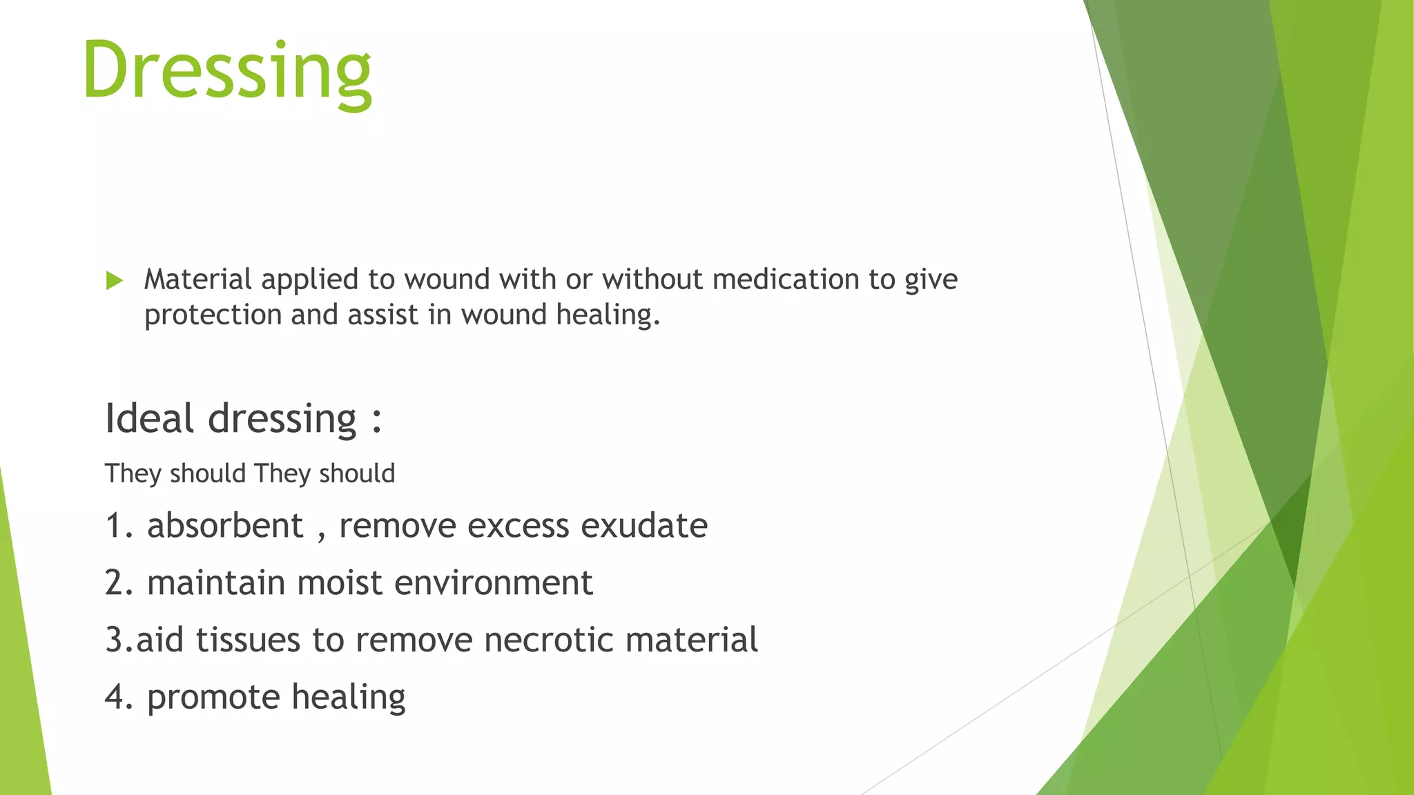
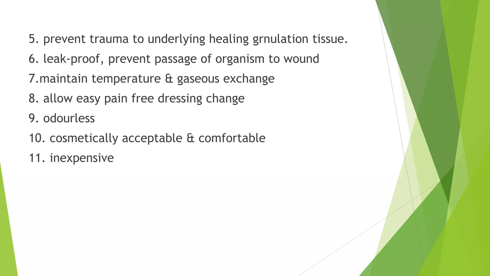
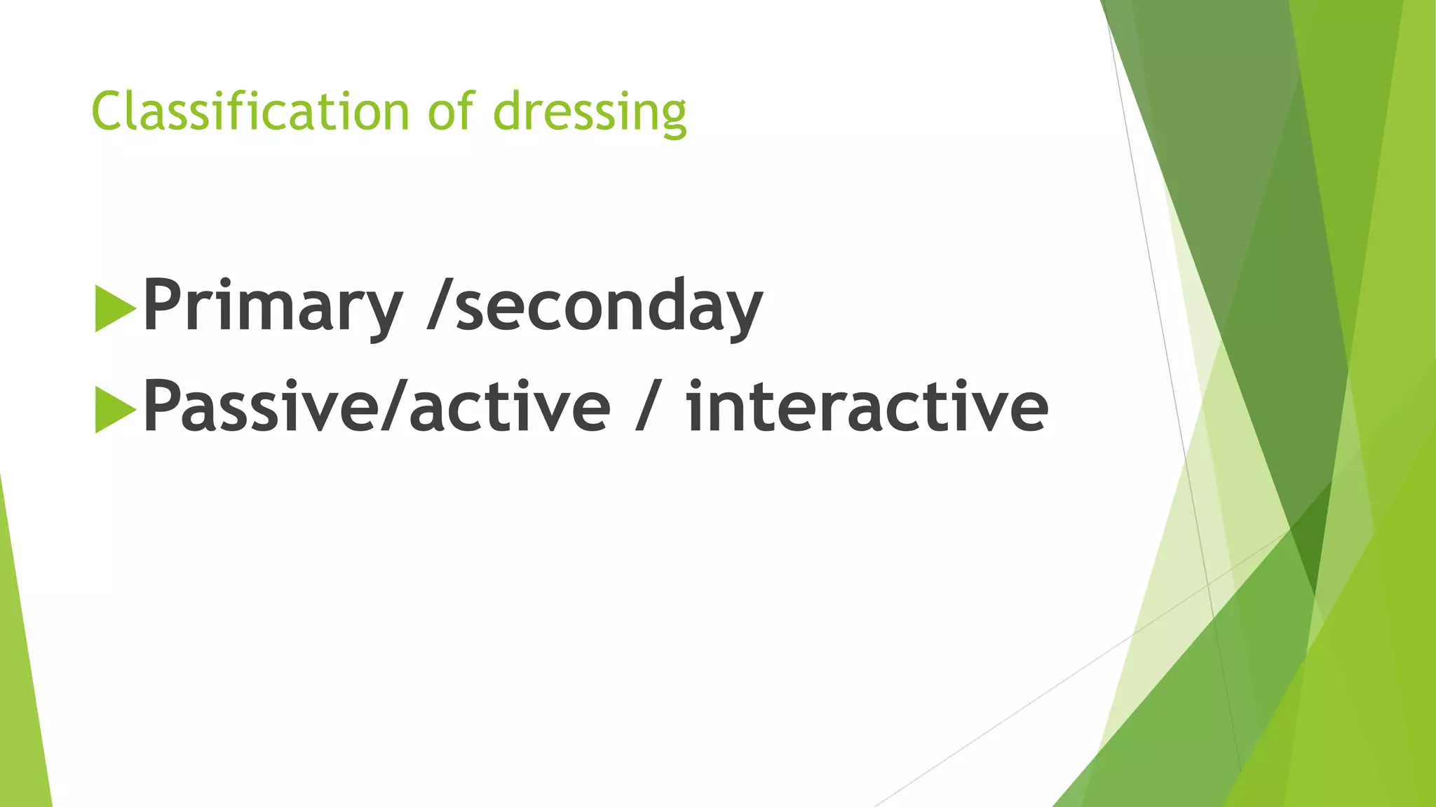
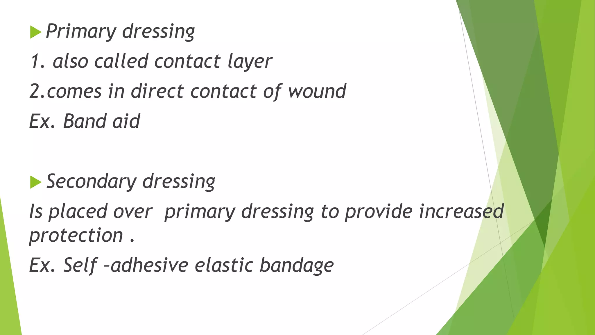
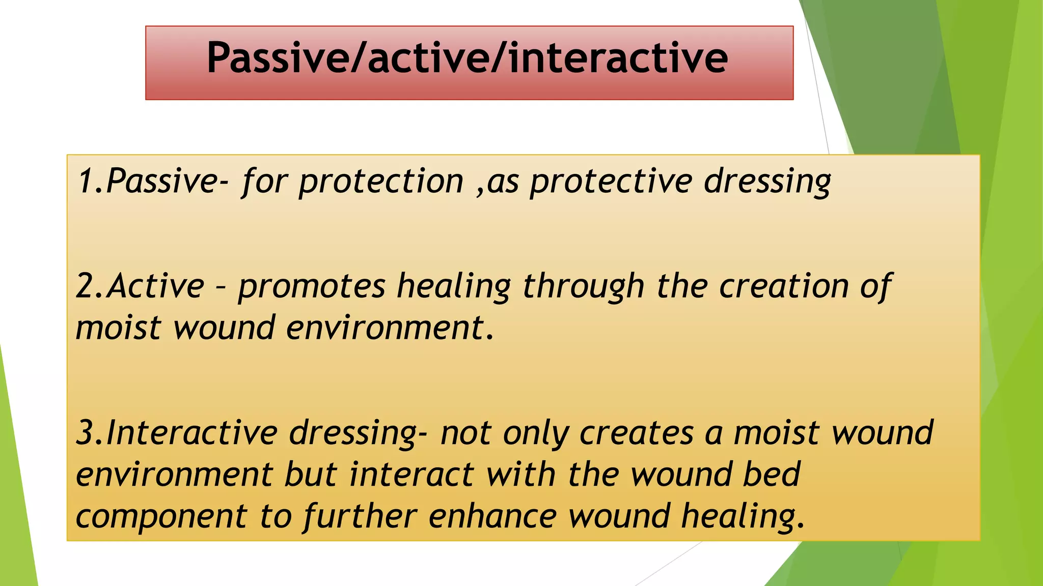
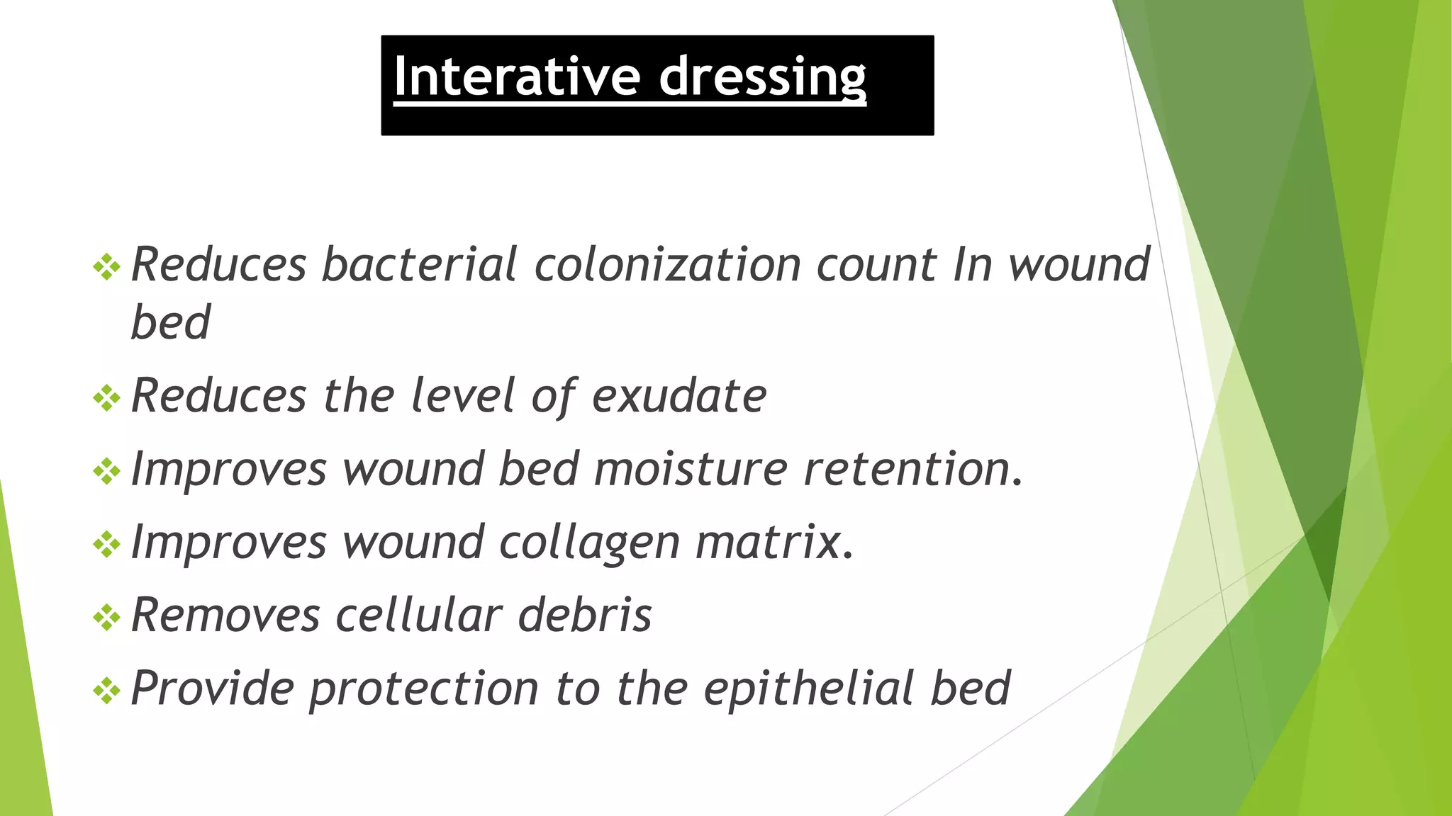
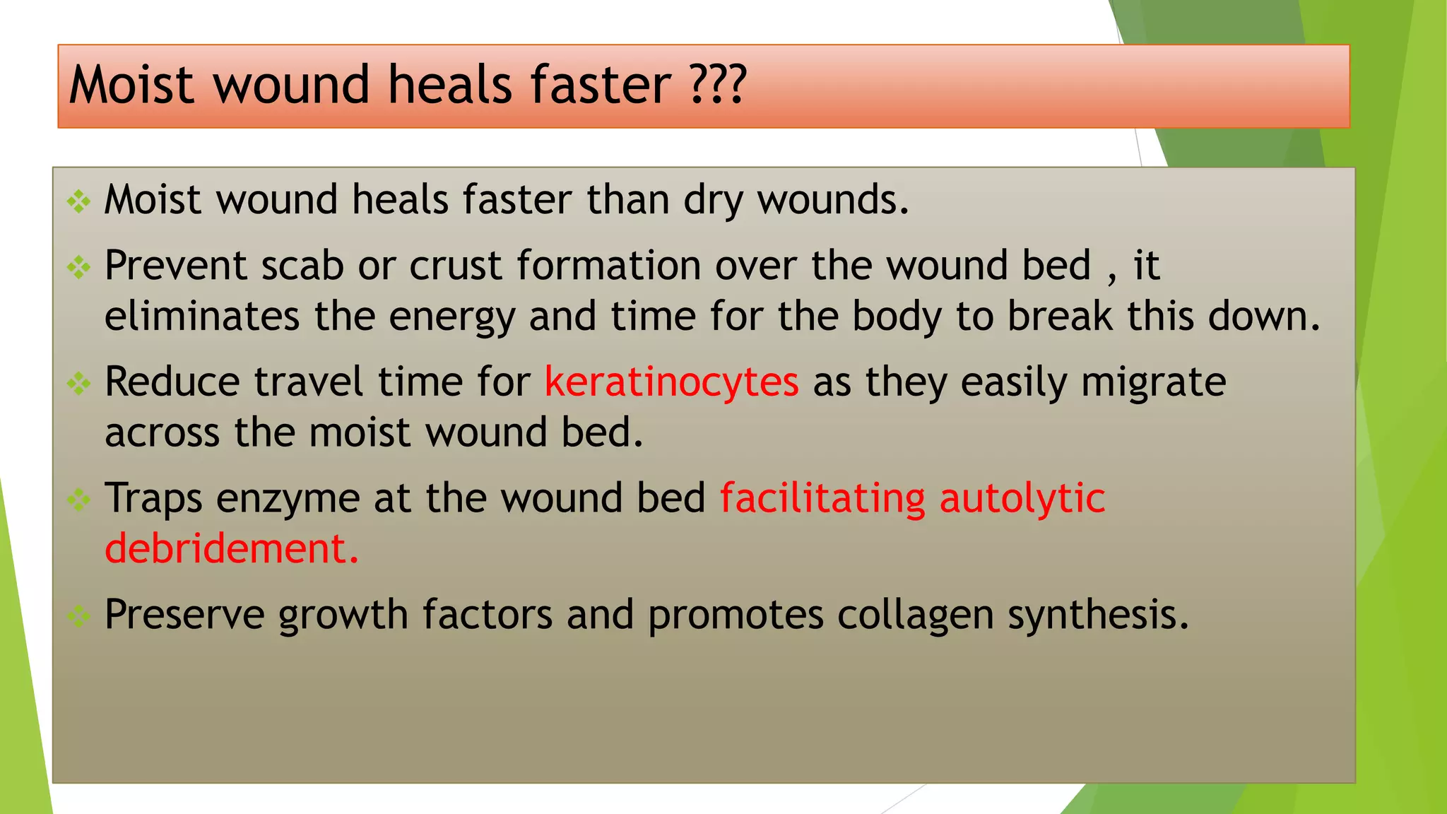
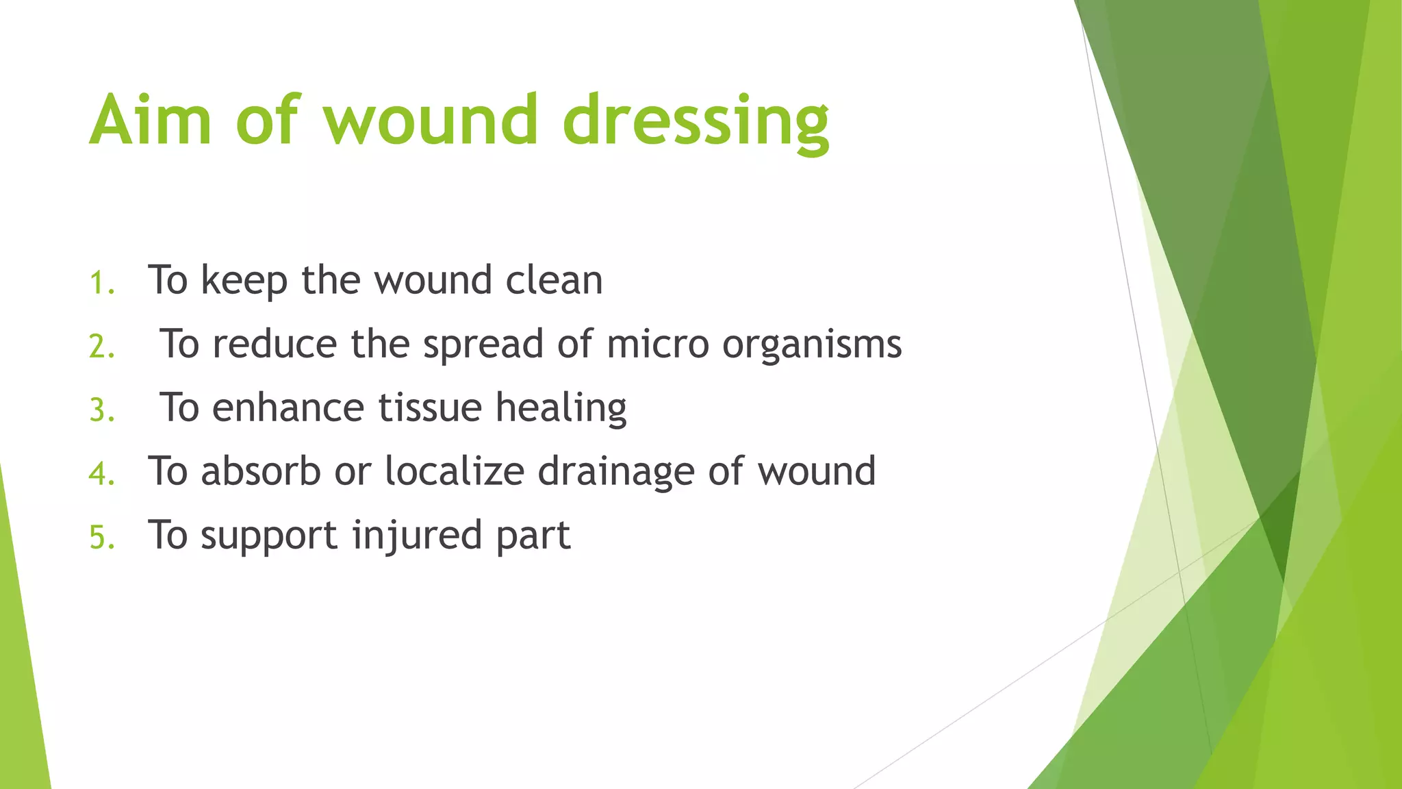
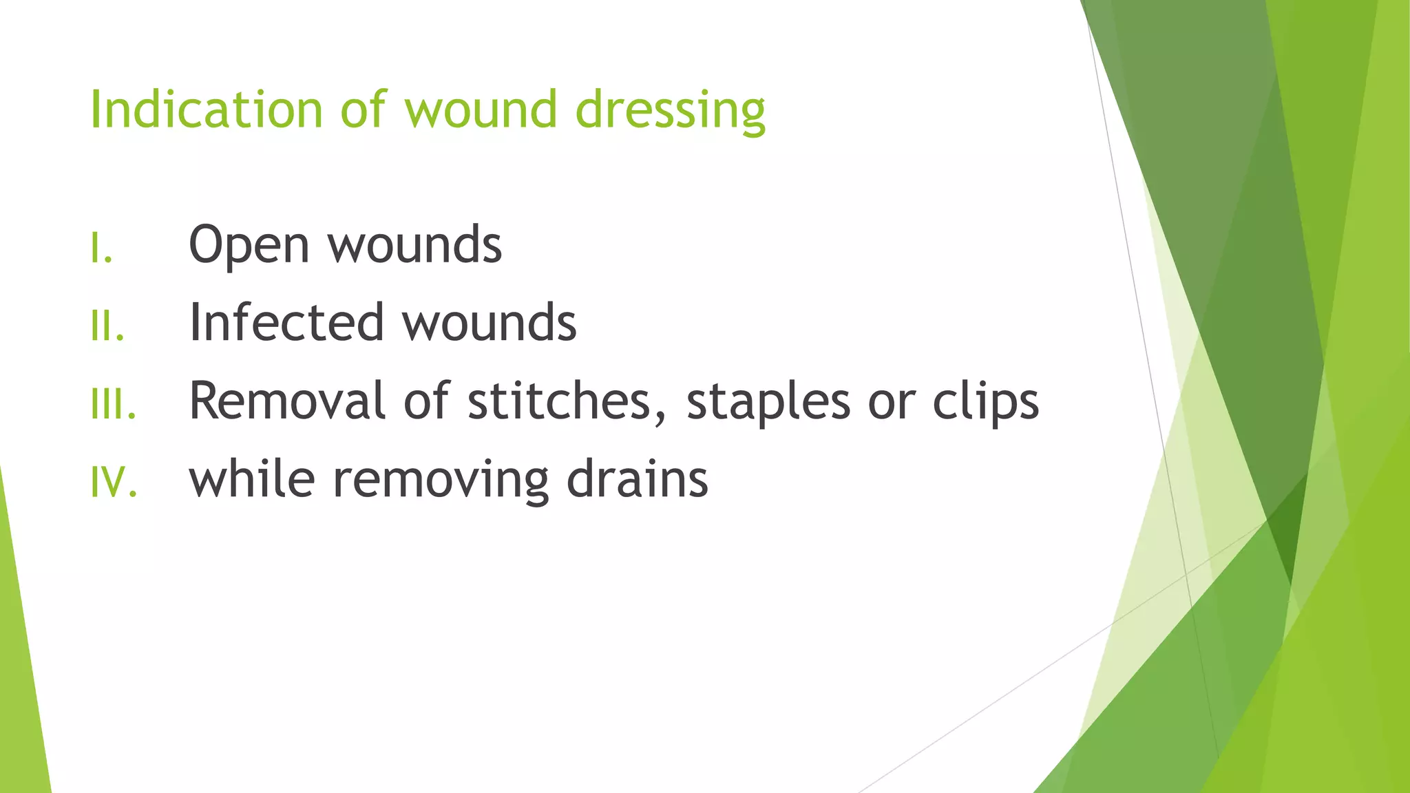
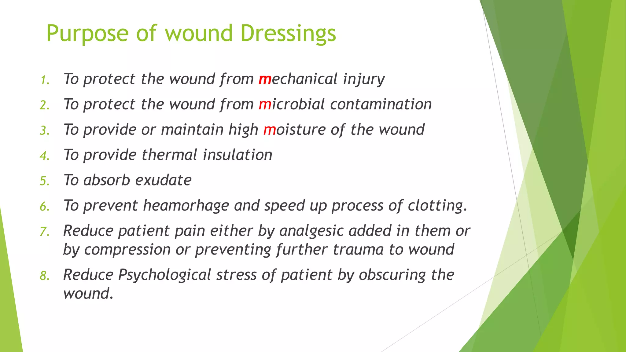
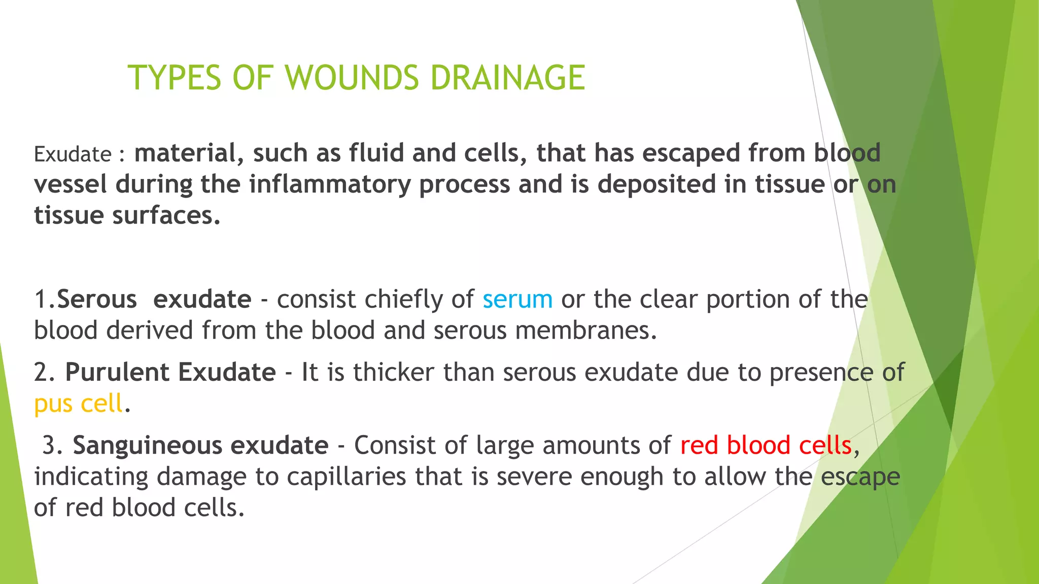
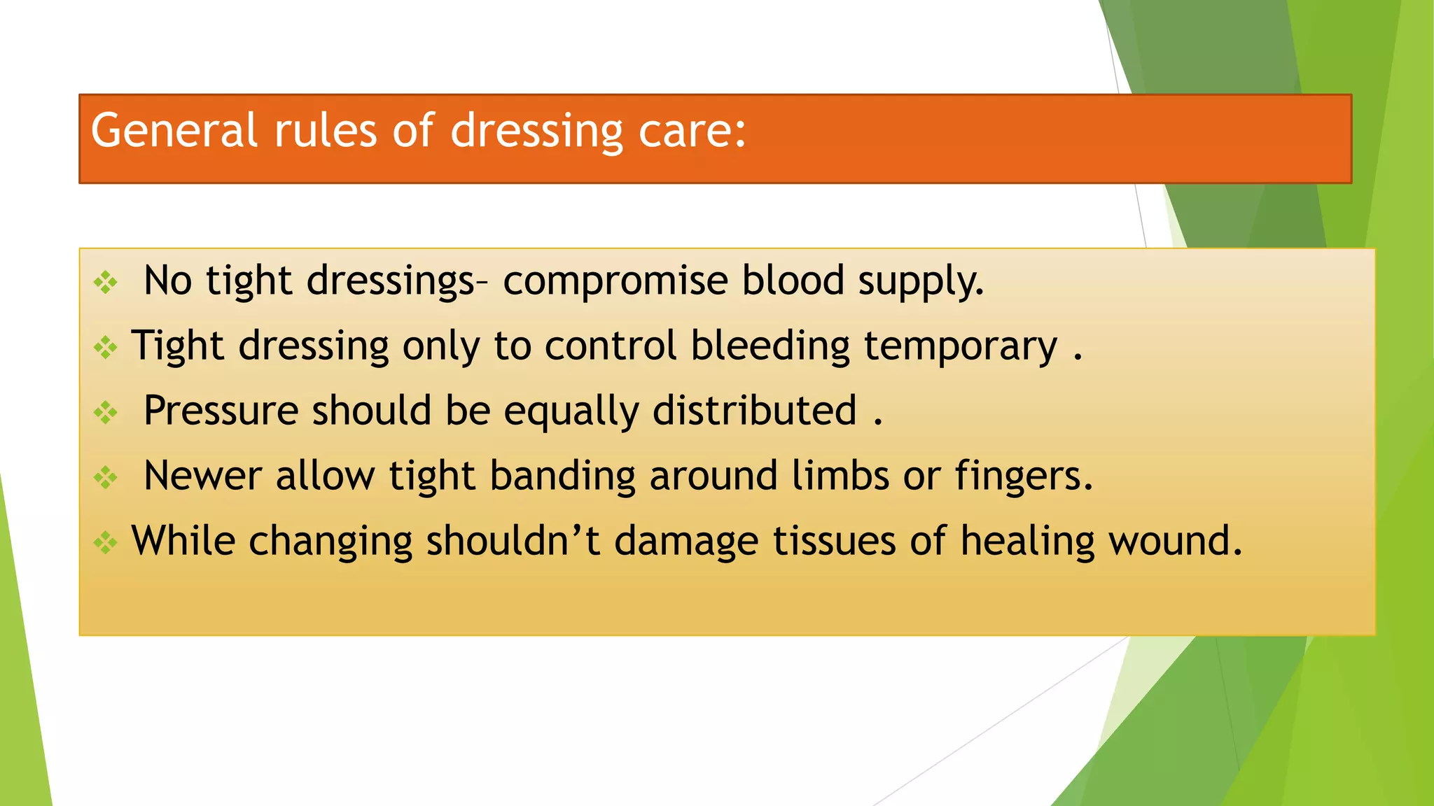
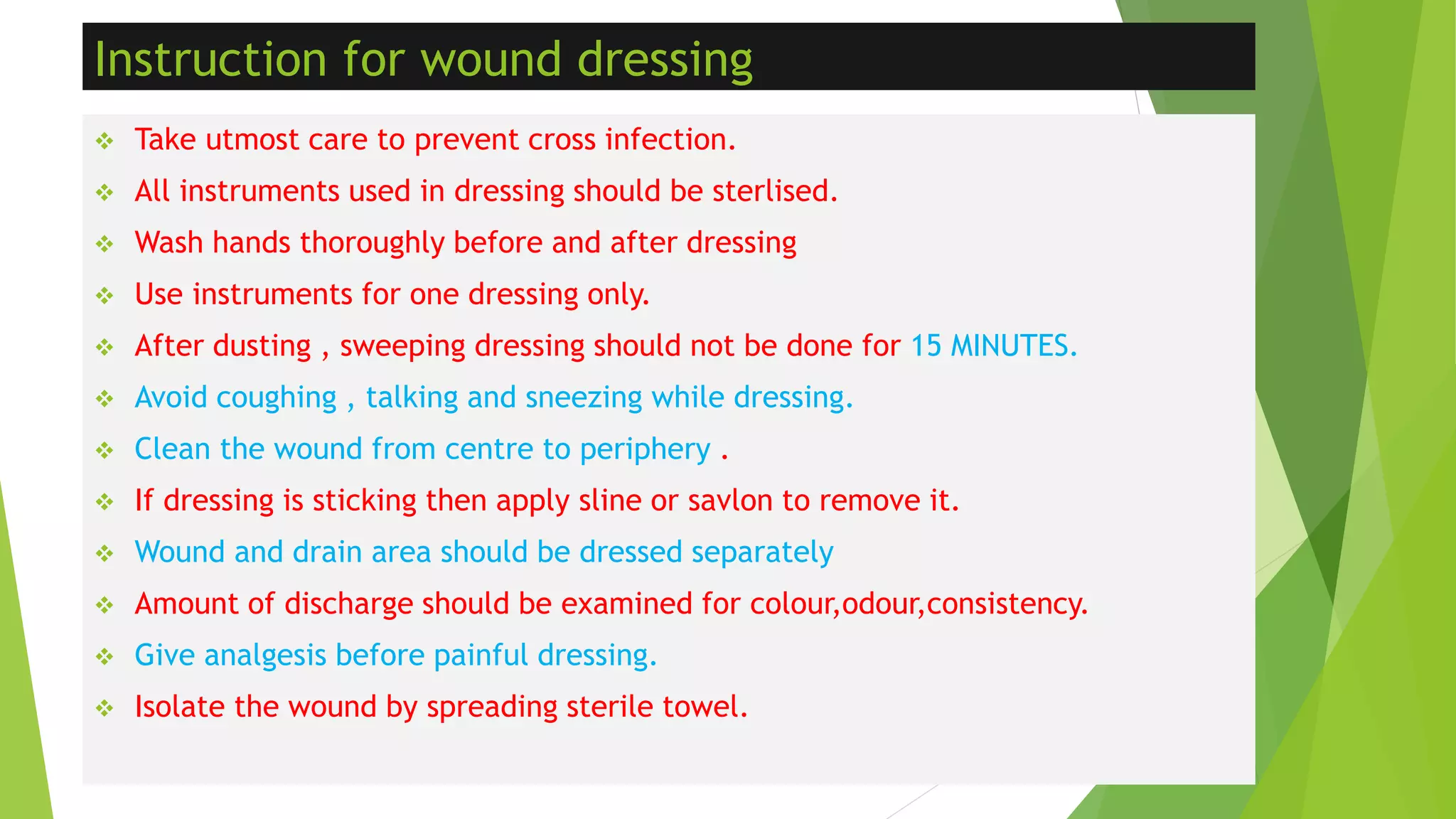
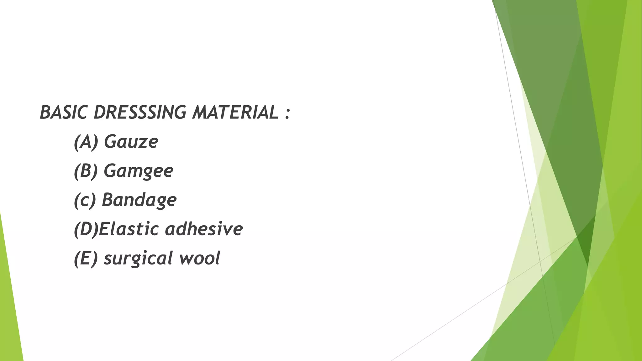
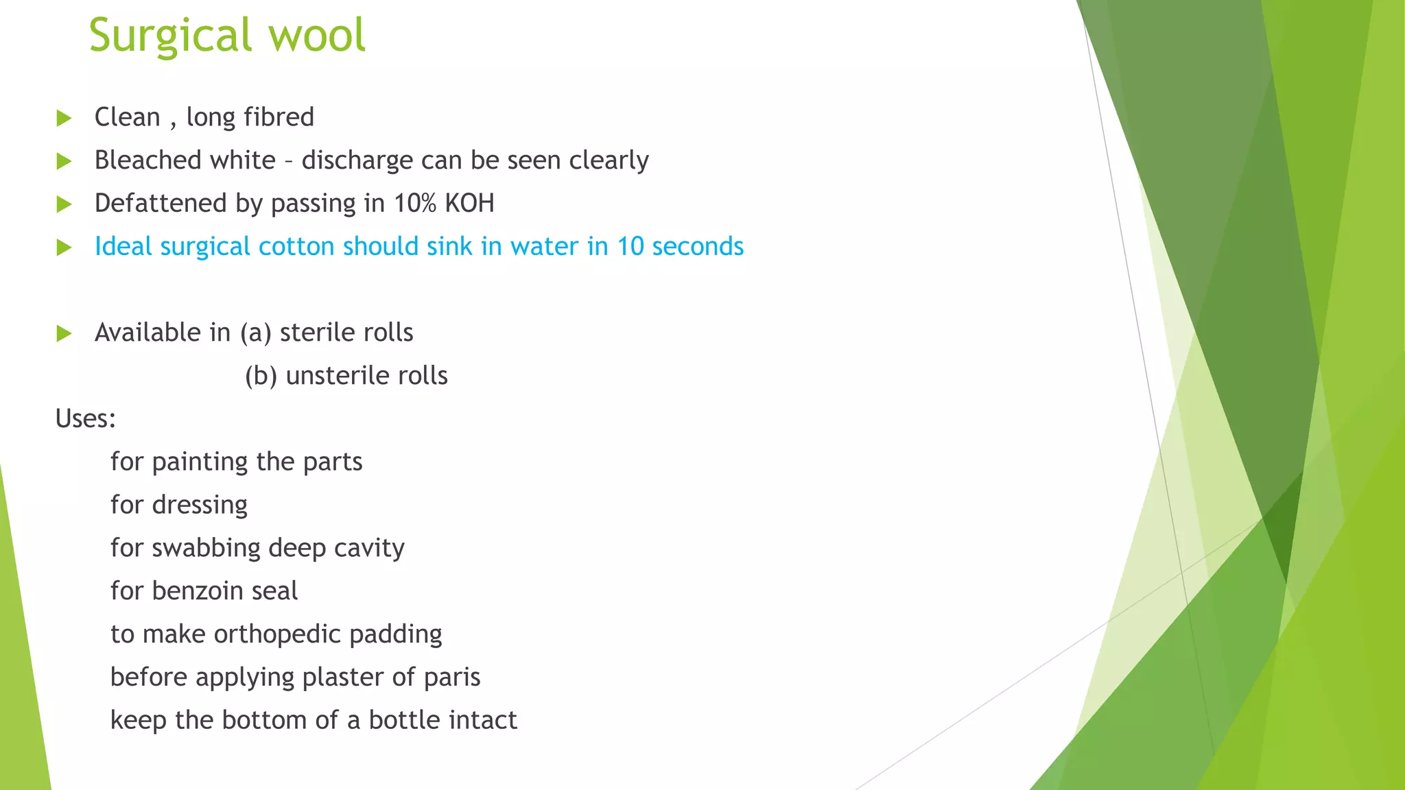
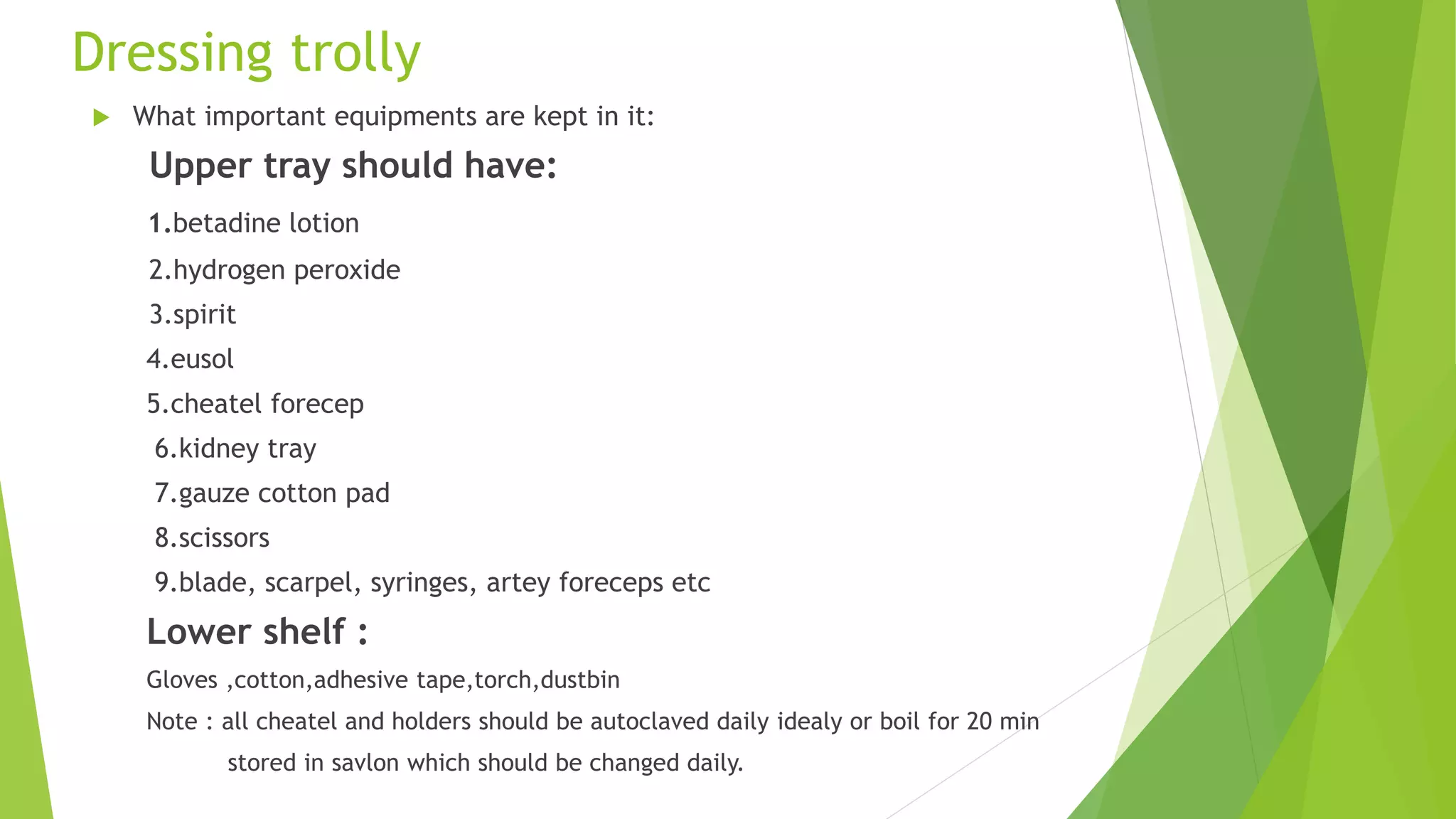
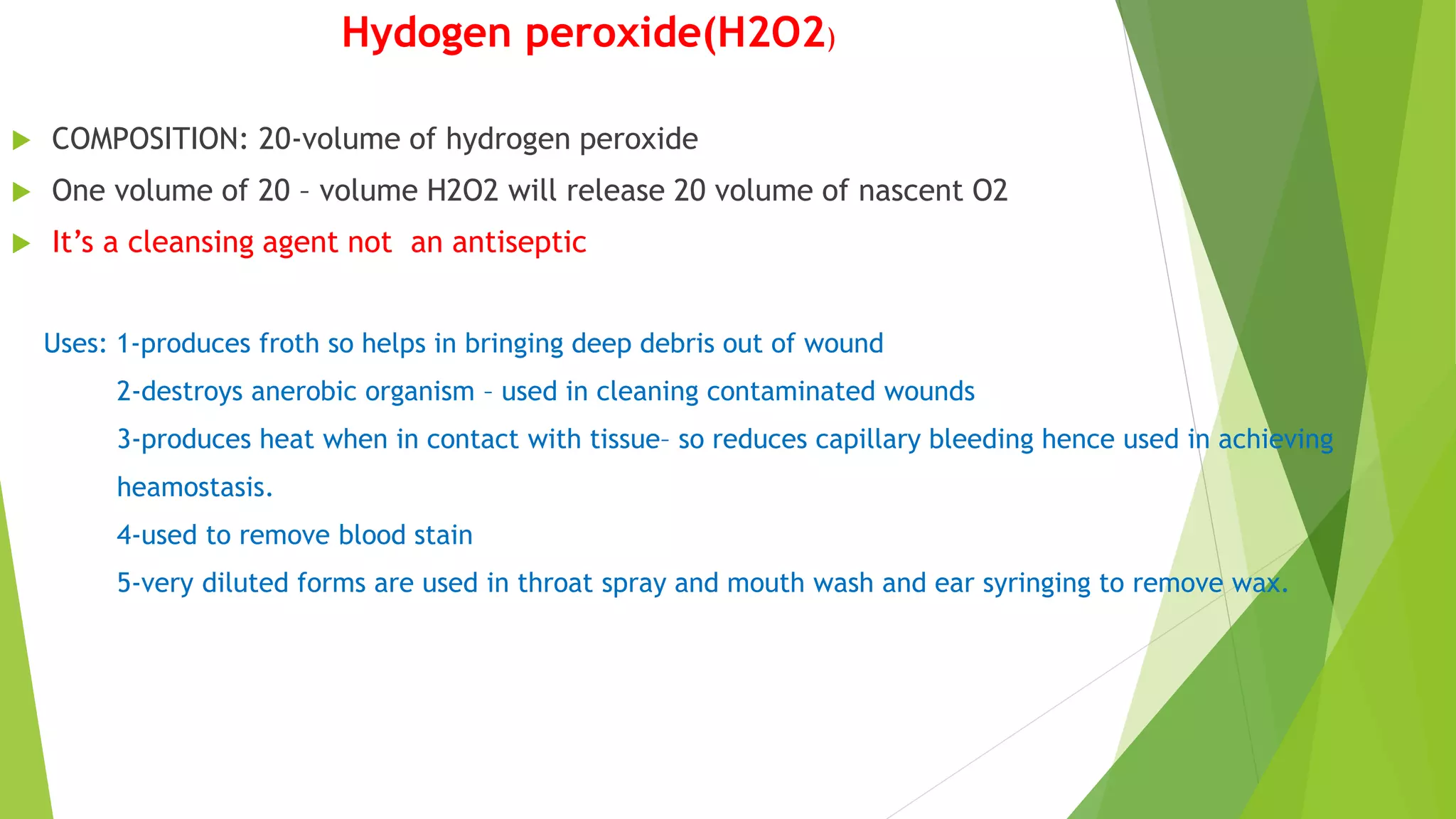
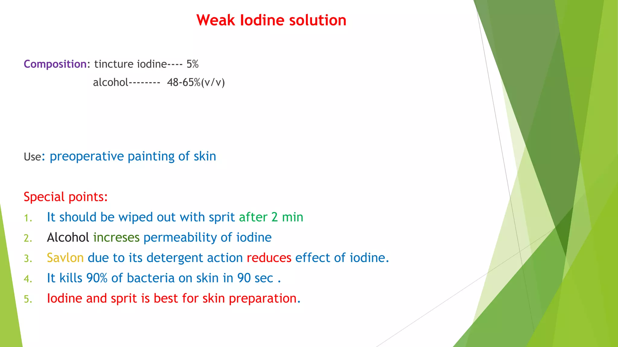
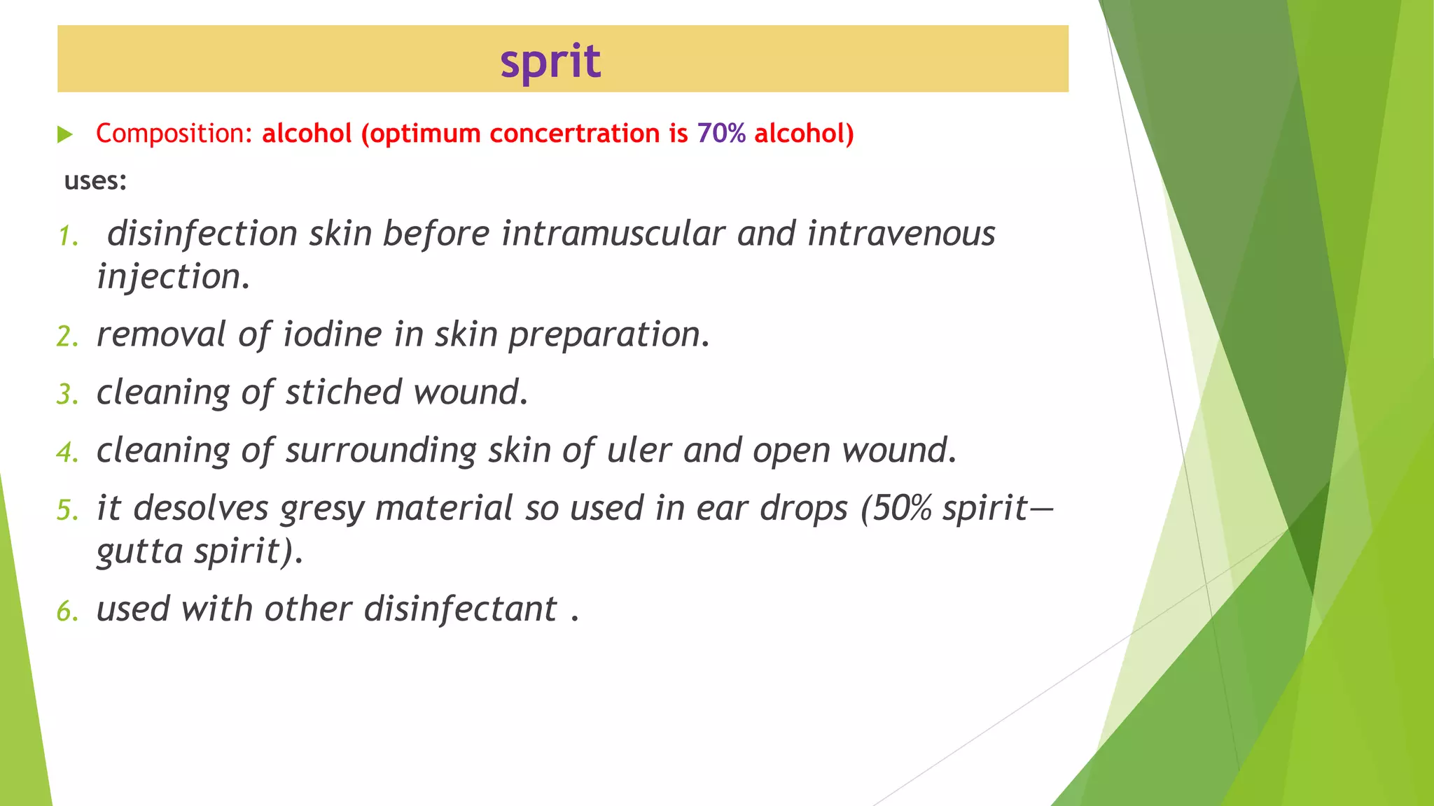
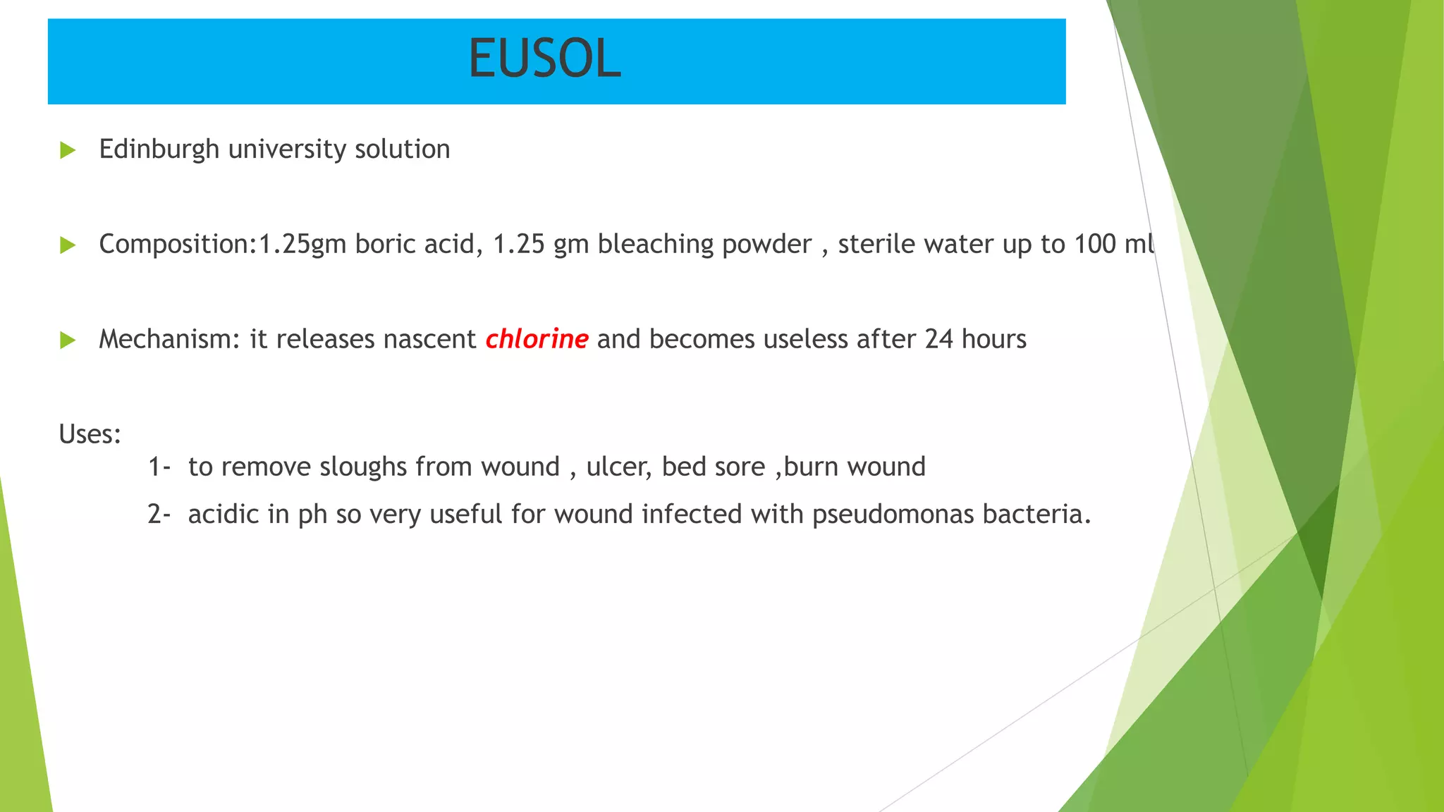
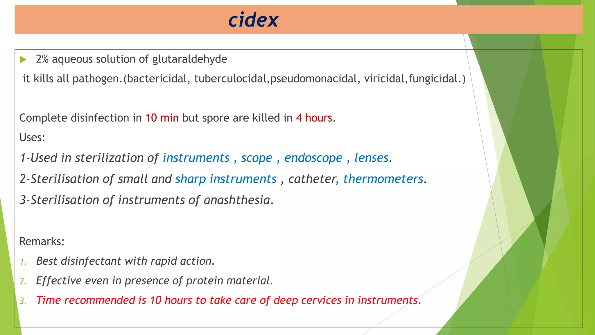
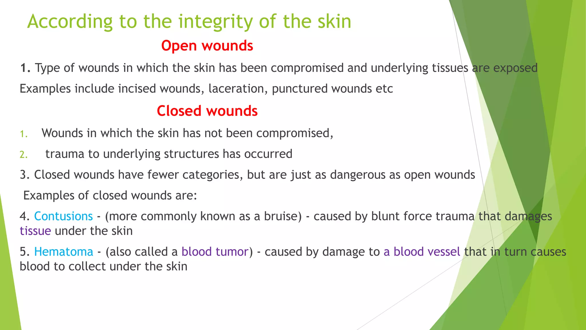
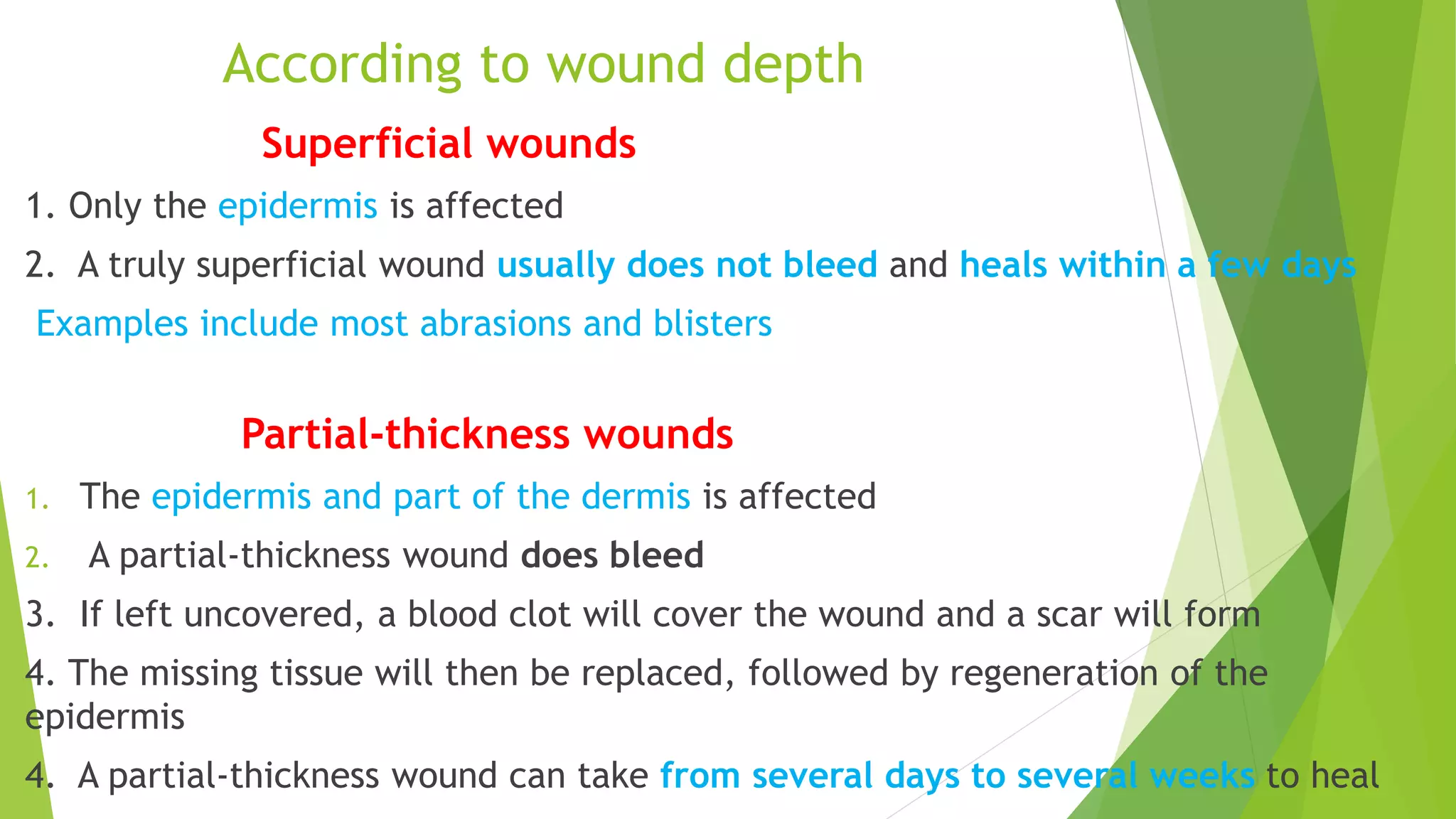
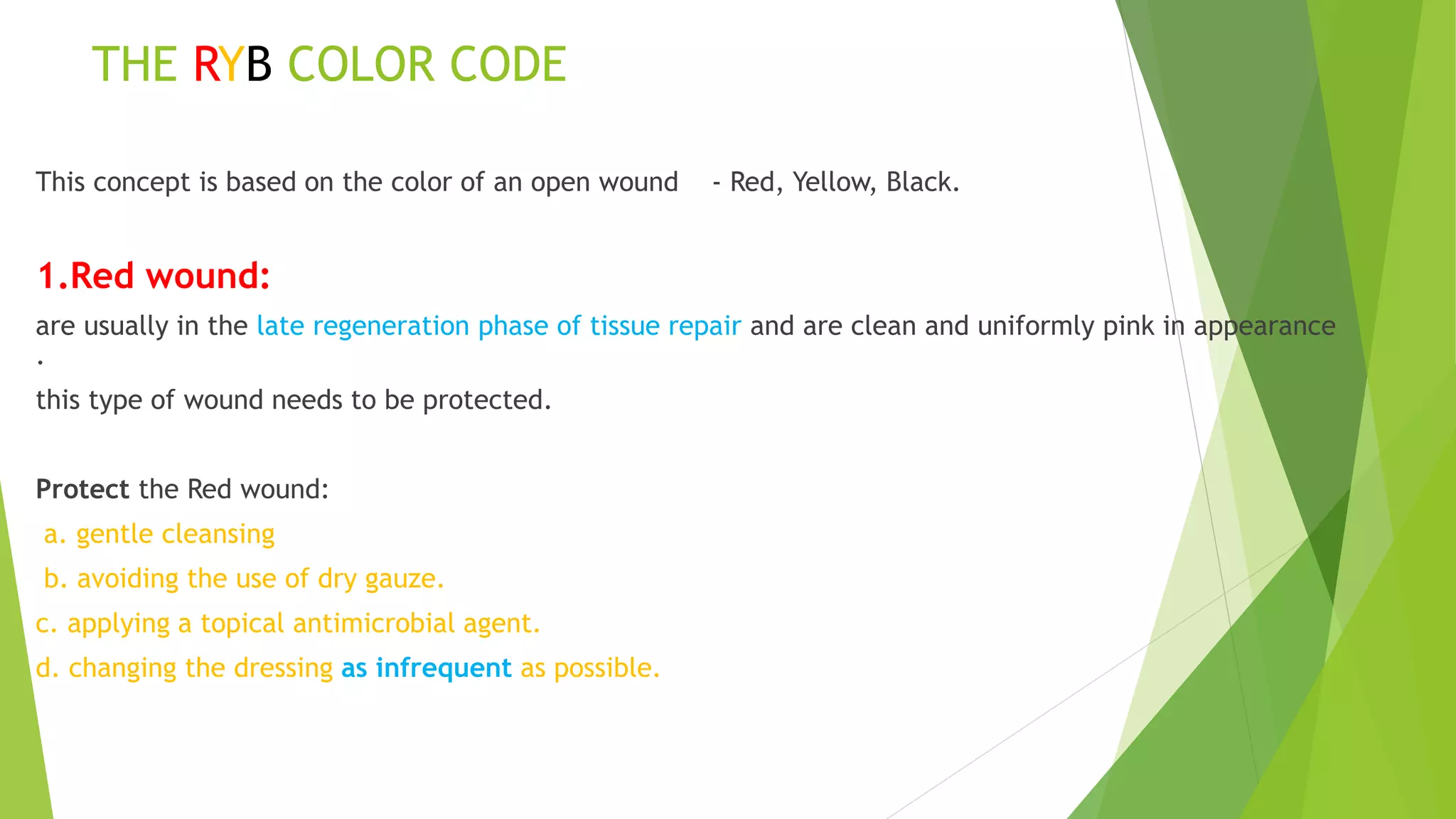
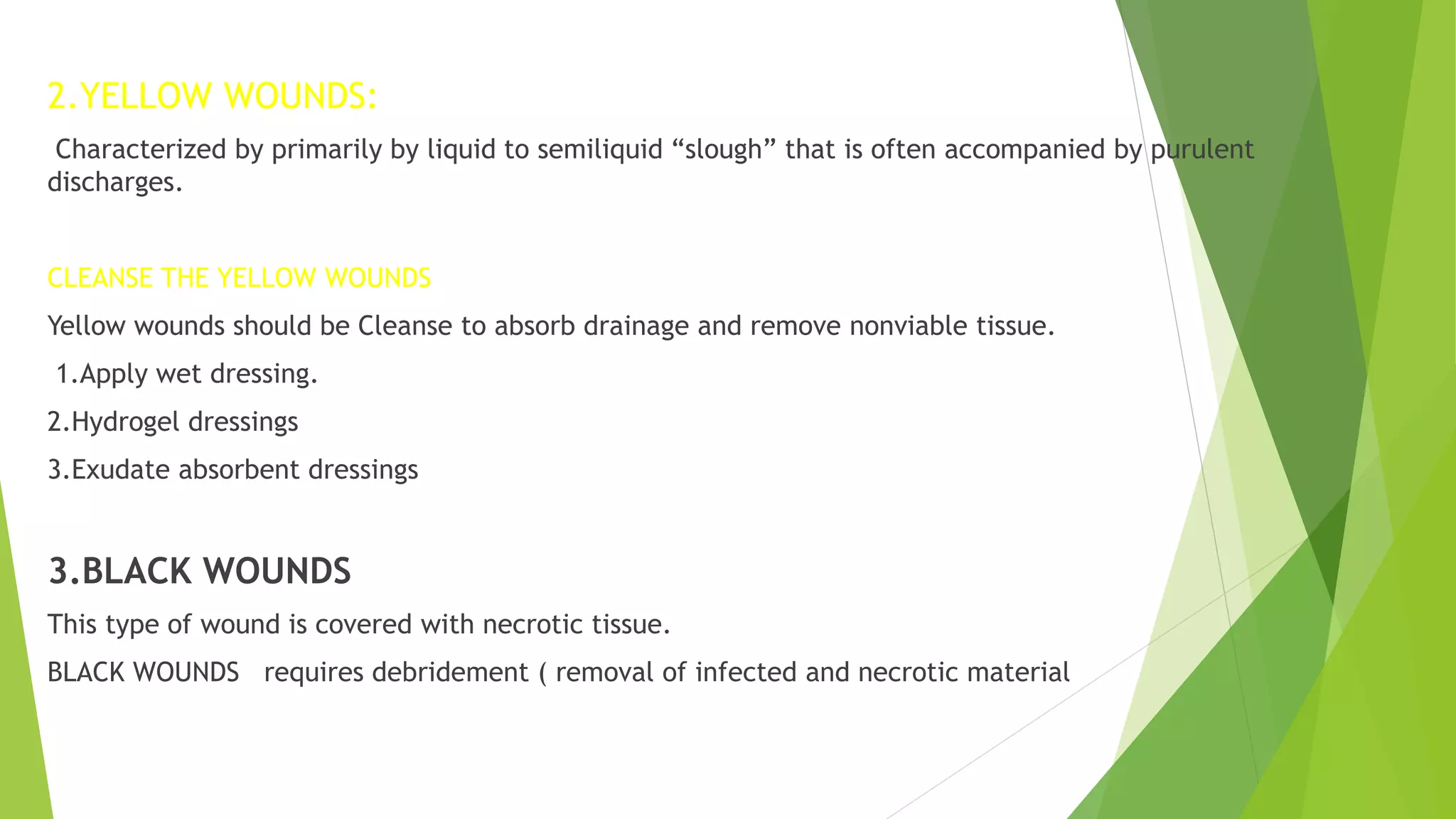
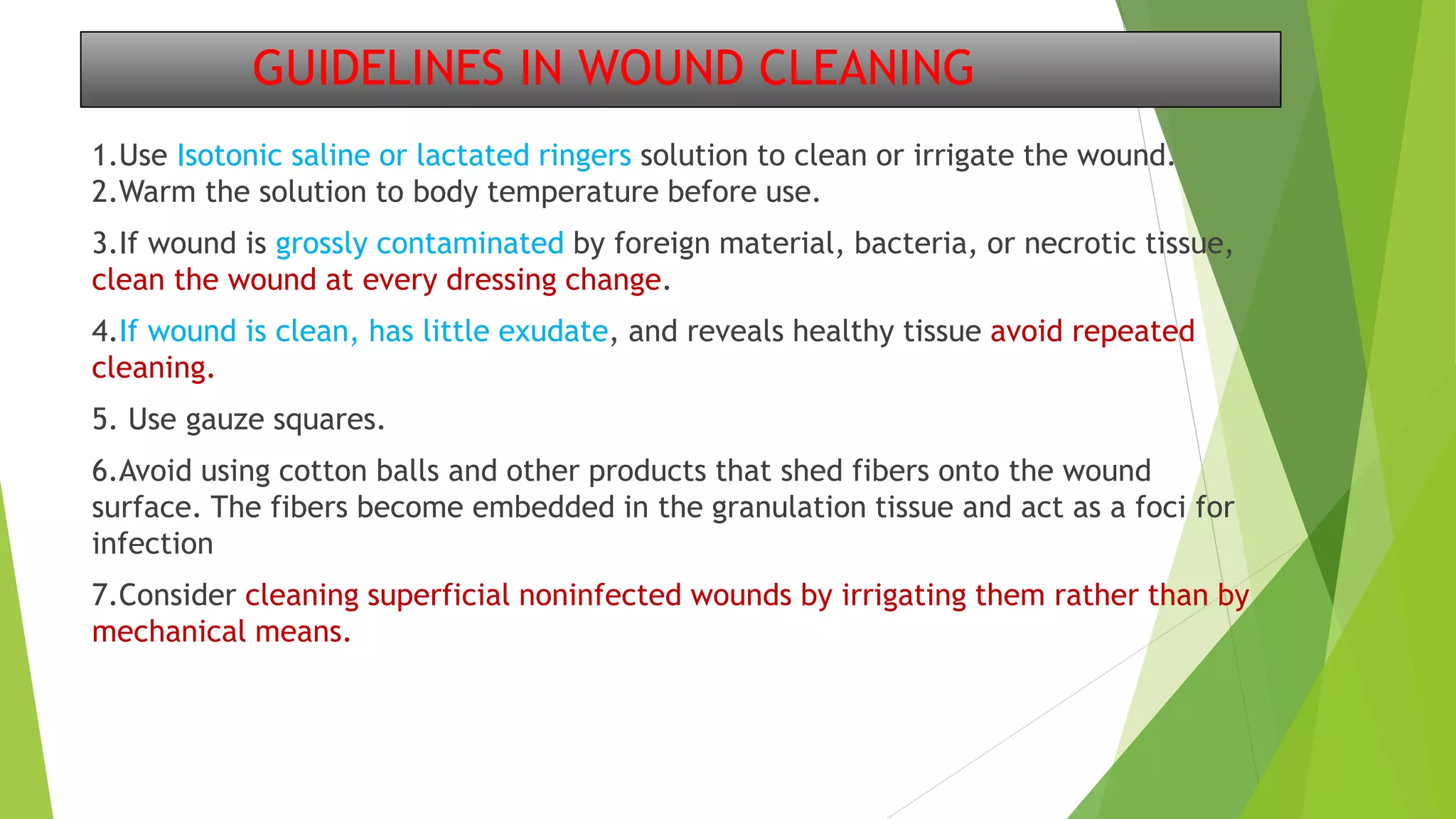
DRESSING
An artificial wound cover
Dr. SUNIL VISHWAKARMA
K.B.B. HOSPITAL , BANDRA WEST
MUMBAI
Introduction
Antisepsis: is a process of destruction of disease causing
microorganism to prevent infection on patient body surface.
it could be bacteriostatic(limiting growth of bacteria) or bacteriocidal procedure (kills bacteria) .
Disinfection: process of killing pathogenic organism from
innominate objects like instruments.
Asepsis: practice to reduce or eliminate infection causing agents from
entering in environment of patient.
Dressing
 Material applied to wound with or without medication to give
protection and assist in wound healing.
Ideal dressing :
They should They should
1. absorbent , remove excess exudate
2. maintain moist environment
3.aid tissues to remove necrotic material
4. promote healing
5. prevent trauma to underlying healing grnulation tissue.
6. leak-proof, prevent passage of organism to wound
7.maintain temperature & gaseous exchange
8. allow easy pain free dressing change
9. odourless
10. cosmetically acceptable & comfortable
11. inexpensive
What is ideal topical agent ?
Ideal topical agent should meet following:
1. Wide antibacterial action
2. Non toxic
3. Non irritant
4. Good penetration
5. economical
Classification of dressing
Primary /seconday
Passive/active / interactive
 Primary dressing
1. also called contact layer
2.comes in direct contact of wound
Ex. Band aid
 Secondary dressing
Is placed over primary dressing to provide increased
protection .
Ex. Self –adhesive elastic bandage
Passive/active/interactive
1.Passive- for protection ,as protective dressing
2.Active – promotes healing through the creation of
moist wound environment.
3.Interactive dressing- not only creates a moist wound
environment but interact with the wound bed
component to further enhance wound healing.
Interative dressing
 Reduces bacterial colonization count In wound
bed
 Reduces the level of exudate
 Improves wound bed moisture retention.
 Improves wound collagen matrix.
 Removes cellular debris
 Provide protection to the epithelial bed
Moist wound heals faster ???
 Moist wound heals faster than dry wounds.
 Prevent scab or crust formation over the wound bed , it
eliminates the energy and time for the body to break this down.
 Reduce travel time for keratinocytes as they easily migrate
across the moist wound bed.
 Traps enzyme at the wound bed facilitating autolytic
debridement.
 Preserve growth factors and promotes collagen synthesis.
Aim of wound dressing
1. To keep the wound clean
2. To reduce the spread of micro organisms
3. To enhance tissue healing
4. To absorb or localize drainage of wound
5. To support injured part
Indication of wound dressing
I. Open wounds
II. Infected wounds
III. Removal of stitches, staples or clips
IV. while removing drains
Purpose of wound Dressings
1. To protect the wound from mechanical injury
2. To protect the wound from microbial contamination
3. To provide or maintain high moisture of the wound
4. To provide thermal insulation
5. To absorb exudate
6. To prevent heamorhage and speed up process of clotting.
7. Reduce patient pain either by analgesic added in them or
by compression or preventing further trauma to wound
8. Reduce Psychological stress of patient by obscuring the
wound.
Layers of dressing
There are three layers in dressing which are fron deep to superficial are as
follows:
First layer: covers the wound and collects blood , discharge, fibrin, debris.
Second layer: collects excess drainage
Third layer: protects the wound from external contamination.
TYPES OF WOUNDS DRAINAGE
Exudate : material, such as fluid and cells, that has escaped from blood
vessel during the inflammatory process and is deposited in tissue or on
tissue surfaces.
1.Serous exudate - consist chiefly of serum or the clear portion of the
blood derived from the blood and serous membranes.
2. Purulent Exudate - It is thicker than serous exudate due to presence of
pus cell.
3. Sanguineous exudate - Consist of large amounts of red blood cells,
indicating damage to capillaries that is severe enough to allow the escape
of red blood cells.
General rules of dressing care:
 No tight dressings– compromise blood supply.
 Tight dressing only to control bleeding temporary .
 Pressure should be equally distributed .
 Newer allow tight banding around limbs or fingers.
 While changing shouldn’t damage tissues of healing wound.
Instruction for wound dressing
 Take utmost care to prevent cross infection.
 All instruments used in dressing should be sterlised.
 Wash hands thoroughly before and after dressing
 Use instruments for one dressing only.
 After dusting , sweeping dressing should not be done for 15 MINUTES.
 Avoid coughing , talking and sneezing while dressing.
 Clean the wound from centre to periphery .
 If dressing is sticking then apply sline or savlon to remove it.
 Wound and drain area should be dressed separately
 Amount of discharge should be examined for colour,odour,consistency.
 Give analgesis before painful dressing.
 Isolate the wound by spreading sterile towel.
BASIC DRESSSING MATERIAL :
(A) Gauze
(B) Gamgee
(c) Bandage
(D)Elastic adhesive
(E) surgical wool
Surgical wool
 Clean , long fibred
 Bleached white – discharge can be seen clearly
 Defattened by passing in 10% KOH
 Ideal surgical cotton should sink in water in 10 seconds
 Available in (a) sterile rolls
(b) unsterile rolls
Uses:
for painting the parts
for dressing
for swabbing deep cavity
for benzoin seal
to make orthopedic padding
before applying plaster of paris
keep the bottom of a bottle intact
Gauze piece
Piece of woven surgical cotton
Types :
(a)closed:16x16 fibers/sq inch
used in making bandage
(b)open: 8x8 fibers/sq inch
in making peanuts- blunt dissection
Uses:
1. dressing
2. heamostatic purpose
3. painting the parts
4. tissue dissection
5. drainage and package of absess cavity.
Surgical pad
Made by wrapping cotton within gauze piece
Usually have size of 4x6 sq inch
Uses:
1. in dressing of drained abscess
2. in dressing of burns and ulcers
3. padding to avoid direct pressure and trauma to wound
Gamgee
 A layer of cotton is sandwiched between gauze piece and rolled
 Uses:
1. in dressing of wound with discharge
2.in dressing of burns patients
Bandage
 Soft ,light, porus
turns:
(a) circular:horizontally across the part
mainly used in begening to secure the bandage
it interfere with circulation so should be avoided in limb .
(b)spiral:spiral fashion across the limb
each turn should cover half to 2/3 of preceding turn
used in parts with uniform thickness e.g. arm , fingers
(c)reverse spiral: reverse turn
useful when thickness is not uniform e.g. forearm
(d)figure of eight : over joints
(e)spica : type of figure of eight bandage but one loop is much larger than other e.g. hip spica
(f)recurrent : series of alternating turns are taken e.g. amputation stump dressing
Dressing trolly
 What important equipments are kept in it:
Upper tray should have:
1.betadine lotion
2.hydrogen peroxide
3.spirit
4.eusol
5.cheatel forecep
6.kidney tray
7.gauze cotton pad
8.scissors
9.blade, scarpel, syringes, artey foreceps etc
Lower shelf :
Gloves ,cotton,adhesive tape,torch,dustbin
Note : all cheatel and holders should be autoclaved daily idealy or boil for 20 min
stored in savlon which should be changed daily.
Hydogen peroxide(H2O2)
 COMPOSITION: 20-volume of hydrogen peroxide
 One volume of 20 – volume H2O2 will release 20 volume of nascent O2
 It’s a cleansing agent not an antiseptic
Uses: 1-produces froth so helps in bringing deep debris out of wound
2-destroys anerobic organism – used in cleaning contaminated wounds
3-produces heat when in contact with tissue– so reduces capillary bleeding hence used in achieving
heamostasis.
4-used to remove blood stain
5-very diluted forms are used in throat spray and mouth wash and ear syringing to remove wax.
Weak Iodine solution
Composition: tincture iodine---- 5%
alcohol-------- 48-65%(v/v)
Use: preoperative painting of skin
Special points:
1. It should be wiped out with sprit after 2 min
2. Alcohol increses permeability of iodine
3. Savlon due to its detergent action reduces effect of iodine.
4. It kills 90% of bacteria on skin in 90 sec .
5. Iodine and sprit is best for skin preparation.
sprit
 Composition: alcohol (optimum concertration is 70% alcohol)
uses:
1. disinfection skin before intramuscular and intravenous
injection.
2. removal of iodine in skin preparation.
3. cleaning of stiched wound.
4. cleaning of surrounding skin of uler and open wound.
5. it desolves gresy material so used in ear drops (50% spirit—
gutta spirit).
6. used with other disinfectant .
EUSOL
 Edinburgh university solution
 Composition:1.25gm boric acid, 1.25 gm bleaching powder , sterile water up to 100 ml
 Mechanism: it releases nascent chlorine and becomes useless after 24 hours
Uses:
1- to remove sloughs from wound , ulcer, bed sore ,burn wound
2- acidic in ph so very useful for wound infected with pseudomonas bacteria.
cidex
 2% aqueous solution of glutaraldehyde
it kills all pathogen.(bactericidal, tuberculocidal,pseudomonacidal, viricidal,fungicidal.)
Complete disinfection in 10 min but spore are killed in 4 hours.
Uses:
1-Used in sterilization of instruments , scope , endoscope , lenses.
2-Sterilisation of small and sharp instruments , catheter, thermometers.
3-Sterilisation of instruments of anashthesia.
Remarks:
1. Best disinfectant with rapid action.
2. Effective even in presence of protein material.
3. Time recommended is 10 hours to take care of deep cervices in instruments.
Glycerine magnesium sulphate
Mixture of mgso4 crystals and glycerine (sterile)
Solution is warmed and then cooled
Uses:
 As it is hygroscopic it is used to reduce edema in cases of
1. cellulitis
2. paraphimosis
3. piles
4. Mgso4 enema in order to reduce intracranial pressure.
Ether
evaporable and highly inflammable , makes skin surface cool.
Uses:
surface and inhalational anashthesia.
Cleaning dirty wound as it dissolve greasy material.
Acriflavin
 available in crystals
0.5-2% solution made with water.
Yellow in colour
Uses.1:mild antiseptic and astringent on ulcers with purulent discharge.
2:effective in wound with gram negative bacteria.
Silver nitrate
Solution with dilution ranging from 1:100 to 1:10000
Kept in dark as on exposure to light it gets destroyed .
Uses:
1.chemical cauterization of warts and hyperpigmented tissue.
2. in dressing of burns patients.
3. in bladder irrigation in heamaturia due to cauterizing effect.
Providone-iodine (betadine)
 Available as : Solution-5-10% ; scrub 7.5% w/v; ointment , mouth wash .
 Iodine is good germicide , effective against viruses, bacteria , protozoa ,yeast , fungi
 But its frequent use is contraindicated due to :
insolubility, staining, irritability, instability--- overcome by mixing it with polyvinyl pyrrolidone
(combination called providone iodine )
 Providone iodine retains bactericidal activity of iodine and lacks toxicity.
 water soluble and eliminates toxic effects of iodine.
 NOTE: Loss of colour OF SOLUTION is accompanied by loss of bactericidal activity .
Advantage: broad spectrum antiseptic
immediate action – nonselective
Film formimg
safe
Uses:
(application Is painful due to its irritant action)
 Used in wound with granulation tissue after eschar sepration .
 mouth rinse and gargles .
 in irrigation of bladder and peritoneum , pyothorax.
 Shuld not be used on superficial burns which are highly painful.
WOUND
A wound is a type of physical trauma whereby the integrity of skin or of any tissue gets
compromised .
Also called break in continuity of mucus membrane, skin , bone or any body tissue caused by
physical chemical and biological insult.
Etiology of wound :
1.Blunt trauma: : RTA
fall
assault
sport injuries and animal bite
2.Penetrating trauma: stab and gun shot injuries
3.Surgical insult: wound caused by surgical procedure
4.Burn injuries: chemical , thermal , electrical , radiation and cold burn
Wound classification
According to the etiology
1. Surgical wounds :These are wounds caused by surgical procedure
2. Penetrating wounds : Wounds caused by penetrating trauma
3. Blunt wounds : Wounds caused by blunt trauma
4. Burn wounds : Wounds caused by burn injuries
According to Rank-Wakefield classification system
Tidy wounds
 These are wounds inflicted by sharp instruments and contain no devitalized tissue
 Such wounds can be closed primarily with the expectation of quite primary healing
 They are usually single with clean cut
 Associated fractures are uncommon
 Examples: surgical incisions, cuts from glass and knife wounds
Untidy wounds
 These are wounds resulting from crushing, avulsion,burns, and contain devitalized tissue
 They are usually multiple and irregular
 Commonly associated with fractures
 Such wounds can not be closed primarily and therefore should be allowed to heal by second
intention
According to the duration of the wound healing
1:Acute wounds
 Acute wounds are wounds that usually heal in the anticipated time frame
 Duration of the wound: days to few weeks
 Examples are wounds acquired as a result of trauma or an operative procedure
2:Chronic wounds
 Wounds that fail to heal in the anticipated time frame and often reoccur
 Duration of the wound ⇒ > 4 weeks to 3 months
 Wounds occur as a result of an underlying condition such as extended pressure on the tissues,
poor circulation, or even poor nutrition
 Pressure ulcers, venous leg ulcers, and diabetic foot ulcers are examples
According to the integrity of the skin
Open wounds
1. Type of wounds in which the skin has been compromised and underlying tissues are exposed
Examples include incised wounds, laceration, punctured wounds etc
Closed wounds
1. Wounds in which the skin has not been compromised,
2. trauma to underlying structures has occurred
3. Closed wounds have fewer categories, but are just as dangerous as open wounds
Examples of closed wounds are:
4. Contusions - (more commonly known as a bruise) - caused by blunt force trauma that damages
tissue under the skin
5. Hematoma - (also called a blood tumor) - caused by damage to a blood vessel that in turn causes
blood to collect under the skin
According to wound depth
Superficial wounds
1. Only the epidermis is affected
2. A truly superficial wound usually does not bleed and heals within a few days
Examples include most abrasions and blisters
Partial-thickness wounds
1. The epidermis and part of the dermis is affected
2. A partial-thickness wound does bleed
3. If left uncovered, a blood clot will cover the wound and a scar will form
4. The missing tissue will then be replaced, followed by regeneration of the
epidermis
4. A partial-thickness wound can take from several days to several weeks to heal
Full-thickness wounds
1. A full-thickness wound involves the epidermis and the dermis
2. The underlying fatty tissue, bones, muscles, or tendons may also be
damaged
3. If full-thickness wounds cannot be sutured, the healing process will create
new tissue to fill the wound, followed by regeneration of the epidermis
4.The full-thickness wound takes longer time to heal than does a partial-
thickness wound, sometimes as long as several months
According to morphological characteristics
1.Bruises / contusion
These are closed wounds
Caused by blunt trauma that damage the tissue under the skin without breaking the skin
Characterized by skin discoloration due to bleeding into the tissues
Ex. Blows to the chest, abdomen, or head with a blunt instrument can cause contusions
2.Hematoma
These are also closed wounds caused by damage to a blood vessel that in turn causes blood to collect
under the skin
Initially this is fluid, but it will clot within minutes or hours ⇒later after few days the hematoma will
again liquefy → increased risk of secondary infection → pus formation
3.Abrasions
An abrasion is a shearing injury of the skin In which the surface is rubbed off
Most are superficial and will heal by epitheliazation
4.Lacerated wound
Caused by tearing of tissues
Wounds have irregular borders
Loss of tissue is limited to skin and s/c tissue
5.Crush wounds
Crush wounds are caused by a great or extreme amount of force applied over a
long period of time .
occur when a heavy object falls onto a person, splitting the skin and shattering or
tearing underlying structures
6.Penetrated wound
1. Caused by sharp pointed object.
2. Have relatively small opening.
3. May be very deep.
4. Infection/ foreign particles might have been carried deep
in to wound.
7.Perforating wound
1.Have two opening one of entrance one of exit ex. Gun shot
According to degree of contamination
1.Clean wounds
 No break in aseptic technique
 Incision is made under sterile condions
 No inflammation is encountered
 The respiratory tract, alimentary, genital or uninfected urinary tracts are
not entered
 Primary closure can be done. ex. Herniorrhaphy
2.Clean Contaminated wounds
 Operative wounds in which the respiratory, alimentary, genital or urinary
tract is entered under controlled conditions and without unusual
contamination .
3.Contaminated wounds
 Open, fresh or accidental wounds; operations with major breaks in sterile
technique or gross spillage from the gastrointestinal tract
4.Dirty or Infected wounds
 Old traumatic wounds with retained devitalized tissue and existing clinical
infection.
THE RYB COLOR CODE
This concept is based on the color of an open wound - Red, Yellow, Black.
1.Red wound:
are usually in the late regeneration phase of tissue repair and are clean and uniformly pink in appearance
.
this type of wound needs to be protected.
Protect the Red wound:
a. gentle cleansing
b. avoiding the use of dry gauze.
c. applying a topical antimicrobial agent.
d. changing the dressing as infrequent as possible.
2.YELLOW WOUNDS:
Characterized by primarily by liquid to semiliquid “slough” that is often accompanied by purulent
discharges.
CLEANSE THE YELLOW WOUNDS
Yellow wounds should be Cleanse to absorb drainage and remove nonviable tissue.
1.Apply wet dressing.
2.Hydrogel dressings
3.Exudate absorbent dressings
3.BLACK WOUNDS
This type of wound is covered with necrotic tissue.
BLACK WOUNDS requires debridement ( removal of infected and necrotic material
GUIDELINES IN WOUND CLEANING
1.Use Isotonic saline or lactated ringers solution to clean or irrigate the wound.
2.Warm the solution to body temperature before use.
3.If wound is grossly contaminated by foreign material, bacteria, or necrotic tissue,
clean the wound at every dressing change.
4.If wound is clean, has little exudate, and reveals healthy tissue avoid repeated
cleaning.
5. Use gauze squares.
6.Avoid using cotton balls and other products that shed fibers onto the wound
surface. The fibers become embedded in the granulation tissue and act as a foci for
infection
7.Consider cleaning superficial noninfected wounds by irrigating them rather than by
mechanical means.
procedure
I. Patient preparation
II. Explain the procedure to the patient to gain
cooperation and consent
III. Screen bed and close nearby windows for privacy
IV. Assist the patient to lie in the most appropriate and
comfortable position considering the site of the wound
and condition of the patient.
V. Expose only the area to be dressed for privacy and
dignity.
CHOICE OF AGENTS
 LOTION:
when wound is increasing and have profuse discharge .ex glycerine mgso4 when you want to reduce
edema by hygroscopic action.
 Ointment:
when discharge from wound has stopped and there is appearance of granulation tissue, ex betadine ,zinc
ointment.
 Powder:
when wound is having scanty discharge and has good granulation tissue, Neosporin powder
 Dry dressing:
in wound without granulation tissue.
 Wet dressing:
when wound is with granulation tissue ex. Eusol, saline,betadine
Vac dressing
 This therapy involves application of controlled sub-atmospheric
pressure to local wound environment, using a sealed wound
dressing connected to a vaccume pump.
 Promotes wound healing –continued fluid is drawn from wound and
and increases blood flow in that area.
 May be applied continuously or intermittent .
 Typically dressing is changed 2-3 times / week .
 Presssure commoly used between negative (75- 125mmHg).
HAND HYGIENE
5 moments of hand washing :
1. Before patient contact
2. Before aseptic procedure
3. After coming in contact with body fluid
4. After patient contact
5. After contact with patient surrounding
HOW TO HAND WASH
 Lather with enough soap
 Scrub your hand vigorously /thoroughly: done for atlesat 20-30seconds
 It is done in 6 stages:
 Make your hand wet with running water
1. palm to palm
2.Back of hand
3.Between the fingers
4.Finger tips
5.Thumbs and its base
6.Nail in the palm of opposite hand
7.Wrist
 Rinse with running tap water dry with clean and dry towel
 Turn off tap with towel or elbow
 Throw the towel
Important events in hand washing
1. Scrubbing with soap : causes 2-4 log germs reduction
2. Rinsing with running water further help in removings germs
3. Drying hand 1 long germs reduction
4. Use of fresh towel – reduction of cross contamination
5. Hand washing removes trasient flora of hand :human hand have
two flora
6. resident : present under cells of stratum corneum and difficult to
remove.
7. Trasient : colonizes the superficial layers of skin more amenebale
to removal by hand wash.
Hand sanitizer
 Useful in clean hands not in grossly contaminated and dirty
hands
 Alcohol based – need atleast 60% alcohol
 Useful in health care setting while handling patients in
between.
 Not useful in killing all microbes
 Must remain in contact for 20 seconds or rub your hands till
they get dry .
 Generally used in non availability of soap and water
THANK YOU
Reference : 1.ward procedure by y.upaydhyay
2.practical orthopedic by Jayant sharma
3.Orthobullet.com

Dressing

  • 1.
    DRESSING An artificial woundcover Dr. SUNIL VISHWAKARMA K.B.B. HOSPITAL , BANDRA WEST MUMBAI
  • 2.
    Introduction Antisepsis: is aprocess of destruction of disease causing microorganism to prevent infection on patient body surface. it could be bacteriostatic(limiting growth of bacteria) or bacteriocidal procedure (kills bacteria) . Disinfection: process of killing pathogenic organism from innominate objects like instruments. Asepsis: practice to reduce or eliminate infection causing agents from entering in environment of patient.
  • 3.
    Dressing  Material appliedto wound with or without medication to give protection and assist in wound healing. Ideal dressing : They should They should 1. absorbent , remove excess exudate 2. maintain moist environment 3.aid tissues to remove necrotic material 4. promote healing
  • 4.
    5. prevent traumato underlying healing grnulation tissue. 6. leak-proof, prevent passage of organism to wound 7.maintain temperature & gaseous exchange 8. allow easy pain free dressing change 9. odourless 10. cosmetically acceptable & comfortable 11. inexpensive
  • 5.
    What is idealtopical agent ? Ideal topical agent should meet following: 1. Wide antibacterial action 2. Non toxic 3. Non irritant 4. Good penetration 5. economical
  • 6.
    Classification of dressing Primary/seconday Passive/active / interactive
  • 7.
     Primary dressing 1.also called contact layer 2.comes in direct contact of wound Ex. Band aid  Secondary dressing Is placed over primary dressing to provide increased protection . Ex. Self –adhesive elastic bandage
  • 8.
    Passive/active/interactive 1.Passive- for protection,as protective dressing 2.Active – promotes healing through the creation of moist wound environment. 3.Interactive dressing- not only creates a moist wound environment but interact with the wound bed component to further enhance wound healing.
  • 9.
    Interative dressing  Reducesbacterial colonization count In wound bed  Reduces the level of exudate  Improves wound bed moisture retention.  Improves wound collagen matrix.  Removes cellular debris  Provide protection to the epithelial bed
  • 10.
    Moist wound healsfaster ???  Moist wound heals faster than dry wounds.  Prevent scab or crust formation over the wound bed , it eliminates the energy and time for the body to break this down.  Reduce travel time for keratinocytes as they easily migrate across the moist wound bed.  Traps enzyme at the wound bed facilitating autolytic debridement.  Preserve growth factors and promotes collagen synthesis.
  • 11.
    Aim of wounddressing 1. To keep the wound clean 2. To reduce the spread of micro organisms 3. To enhance tissue healing 4. To absorb or localize drainage of wound 5. To support injured part
  • 12.
    Indication of wounddressing I. Open wounds II. Infected wounds III. Removal of stitches, staples or clips IV. while removing drains
  • 13.
    Purpose of woundDressings 1. To protect the wound from mechanical injury 2. To protect the wound from microbial contamination 3. To provide or maintain high moisture of the wound 4. To provide thermal insulation 5. To absorb exudate 6. To prevent heamorhage and speed up process of clotting. 7. Reduce patient pain either by analgesic added in them or by compression or preventing further trauma to wound 8. Reduce Psychological stress of patient by obscuring the wound.
  • 14.
    Layers of dressing Thereare three layers in dressing which are fron deep to superficial are as follows: First layer: covers the wound and collects blood , discharge, fibrin, debris. Second layer: collects excess drainage Third layer: protects the wound from external contamination.
  • 15.
    TYPES OF WOUNDSDRAINAGE Exudate : material, such as fluid and cells, that has escaped from blood vessel during the inflammatory process and is deposited in tissue or on tissue surfaces. 1.Serous exudate - consist chiefly of serum or the clear portion of the blood derived from the blood and serous membranes. 2. Purulent Exudate - It is thicker than serous exudate due to presence of pus cell. 3. Sanguineous exudate - Consist of large amounts of red blood cells, indicating damage to capillaries that is severe enough to allow the escape of red blood cells.
  • 16.
    General rules ofdressing care:  No tight dressings– compromise blood supply.  Tight dressing only to control bleeding temporary .  Pressure should be equally distributed .  Newer allow tight banding around limbs or fingers.  While changing shouldn’t damage tissues of healing wound.
  • 17.
    Instruction for wounddressing  Take utmost care to prevent cross infection.  All instruments used in dressing should be sterlised.  Wash hands thoroughly before and after dressing  Use instruments for one dressing only.  After dusting , sweeping dressing should not be done for 15 MINUTES.  Avoid coughing , talking and sneezing while dressing.  Clean the wound from centre to periphery .  If dressing is sticking then apply sline or savlon to remove it.  Wound and drain area should be dressed separately  Amount of discharge should be examined for colour,odour,consistency.  Give analgesis before painful dressing.  Isolate the wound by spreading sterile towel.
  • 18.
    BASIC DRESSSING MATERIAL: (A) Gauze (B) Gamgee (c) Bandage (D)Elastic adhesive (E) surgical wool
  • 19.
    Surgical wool  Clean, long fibred  Bleached white – discharge can be seen clearly  Defattened by passing in 10% KOH  Ideal surgical cotton should sink in water in 10 seconds  Available in (a) sterile rolls (b) unsterile rolls Uses: for painting the parts for dressing for swabbing deep cavity for benzoin seal to make orthopedic padding before applying plaster of paris keep the bottom of a bottle intact
  • 20.
    Gauze piece Piece ofwoven surgical cotton Types : (a)closed:16x16 fibers/sq inch used in making bandage (b)open: 8x8 fibers/sq inch in making peanuts- blunt dissection Uses: 1. dressing 2. heamostatic purpose 3. painting the parts 4. tissue dissection 5. drainage and package of absess cavity.
  • 21.
    Surgical pad Made bywrapping cotton within gauze piece Usually have size of 4x6 sq inch Uses: 1. in dressing of drained abscess 2. in dressing of burns and ulcers 3. padding to avoid direct pressure and trauma to wound
  • 22.
    Gamgee  A layerof cotton is sandwiched between gauze piece and rolled  Uses: 1. in dressing of wound with discharge 2.in dressing of burns patients
  • 23.
    Bandage  Soft ,light,porus turns: (a) circular:horizontally across the part mainly used in begening to secure the bandage it interfere with circulation so should be avoided in limb . (b)spiral:spiral fashion across the limb each turn should cover half to 2/3 of preceding turn used in parts with uniform thickness e.g. arm , fingers (c)reverse spiral: reverse turn useful when thickness is not uniform e.g. forearm (d)figure of eight : over joints (e)spica : type of figure of eight bandage but one loop is much larger than other e.g. hip spica (f)recurrent : series of alternating turns are taken e.g. amputation stump dressing
  • 24.
    Dressing trolly  Whatimportant equipments are kept in it: Upper tray should have: 1.betadine lotion 2.hydrogen peroxide 3.spirit 4.eusol 5.cheatel forecep 6.kidney tray 7.gauze cotton pad 8.scissors 9.blade, scarpel, syringes, artey foreceps etc Lower shelf : Gloves ,cotton,adhesive tape,torch,dustbin Note : all cheatel and holders should be autoclaved daily idealy or boil for 20 min stored in savlon which should be changed daily.
  • 25.
    Hydogen peroxide(H2O2)  COMPOSITION:20-volume of hydrogen peroxide  One volume of 20 – volume H2O2 will release 20 volume of nascent O2  It’s a cleansing agent not an antiseptic Uses: 1-produces froth so helps in bringing deep debris out of wound 2-destroys anerobic organism – used in cleaning contaminated wounds 3-produces heat when in contact with tissue– so reduces capillary bleeding hence used in achieving heamostasis. 4-used to remove blood stain 5-very diluted forms are used in throat spray and mouth wash and ear syringing to remove wax.
  • 26.
    Weak Iodine solution Composition:tincture iodine---- 5% alcohol-------- 48-65%(v/v) Use: preoperative painting of skin Special points: 1. It should be wiped out with sprit after 2 min 2. Alcohol increses permeability of iodine 3. Savlon due to its detergent action reduces effect of iodine. 4. It kills 90% of bacteria on skin in 90 sec . 5. Iodine and sprit is best for skin preparation.
  • 27.
    sprit  Composition: alcohol(optimum concertration is 70% alcohol) uses: 1. disinfection skin before intramuscular and intravenous injection. 2. removal of iodine in skin preparation. 3. cleaning of stiched wound. 4. cleaning of surrounding skin of uler and open wound. 5. it desolves gresy material so used in ear drops (50% spirit— gutta spirit). 6. used with other disinfectant .
  • 28.
    EUSOL  Edinburgh universitysolution  Composition:1.25gm boric acid, 1.25 gm bleaching powder , sterile water up to 100 ml  Mechanism: it releases nascent chlorine and becomes useless after 24 hours Uses: 1- to remove sloughs from wound , ulcer, bed sore ,burn wound 2- acidic in ph so very useful for wound infected with pseudomonas bacteria.
  • 29.
    cidex  2% aqueoussolution of glutaraldehyde it kills all pathogen.(bactericidal, tuberculocidal,pseudomonacidal, viricidal,fungicidal.) Complete disinfection in 10 min but spore are killed in 4 hours. Uses: 1-Used in sterilization of instruments , scope , endoscope , lenses. 2-Sterilisation of small and sharp instruments , catheter, thermometers. 3-Sterilisation of instruments of anashthesia. Remarks: 1. Best disinfectant with rapid action. 2. Effective even in presence of protein material. 3. Time recommended is 10 hours to take care of deep cervices in instruments.
  • 30.
    Glycerine magnesium sulphate Mixtureof mgso4 crystals and glycerine (sterile) Solution is warmed and then cooled Uses:  As it is hygroscopic it is used to reduce edema in cases of 1. cellulitis 2. paraphimosis 3. piles 4. Mgso4 enema in order to reduce intracranial pressure.
  • 31.
    Ether evaporable and highlyinflammable , makes skin surface cool. Uses: surface and inhalational anashthesia. Cleaning dirty wound as it dissolve greasy material. Acriflavin  available in crystals 0.5-2% solution made with water. Yellow in colour Uses.1:mild antiseptic and astringent on ulcers with purulent discharge. 2:effective in wound with gram negative bacteria.
  • 32.
    Silver nitrate Solution withdilution ranging from 1:100 to 1:10000 Kept in dark as on exposure to light it gets destroyed . Uses: 1.chemical cauterization of warts and hyperpigmented tissue. 2. in dressing of burns patients. 3. in bladder irrigation in heamaturia due to cauterizing effect.
  • 33.
    Providone-iodine (betadine)  Availableas : Solution-5-10% ; scrub 7.5% w/v; ointment , mouth wash .  Iodine is good germicide , effective against viruses, bacteria , protozoa ,yeast , fungi  But its frequent use is contraindicated due to : insolubility, staining, irritability, instability--- overcome by mixing it with polyvinyl pyrrolidone (combination called providone iodine )  Providone iodine retains bactericidal activity of iodine and lacks toxicity.  water soluble and eliminates toxic effects of iodine.  NOTE: Loss of colour OF SOLUTION is accompanied by loss of bactericidal activity .
  • 34.
    Advantage: broad spectrumantiseptic immediate action – nonselective Film formimg safe Uses: (application Is painful due to its irritant action)  Used in wound with granulation tissue after eschar sepration .  mouth rinse and gargles .  in irrigation of bladder and peritoneum , pyothorax.  Shuld not be used on superficial burns which are highly painful.
  • 35.
    WOUND A wound isa type of physical trauma whereby the integrity of skin or of any tissue gets compromised . Also called break in continuity of mucus membrane, skin , bone or any body tissue caused by physical chemical and biological insult. Etiology of wound : 1.Blunt trauma: : RTA fall assault sport injuries and animal bite 2.Penetrating trauma: stab and gun shot injuries 3.Surgical insult: wound caused by surgical procedure 4.Burn injuries: chemical , thermal , electrical , radiation and cold burn
  • 36.
    Wound classification According tothe etiology 1. Surgical wounds :These are wounds caused by surgical procedure 2. Penetrating wounds : Wounds caused by penetrating trauma 3. Blunt wounds : Wounds caused by blunt trauma 4. Burn wounds : Wounds caused by burn injuries
  • 37.
    According to Rank-Wakefieldclassification system Tidy wounds  These are wounds inflicted by sharp instruments and contain no devitalized tissue  Such wounds can be closed primarily with the expectation of quite primary healing  They are usually single with clean cut  Associated fractures are uncommon  Examples: surgical incisions, cuts from glass and knife wounds Untidy wounds  These are wounds resulting from crushing, avulsion,burns, and contain devitalized tissue  They are usually multiple and irregular  Commonly associated with fractures  Such wounds can not be closed primarily and therefore should be allowed to heal by second intention
  • 38.
    According to theduration of the wound healing 1:Acute wounds  Acute wounds are wounds that usually heal in the anticipated time frame  Duration of the wound: days to few weeks  Examples are wounds acquired as a result of trauma or an operative procedure 2:Chronic wounds  Wounds that fail to heal in the anticipated time frame and often reoccur  Duration of the wound ⇒ > 4 weeks to 3 months  Wounds occur as a result of an underlying condition such as extended pressure on the tissues, poor circulation, or even poor nutrition  Pressure ulcers, venous leg ulcers, and diabetic foot ulcers are examples
  • 39.
    According to theintegrity of the skin Open wounds 1. Type of wounds in which the skin has been compromised and underlying tissues are exposed Examples include incised wounds, laceration, punctured wounds etc Closed wounds 1. Wounds in which the skin has not been compromised, 2. trauma to underlying structures has occurred 3. Closed wounds have fewer categories, but are just as dangerous as open wounds Examples of closed wounds are: 4. Contusions - (more commonly known as a bruise) - caused by blunt force trauma that damages tissue under the skin 5. Hematoma - (also called a blood tumor) - caused by damage to a blood vessel that in turn causes blood to collect under the skin
  • 40.
    According to wounddepth Superficial wounds 1. Only the epidermis is affected 2. A truly superficial wound usually does not bleed and heals within a few days Examples include most abrasions and blisters Partial-thickness wounds 1. The epidermis and part of the dermis is affected 2. A partial-thickness wound does bleed 3. If left uncovered, a blood clot will cover the wound and a scar will form 4. The missing tissue will then be replaced, followed by regeneration of the epidermis 4. A partial-thickness wound can take from several days to several weeks to heal
  • 41.
    Full-thickness wounds 1. Afull-thickness wound involves the epidermis and the dermis 2. The underlying fatty tissue, bones, muscles, or tendons may also be damaged 3. If full-thickness wounds cannot be sutured, the healing process will create new tissue to fill the wound, followed by regeneration of the epidermis 4.The full-thickness wound takes longer time to heal than does a partial- thickness wound, sometimes as long as several months
  • 42.
    According to morphologicalcharacteristics 1.Bruises / contusion These are closed wounds Caused by blunt trauma that damage the tissue under the skin without breaking the skin Characterized by skin discoloration due to bleeding into the tissues Ex. Blows to the chest, abdomen, or head with a blunt instrument can cause contusions 2.Hematoma These are also closed wounds caused by damage to a blood vessel that in turn causes blood to collect under the skin Initially this is fluid, but it will clot within minutes or hours ⇒later after few days the hematoma will again liquefy → increased risk of secondary infection → pus formation
  • 43.
    3.Abrasions An abrasion isa shearing injury of the skin In which the surface is rubbed off Most are superficial and will heal by epitheliazation 4.Lacerated wound Caused by tearing of tissues Wounds have irregular borders Loss of tissue is limited to skin and s/c tissue 5.Crush wounds Crush wounds are caused by a great or extreme amount of force applied over a long period of time . occur when a heavy object falls onto a person, splitting the skin and shattering or tearing underlying structures
  • 44.
    6.Penetrated wound 1. Causedby sharp pointed object. 2. Have relatively small opening. 3. May be very deep. 4. Infection/ foreign particles might have been carried deep in to wound. 7.Perforating wound 1.Have two opening one of entrance one of exit ex. Gun shot
  • 45.
    According to degreeof contamination 1.Clean wounds  No break in aseptic technique  Incision is made under sterile condions  No inflammation is encountered  The respiratory tract, alimentary, genital or uninfected urinary tracts are not entered  Primary closure can be done. ex. Herniorrhaphy 2.Clean Contaminated wounds  Operative wounds in which the respiratory, alimentary, genital or urinary tract is entered under controlled conditions and without unusual contamination .
  • 46.
    3.Contaminated wounds  Open,fresh or accidental wounds; operations with major breaks in sterile technique or gross spillage from the gastrointestinal tract 4.Dirty or Infected wounds  Old traumatic wounds with retained devitalized tissue and existing clinical infection.
  • 47.
    THE RYB COLORCODE This concept is based on the color of an open wound - Red, Yellow, Black. 1.Red wound: are usually in the late regeneration phase of tissue repair and are clean and uniformly pink in appearance . this type of wound needs to be protected. Protect the Red wound: a. gentle cleansing b. avoiding the use of dry gauze. c. applying a topical antimicrobial agent. d. changing the dressing as infrequent as possible.
  • 48.
    2.YELLOW WOUNDS: Characterized byprimarily by liquid to semiliquid “slough” that is often accompanied by purulent discharges. CLEANSE THE YELLOW WOUNDS Yellow wounds should be Cleanse to absorb drainage and remove nonviable tissue. 1.Apply wet dressing. 2.Hydrogel dressings 3.Exudate absorbent dressings 3.BLACK WOUNDS This type of wound is covered with necrotic tissue. BLACK WOUNDS requires debridement ( removal of infected and necrotic material
  • 49.
    GUIDELINES IN WOUNDCLEANING 1.Use Isotonic saline or lactated ringers solution to clean or irrigate the wound. 2.Warm the solution to body temperature before use. 3.If wound is grossly contaminated by foreign material, bacteria, or necrotic tissue, clean the wound at every dressing change. 4.If wound is clean, has little exudate, and reveals healthy tissue avoid repeated cleaning. 5. Use gauze squares. 6.Avoid using cotton balls and other products that shed fibers onto the wound surface. The fibers become embedded in the granulation tissue and act as a foci for infection 7.Consider cleaning superficial noninfected wounds by irrigating them rather than by mechanical means.
  • 50.
    procedure I. Patient preparation II.Explain the procedure to the patient to gain cooperation and consent III. Screen bed and close nearby windows for privacy IV. Assist the patient to lie in the most appropriate and comfortable position considering the site of the wound and condition of the patient. V. Expose only the area to be dressed for privacy and dignity.
  • 51.
    CHOICE OF AGENTS LOTION: when wound is increasing and have profuse discharge .ex glycerine mgso4 when you want to reduce edema by hygroscopic action.  Ointment: when discharge from wound has stopped and there is appearance of granulation tissue, ex betadine ,zinc ointment.  Powder: when wound is having scanty discharge and has good granulation tissue, Neosporin powder  Dry dressing: in wound without granulation tissue.  Wet dressing: when wound is with granulation tissue ex. Eusol, saline,betadine
  • 52.
    Vac dressing  Thistherapy involves application of controlled sub-atmospheric pressure to local wound environment, using a sealed wound dressing connected to a vaccume pump.  Promotes wound healing –continued fluid is drawn from wound and and increases blood flow in that area.  May be applied continuously or intermittent .  Typically dressing is changed 2-3 times / week .  Presssure commoly used between negative (75- 125mmHg).
  • 53.
    HAND HYGIENE 5 momentsof hand washing : 1. Before patient contact 2. Before aseptic procedure 3. After coming in contact with body fluid 4. After patient contact 5. After contact with patient surrounding
  • 54.
    HOW TO HANDWASH  Lather with enough soap  Scrub your hand vigorously /thoroughly: done for atlesat 20-30seconds  It is done in 6 stages:  Make your hand wet with running water 1. palm to palm 2.Back of hand 3.Between the fingers 4.Finger tips 5.Thumbs and its base 6.Nail in the palm of opposite hand 7.Wrist  Rinse with running tap water dry with clean and dry towel  Turn off tap with towel or elbow  Throw the towel
  • 55.
    Important events inhand washing 1. Scrubbing with soap : causes 2-4 log germs reduction 2. Rinsing with running water further help in removings germs 3. Drying hand 1 long germs reduction 4. Use of fresh towel – reduction of cross contamination 5. Hand washing removes trasient flora of hand :human hand have two flora 6. resident : present under cells of stratum corneum and difficult to remove. 7. Trasient : colonizes the superficial layers of skin more amenebale to removal by hand wash.
  • 56.
    Hand sanitizer  Usefulin clean hands not in grossly contaminated and dirty hands  Alcohol based – need atleast 60% alcohol  Useful in health care setting while handling patients in between.  Not useful in killing all microbes  Must remain in contact for 20 seconds or rub your hands till they get dry .  Generally used in non availability of soap and water
  • 57.
    THANK YOU Reference :1.ward procedure by y.upaydhyay 2.practical orthopedic by Jayant sharma 3.Orthobullet.com