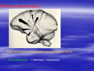
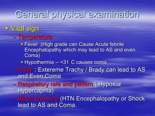
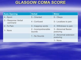
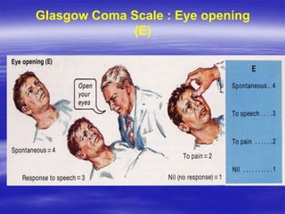
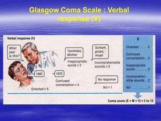
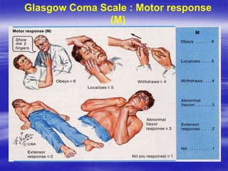
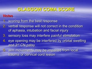
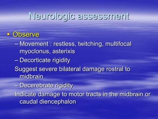
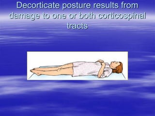
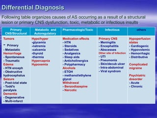
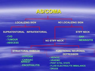
2. Recognize the importance of historical factors in
diagnosing causes of AS
Identify dementia, delirium and psychosis as the three
most common classifications of AS
Articulate a differential diagnosis of AS
Construct an approach to the diagnostic workup and
management of a patient with AS
Describe initial management of many causes of AS
Discuss the disposition of a patient with AS
Objectives:
3. What is Sensorium?
Ability of the brain to receive and interpret sensory stimuli
Good Sensorium = Alertness + Awareness
4. Altered Sensorium is not a disease:
It is a symptom.
Causes could be easily reversible (hypoglycemia) to
permanent (stroke) and from the relatively benign (alcohol
intoxication) to life threatening (meningitis or encephalitis).
Altered Sensorium
5. Unfortunately, there is no classic presentation for a patient
with Altered Sensorium. Presentations can range from
CNS depression to confusion, agitation, etc. Altered
sensorium can be determined by evaluating level of
Consciousness
Presentation of Patient with AS:
6. Level of Consciousness
Alert : Normal awake and responsive state
Drowsiness : State of apparent sleep, briefly
arousal with oral command
Lethargic : Resembles sleepiness, but not
becoming fully alert, slow verbal response
and inattentive. Unable to adequately
perform simple concentration task (such as
counting 20 to 1)
7. Level of Consciousness
Somnolent : Easily aroused by voice or touch;
awakens and follows commands; required
stimulation to maintain arousal
Obtunded/Stuporous : Arousable only with
repeated and painful stimulation; verbal output is
unintelligible or nil; some purposeful movement to
noxious stimulation
Comatose : No arousal despite vigorous
stimulation, no purposeful movement- only
posturing, brainstem reflexes often absent
8. Some Common Terms in Altered Sensorium
Confusion :
– impaired attention and concentration, manifest
disorientation in time, place and person, impersistent
thinking, speech and performance, reduced
comprehension and capacity to reason
– Fluctuate in severity, typically worse at night
„sundowning‟
– Perceptual disturbances and misinterpret voices,
common objects and actions of other persons
9. Delirium : confusion and associated agitation,
hallucination, convulsion and tremor
Amnesia : a loss of past memories and to an
ability to form new ones, despite alert and
normal attentiveness
Dementia the progressive deterioration in
cognitive function - the ability to process
thought (intelligence).
Phychosis refers to a mental state often
described as involving a "loss of contact with
reality".
10. Differentiating Delirium, Dementia and Phychosis
Finding Delirium Dementia Psychosis
Onset Rapid Slow Variable
Course Fluctuating Progressive Variable
Vital signs Often abnormal Usually normal Usually normal
Level of
consciousness
Altered Normal Variable
Hallucinations
Visual (related to
external stimuli)
Rare
Auditory (related
to internal stimuli)
Physical exam Often abnormal Often normal Often normal
Prognosis
Poor if cause not
treated
Progressive Variable
Underlying cause Organic (myriad)
Organic
(degenerative)
Functional
11. Dementia VS Confusional state
Dementia
– Longstanding nature
– Varies little from time to
time
– Memory problem
Confusional state
– Acute
– Fluctuate
– Clouding of
consciousness
13. Initial Actions and Primary Survey
A- Check to see that the airway is open and protected. Hypoxia is a potentially
reversible cause of Altered Sensorium.
B- Assess breathing. Inadequate ventilation will lead to elevated levels of CO2
(respiratory acidosis) and can cause AS.
C- Assess circulatory status. Hypoperfusion starves the brain of oxygen and glucose and
leads to AS.
D- Check for neurologic disability. Use GCS or AVPU scale for a quick assessment of
level of consciousness. Look for seizure activity. Are the pupils equal and reactive? Pay
attention to spontaneous movements. Lack of movement on one side of the body night
indicate stroke while lack of movement below a certain level of the body could indicate
spinal cord injury. If there is any suspicion of trauma the cervical spine should be
stabilized.
E- Expose (fully undress) and perform a rapid head to toe look for signs of trauma,
transdermal drug patches, dialysis access, infectious sources (such as catheters)
All emergency department patients require an initial assessment for immediate
threats. The “ABCDE approach” also provides a good opportunity to check for
quickly reversible causes of Altered sensorium.
14. As we proceed through ABCDE , keep in mind rapidly reversible
causes for the Altered Sensorium . Hypoglycemia and narcotic
overdose are very common causes of Altered Sensorium and can
easily be managed with dextrose and naloxone respectively.
At a minimum, all Altered Sensorium patients deserve:
Assessment of the ABC's
Cardiac monitoring and pulse oximetry
Supplemental oxygen
Bedside glucose testing
Intravenous access
Evaluation for signs of trauma and consider c-spine stabilization
Consider naloxone administration if narcotic overdose is suspected
15. Detailed History and Physical Exam
Patients with an AS are difficult to derive a comprehensive and detailed
history from. Family, friends, caretakers, nursing home workers, witnesses
are all invaluable sources of information. Make the effort to contact them to
ascertain the nature of the change in mental status.
Many medical conditions manifest as AS when decompensated.
Look for a history of:
diabetes (DKA, HONK),
hypertension (hypertensive encephalopathy or medication overdose)
endocrine disease (thyroid, Addisons)
renal failure
cancer (paraneoplastic syndromes, Na+, Ca++)
cardiovascular and cerebrovascular disease
seizure (atypical?)
psychiatric issues
Medication effects are also very common causes of AS in the elderly. A
detailed review of medications (including non prescription, health
supplements, home remedies) is critical. Has the patient recently started or
stopped any medications?
16. Vital signs
Neurologic status
Level of alertness GCS score or AVPU
Content of thought and speech
Does the patient stay focused?
Is their speech tangential?
Is the patient appropriately oriented?
Does the patient keep asking the same questions over and over
(perseveration)?
Are they reacting to internal stimuli?
Assess for focal motor findings
Is there weakness or pronator drift?
Cranial nerve exam (especially pupils)
Evaluate for tremulousness or abnormal reflexes
Common in withdrawal states or metabolic derangements
Physical Exam
17. Cardiovascular exam
Are there arrhythmias (a-fib) that predispose to embolic strokes?
Is there a murmur? endocarditis?
Is there evidence of good peripheral circulation?
Are there pulmonary findings that indicate pneumonia (sepsis) or
pulmonary edema
(hypoxia)?
Are there bruits over the carotid arteries?
Abdominal exam
Is there ascites, caput medusa, liver enlargement or tenderness (hepatic
encephalopathy)?
Is the abdomen tender (appendicitis, intussusception, abdominal sepsis
source, mesenteric ischemia)?
Genitourinary and rectal exam
Is the patient making urine (uremic encephalopathy)?
Are there signs or urinary, vaginal, prostatic or perineal infection?
Is there melena or blood in the stool?
18. Skin, extremity, musculoskeletal exam Are there petechiae
(meningococcemia)?
Is there a dialysis graft (uremic encephalopathy)?
Are there track marks from injection drug abuse?
Are there transdermal drug patches?
Is the skin jaundiced (hepatic encephalopathy)?
Is there nuchal rigidity or meningismus (CNS infection)?
Are there signs of trauma (raccoon's eyes, Battle „s sign,
hemotympanum)?
Are there infectious sources noted (decubitus ulcers, cellulitis,
abscesses)?
Are there masses or lymphadenopathy that might indicate cancer
History and physical exam findings are usually enough to help you
categorize the change in mental status.
20. General physical examination
Vital sign
– Temperature
Fever (High grade can Cause Acute febrile
Encephalopathy which may lead to AS and even
Coma)
Hypothermia -- <31 C causes coma
– Pulse : Extereme Trachy / Brady can lead to AS
and Even Coma
– Respiratory rate and pattern ( Hypoxia/
Hypercapnia)
– Blood pressure (HTN Encephalopathy or Shock
lead to AS and Coma.
21. GLASGOW COMA SCORE
Eyes Opening Verbal Motor
4 - Spont 5 - Oriented 6 - Obeys
3 – Response Verbal
command
4 - Confused 5 - Localizes to pain
2 - To Pain 3 - Inapprop words 4 - Withdraws to pain
1- None
2 - Incomprehensible
sounds
3 - Abnormal flexion
posturing
1 - No Sounds
2 - Abnormal extension
posturing
1 - None
25. Notes
1. scoring from the best response
2. verbal response will not correct in the condition
of aphasia, intubation and facial injury
3. sensory loss may interfere painful stimulation
4. eye opening may be interfered by orbital swelling
and 3rd CN palsy
5. arm movements may be impaired from local
trauma or cervical cord lesion
GLASGOW COMA SCORE
26. Neurologic assessment
Observe
– Movement : restless, twitching, multifocal
myoclonus, asterixis
– Decorticate rigidity
Suggest severe bilateral damage rostral to
midbrain
– Decerebrate rigidity
Indicate damage to motor tracts in the midbrain or
caudal diencephalon
34. Respiratory pattern
Cheyne-Stokes respiration : abnormal respiration in which periods of
shallow and deep breathing alternate. a/w bilateral cortical or bilateral
thalamic lesions, metabolic disturbances, incipient transtentorial
herniation
Hyperventilation : midbrain or pons lesions
Apneusis : lateral tegmentum of lower half of pons
Cluster : a breathing pattern in which a closely grouped series of
respirations is followed by apnea. a/w lower pontine or high medullary
lesions
Ataxic : is an abnormal pattern of breathing characterized by complete
irregularity of breathing, with irregular pauses and increasing periods of
apnea . a?/w dorsomedial medulla lesion
Kussmaul breathing is a deep and labored breathing pattern often
associated with severe metabolic acidosis, particularly DKA
35. Conditions mimic AS/Coma
Brain death
Locked-in syndrome
Vegetative state
Frontal lobe disease
Non-convulsive status epilepticus
Psychiatric disorder (catatonia,
depression)
36. Vegetative state
An awake but unresponsive state
Extensive damage in both cerebral
hemisphere
Retained respiratory and autonomic
functions
Cardiac arrest and head injury are the most
common causes.
37. Locked-in state
Awake patient has no means of
producing speech or volitional limb,
face and pharyngeal movements
Vertical eye movement and lid
elevation remain unimpaired
Infarction or hemorrhage of the ventral
pons
38. Differential Diagnosis
Following table organizes causes of AS occurring as a result of a structural
lesion or primary CNS dysfunction, toxic, metabolic or infectious insults.
Primary
CNS/Structural
Metabolic and
Autoregulatory
Pharmacologic/Toxic Infectious others
Tumors
- Primary
- Metastatic
Hemorrhage
- Spontaneous
- Traumatic
Edema
- HTN enceph
- Obstructive
hydrocephalus
Seizure
- Post-ictal state
- Todd's
paralysis
Dementia
- Degenerative
- Multi-infarct
Hypo/hyper
-glycemia
-natremia
-calcemia
-thyroid
-thermia
Hypercapnia
Hypoxemia
Medication effects
- HTN
- Steroids
- Sedatives
- Analgesics
- Sleep aids
- Anticholinergics
- Polypharmacy
Alcohols
- ETOH
- methanol/ethylene
glycol
Withdrawal
- Benzodiazepine
- Narcotic
Primary CNS
- Meningitis
- Encephalitis
- Abscesses
Other site of Infection
- UTI
- Pneumonia
- Skin/decub ulcer
- Intra-abdominal
- Viral syndrom
Hypoperfusion
states
- Cardiogenic
- Hypovolemic
- Hemorrhagic
- Distributive
Complicated
migraine
Psychiatric
dosorder
- Acute
- Chronic
40. Alternatively, a mnemonic that is commonly used to
help generate a differential diagnosis of AMS is:
AS = AEIOU TIPS
A Alcohol
E Epilepsy, Electrolytes, and Encephalopathy
I Insulin
O Opiates and Oxygen
U Uremia
T Trauma and Temperature
I Infection
P Poisons and Psychogenic
S
Shock, Stroke, Subarachnoid Hemorrhage and Space-
Occupying Lesion
41. Diagnostic Testing
Metabolic or Endocrine causes
– Rapid glucose
– Serum electrolytes (Na+, Ca+)
– ABG or VBG (with co-oxymetry for carboxy- or met-hemoglobinemia)
– BUN/Creatinine
– Thyroid function tests
– Ammonia level
– Serum cortisol level
Toxic or medication causes
– Levels of medications (anticonvulsants, digoxin, theophylline, lithium, etc.)
– Drug screen (benzodiazepines, opioids, barbiturates, etc.)
– Alcohol level
– Serum osmolality (toxic alcohols)
42. Infectious causes
– CBC with differential
– Urinalysis and culture
– Blood cultures
– Chest X-ray
– Lumbar puncture (with opening pressure)
– Always CT first if you suspect increased ICP.
Traumatic causes
– Head CT/ cervical spine CT
Neurologic causes
– Head CT (usually start without contrast for trauma or CVA)
– MRI (if brainstem/posterior fossa pathology suspected)
– EEG (if non-convulsive status epileptics suspected)
Hemodynamic instability causes
– ECG
– Cardiac enzymes (silent MI)
– Echocardiogram
– Carotid/vertebral artery ultrasound
43. Prognosis of AS/Coma
Recovery depends primarily on the causes
Intoxication and metabolic causes carry the best
prognosis
Coma from traumatic head injury far better than
those with coma from other structural causes
Coma from global hypoxic-ischemic carries least
favorable prognosis
At 3rd day, no papillary light reflex or GCS < 5 is
associated with poor prognosis
44. Treatment
Beyond interventions required for the immediate life threats such as
impending cardiopulmonary collapse, treatment should be geared towards
correcting / treating the underlying pathology
If the Cause of Coma/AS is unknown, what is often called a "coma cocktail"
is given to the patient. This cocktail consists of T=Thiamine, O=oxygen
N= Naloxene G= Glucose
The „‟TONG‟‟ describes the sequence the cocktail should be given.
Thiamine: Thiamine converts pyruvic acid to acetyl coenzyme. Without
Thiamine the energy contained in glucose couldn‟t be obtained. Alcohol
intake interferes with absorption of thiamine. Hence thiamine should always
be given prior to glucose if alcohol is suspected cause of coma.
45. Oxygen: Oxygen is essential for cellular functioning. Indication for
oxygen would be any clinical situation in which ventilation is not
adequate or oxygen carrying capacity is diminished.
Naloxene: If opoid toxicity is suspected, naloxene can be used. It
acts as competitive antagonist at opoid receptor and is indicated for
the reversal of CNS and Respiratory system depression caused by
opoids. It is less common in Nepal. Recomended dose is mg and
increasing the dose slowly at - minutes interval. At total of
mg can be goven. If there is no response then opoid toxity is ruled
out.
Glucose: Glucose is essential energy source and primary source of
braid. If hypoglycemia is indicated then glucose should be used.
46. Rx Contd.....
•Supportive care and sedation for agitated withdrawal states
•Intravenous fluids for dehydration, hypovolemia, hypotension
or hyperosmolar states or hypernatremia
•Empiric antibiotics for suspected meningitis, urosepsis,
pneumonia, etc.
•Rewarming or aggressive cooling for temperature extremes
•Fomepazole, pyridoxine, digoxin-fab fragments or other
antidotes for specific toxins
•Controlled reduction of blood pressure with nitroprusside,
labetolol or fenoldepam for hypertensive encephalopathy
•Hypertonic saline for profound hyponatremia with seizures or
AS
•Glucocorticoids for metastatic CNS lesions with vasogenic
edema
47. Disposition
The majority of patients with an AS will require hospitalization. Sometimes,
however, patients with acute alterations in consciousness that are easily
reversed and observed to be stable in the emergency department can safely
be discharged home.
The decision to admit the patient to the hospital ward may be based on
hemodynamic stability, etiology of the AS