Download to read offline


![Why deep learning for medical-image
analysis?
• Big data
• High complexity
• High variability (patients, surgeons,
devices, …)
à Deep learning learns rule from
data! We do not have to write
computationally-expensive
equations!
Sara Moccia, sara.moccia@santannapisa.it
[Maier-Hein et al., Nat. Biomed. Eng, 2017]](https://image.slidesharecdn.com/dli-meetupmoccia-211102085524/85/Dli-meetup-moccia-10-320.jpg)
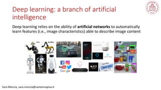
![Different opportunities
Sara Moccia, sara.moccia@santannapisa.it
[Litjens et al., Med. Image Anal., 2017]](https://image.slidesharecdn.com/dli-meetupmoccia-211102085524/85/Dli-meetup-moccia-11-320.jpg)
![A huge variety of medical images
Sara Moccia, sara.moccia@santannapisa.it
• Ionizing / not-ionizing radiation
• Pre-operative / intra-operative
• Anatomical / functional
• Contrasted tissue (bones, vessels,
muscles, organs, …)
• Signal-to-noise ratio
• Depth of the contrasted tissues
• Static / dynamic
• …
[Litjens et al., Med. Image Anal., 2017]](https://image.slidesharecdn.com/dli-meetupmoccia-211102085524/85/Dli-meetup-moccia-12-320.jpg)
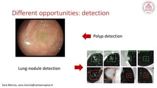
![Sara Moccia, sara.moccia@santannapisa.it
Different opportunities: classification
Tumor staging [Moccia et al., 2017]
Image tagging with tissues in the
field of view [Moccia et al., 2018]](https://image.slidesharecdn.com/dli-meetupmoccia-211102085524/85/Dli-meetup-moccia-13-320.jpg)
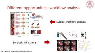
![Sara Moccia, sara.moccia@santannapisa.it
Different opportunities: segmentation
Scar tissue segmentation [Moccia et
al., 2019]
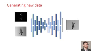
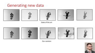
Surgical instrument segmentation
and generation [Colleoni, Moccia et al.,
2019]](https://image.slidesharecdn.com/dli-meetupmoccia-211102085524/85/Dli-meetup-moccia-14-320.jpg)
![Sara Moccia, sara.moccia@santannapisa.it
Different opportunities: informative frame
selection
Laryngoscopy [Moccia et al., 2019]
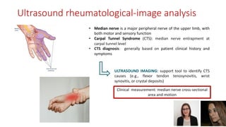
Ultrasound in rheumatology
[Fiorentino, Moccia et al., 2020]](https://image.slidesharecdn.com/dli-meetupmoccia-211102085524/85/Dli-meetup-moccia-16-320.jpg)
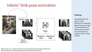
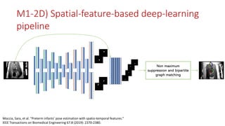
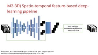
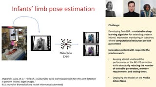
![Sara Moccia, sara.moccia@santannapisa.it
Different opportunities: pose estimation
Preterm-infants’ pose estimation
[Moccia et al., 2020]
Pose estimation in subjects using
smart walkers [Palermo, Moccia et al., 2021]](https://image.slidesharecdn.com/dli-meetupmoccia-211102085524/85/Dli-meetup-moccia-17-320.jpg)
![A gap between clinics and DL research
Sara Moccia, sara.moccia@santannapisa.it
Only few commercially-available
solutions
GI Genius™ intelligent endoscopy
module.
The first-to-market,
deep learning, computer-aided
polyp detection system.
99.7% sensitivity [Hassan et al.,
2020]](https://image.slidesharecdn.com/dli-meetupmoccia-211102085524/85/Dli-meetup-moccia-24-320.jpg)


![Mask R-CNN
Median Nerve
segmentation
- Input images size: 512x512 pixels
- Data augmentation on-the-fly à scale: [0.8, 1.2] rotation : [-10, 10] translation: [-20, 20] shearing : [-2, 2]
- Transfer learning with COCO dataset weights, initializing all layers of the model except for input layers of the network heads
- 256 anchors per image, with varying size (32, 64, 128, 256 and 512) and aspect ratios (1:1, 2:1, 1:2)
- Architectural changes: two more transposed layers inserted at the segmentation head
Backbone
(ResNet101 and Feature
Pyramidal Network)
Region
Proposal
Network
Segmentation head
Classification scores : C
(Median Nerve – Background)
Bounding box coordinates: 4*C
Fully
Connected
layers
Classification and bounding box regression heads
MASKS
US IMAGES
Roi Align
Mask for each of C classes
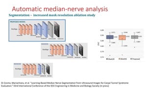
Automatic median-nerve analysis
Di Cosmo, Mariachiara, et al. "Learning-Based Median Nerve Segmentation from Ultrasound Images for Carpal Tunnel Syndrome
Evaluation.” 43rd International Conference of the IEEE Engineering in Medicine and Biology Society (in press)](https://image.slidesharecdn.com/dli-meetupmoccia-211102085524/85/Dli-meetup-moccia-30-320.jpg)


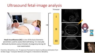
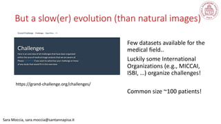
![Developing an algorithm for automatic HC delineation
Grand Challenge HC18
TRAIN
999 images
800 x 540 pixel
pixel range [0.052,0.326] mm
TEST
335 images
800 x 540 pixels
https://hc18.grand-challenge.org/
AIM and dataset](https://image.slidesharecdn.com/dli-meetupmoccia-211102085524/85/Dli-meetup-moccia-35-320.jpg)

![[1]
[2]
[3]
[4]
[5]
[1] T. L. van den Heuvel et al, Automated measurement of fetal head circumference using 2D ultrasound images, PloS One 13 (8) (2018) e0200412.
[2] Z. Sobhaninia, et al. Fetal ultrasound image segmentation for measuring biometric parameters using multi-task deep learning, in: 41st Annual In- ternational Conference of the IEEE Engineering in
Medicine and Biology Society, IEEE, 2019, pp. 6545–6548
[3] Z. Sobhaninia et al, Localization of fetal head in ultrasound images by multiscale view and deep neural networks, Available online at: arXiv preprint arXiv:1911.00908 (2019)
[4] Y. Rong et al, Deriving external forces via convolutional neural networks for biomedical image segmentation, Biomedical Optics Express 10 (8) (2019) 3800–3814.
[5] B. Al-Bander, et al,Improving fetal head contour detection by object localisation with deep learning, in: Annual Conference on Medical Image Understanding and Analysis, Springer, 2019, pp. 142–150
Results](https://image.slidesharecdn.com/dli-meetupmoccia-211102085524/85/Dli-meetup-moccia-37-320.jpg)
![• Birth before 37 gestational weeks
• Preterm births account for 11.1 % of the world’s births
[WHO, 2020]
Long-term complications
• Delayed language, cognitive deficits, behavioral and
motor disorders [Tucker et al., 2004]
Clinical need:
• Crucial necessity of continuously monitoring preterm
infants’ movement for evaluating the evolution of long-
term complications [Zuzarte et al., 2019]
Preterm birth](https://image.slidesharecdn.com/dli-meetupmoccia-211102085524/85/Dli-meetup-moccia-39-320.jpg)
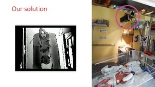
![Actual monitoring procedure:
Clinicians’ visual inspections at the crib-side in the NICUs
Drawbacks: discontinuous, inter-clinician-variable, time-consuming
Computer-based assisted methodologies:
Wearable sensors [Redd et al., 2019]
Drawbacks: pain, itch, hindering infants’ spontaneous motility
Camera sensors [Tsuji, et al., 2019, Marchi et al., 2020]
Drawbacks: semi-automatic approach, RGB-based
Movement monitoring solutions](https://image.slidesharecdn.com/dli-meetupmoccia-211102085524/85/Dli-meetup-moccia-41-320.jpg)

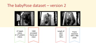
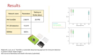
![M1-2D-detection M4-TwinEDA EDANet [Lo et al., 2019]
Migliorelli, Lucia, et al. "TwinEDA: a sustainable deep-learning approach for limb-joint detection
in preterm infants’ depth images."
IEEE Journal of Biomedical and Health Informatics (submitted)
M4-
TwinEDA
pipeline](https://image.slidesharecdn.com/dli-meetupmoccia-211102085524/85/Dli-meetup-moccia-51-320.jpg)
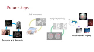


The document discusses deep learning applications for medical image analysis, including for diagnosis, surgical planning and guidance, and risk assessment. Specifically, it presents examples of using deep learning for tasks like classification, segmentation, detection, and pose estimation using medical images from modalities like ultrasound, X-ray, and video. Challenges in the field include limited datasets, variability in medical images, and privacy concerns, but deep learning methods are able to learn features directly from data to help with complex medical image analysis problems.


![[DSC Europe 25] Dubravko Culibrk - Deep Learning for Mammography.pptx](https://cdn.slidesharecdn.com/ss_thumbnails/yiscimuktacgqoiu4dkp-deep-learning-for-mammography-260119121559-aad59182-thumbnail.jpg?width=640&height=640&fit=bounds)
![[DSC Europe 25] Milos Belcevic - Product Professional's Journey to Full-Stack...](https://cdn.slidesharecdn.com/ss_thumbnails/1zovd6fgsycdg4wvgvls-milos-belcevic-product-professionals-journey-to-full-stack-product-developer-260123083019-d993120d-thumbnail.jpg?width=640&height=640&fit=bounds)


![[DSC Europe 25] Andrzej Kowalczyk - AI - how to start small and grow in the f...](https://cdn.slidesharecdn.com/ss_thumbnails/oy1zmo94qv6vpcqjvno2-andrzej-kowalczyk-ai-how-to-start-small-and-grow-in-the-future-1-260119121559-cf093b23-thumbnail.jpg?width=640&height=640&fit=bounds)


![[DSC Europe 25] Tamas Srancsik - How To Teach Your AI Football? An Argument f...](https://cdn.slidesharecdn.com/ss_thumbnails/bcjh1m9xtbosv20ucftb-tamas-srancsik-how-to-teach-your-ai-football-260121115910-08b53e9e-thumbnail.jpg?width=640&height=640&fit=bounds)


![[DSC Europe 25] Josip Saban - Career building for data professionals.pptx](https://cdn.slidesharecdn.com/ss_thumbnails/zroflcttkm1vmli0txea-josip-saban-career-building-for-data-professionals-260123083019-587cdb8c-thumbnail.jpg?width=640&height=640&fit=bounds)