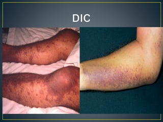
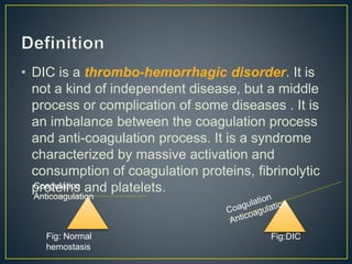
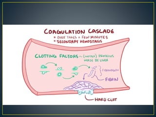
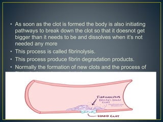
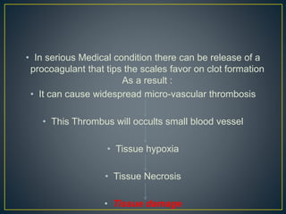
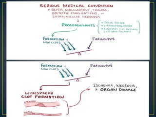
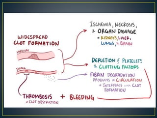
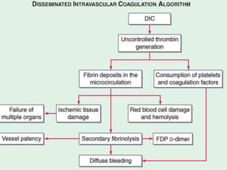
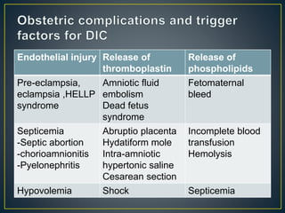
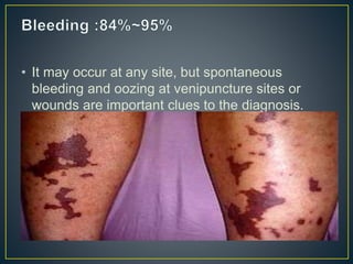
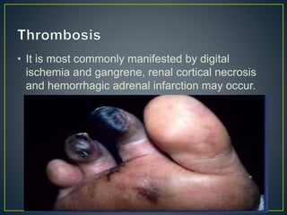
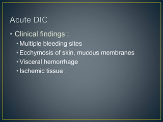
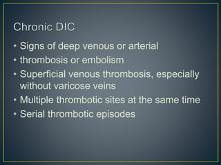
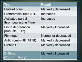
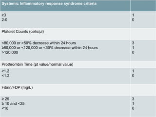
Dr.Ashraful Haque Oly
Dept.Of Medicine
Enam Medical College
โ€ขSynonyms :
โ€ขConsumptive
Coagulopathy
โ€ขDefibrination Syndrome
โ€ข DIC is a thrombo-hemorrhagic disorder. It is
not a kind of independent disease, but a middle
process or complication of some diseases . It is
an imbalance between the coagulation process
and anti-coagulation process. It is a syndrome
characterized by massive activation and
consumption of coagulation proteins, fibrinolytic
proteins and platelets.Coagulation
Anticoagulation
Fig: Normal
hemostasis
Fig:DIC
โ€ข As it is thrombo-hemorrhagic disorder . It has 2 Phases .
One is thrombotic phase and another one is Hemorrhagic
phase.
โ€ข Major Mechanisms which Trigger DIC , are :
โ€ข Release Procoagulant (Ex.TF , Lipopolysaccharide ,Enzymes that
active Clotting factors)
โ€ข Widespread Injury to the Endothelial cell
โ€ข Normally , after a cut or damage to endothelium or inner
lining of blood vessel walls , there is an immediate
vasoconstriction of the blood vessel which limit the
amount of blood flow.
โ€ข After that some platelet adhere to the damaged vessel
wall and become activated and then recruit additional
platelet to form a plug
โ€ข Formation of platelet plug is also known as primary
hemostasis
โ€ข After that coagulation cascades are activate. First off in the
blood there is a set of clotting factors most of which are
proteins synthesized by the liver and usually these are inactive
and floating around in the blood
โ€ข The coagulation cascade starts when one of these proteins
gets proteolytically cleaved .This active protein then
proteolytically cleaves and activates the next clotting factor
and so on .
โ€ข This cascade has a huge degree of amplification and takes
only a few mint from injury to clot formation .
โ€ข The final step is activation of the protein fibrinogen to fibrin
which deposits and polymerizes to form a mesh around the
platelet .
โ€ข This is called secondary hemostasis and result a hard clot at
โ€ข As soon as the clot is formed the body is also initiating
pathways to break down the clot so that it doesnot get
bigger than it needs to be and dissolves when itโ€™s not
needed any more
โ€ข This process is called fibrinolysis.
โ€ข This process produce fibrin degradation products.
โ€ข Normally the formation of new clots and the process of
fibrinolysis are in steady balance .
โ€ข In serious Medical condition there can be release of a
procoagulant that tips the scales favor on clot formation
As a result :
โ€ข It can cause widespread micro-vascular thrombosis
โ€ข This Thrombus will occults small blood vessel
โ€ข Tissue hypoxia
โ€ข Tissue Necrosis
โ€ข Tissue damage
โ€ข Due to increase clot formation
โ€ข Consumption of Clotting factors and platelets
โ€ข Deficiency of platelet and clotting factors on
circulation
โ€ข As a result it will Prevent further clot formation
โ€ข So , Bleeding will increase
โ€ข Another reason of bleeding is activating
plasmin
โ€ข Itโ€™s a fibrinolytic agent , it degrades fibrin and
remove unwanted elements from our circulation
โ€ข When there is widespread micro vascular
thrombosis there will be more plasmin activation
as a result fibrin polymerization will inhibit which
ultimately result in abnormal bleeding
โ€ข Plasmin also destroying clotting factor which also
result in increase bleeding.
โ€ข Acute DIC :It happened rapidly, the
coagulopathy is dominant and major
symptoms are bleeding and shock, mainly
seen in severe infection, amniotic fluid
embolism.
โ€ข Chronic DIC: it happened slowly and last
several weeks, thrombosis and clotting
may predominate mainly seen in cancer.
โ€ข DIC is not a primary disease, but a disorder
secondary to numerous triggering events such
as serious illnesses like :
โ€ข Infectious disease 31%~43%
โ€ข Cancer 24%~34%
โ€ข Obstetric complications 4%~12%
โ€ข Severe tissue injury 1%~5%
โ€ข Systemic disease
โ€ข (Bacterial, viral, rickettsial, parasitic
diseases and so on) Bacterial infection, in
particular septicemia, is commonly
associated with DIC. However, systemic
infections with other microorganisms, such
as viruses and parasites, also may lead to
DIC.
โ€ข (Acute promyelocytic leukemia, acute
myelomonocytic or monocytic
leukemia, disseminated prostatic
carcinoma ,Lung, breast,
gastrointestinal malignancy)
โ€ข (Burn, heart shock, fracture and so on)
Head trauma in particular is strongly
associated with DIC; both local and
systemic activation of coagulation may be
detected after such an event. The
increased risk of DIC after head trauma is
understandable in view of the relatively
large amount of tissue factor in the
cerebral compartment.
โ€ข (Malignant hypertension ,Acute respiratory
distress syndrome<ARDS>, hemolytic
transfusion reaction)
โ€ข a. Abruptio placentae.
โ€ข b. Amniotic fluid embolism.
โ€ข c. Endotoxic shock.
โ€ข d. Eclampsia and pre-eclampsia.
โ€ข e. Hydatidiform mole.
โ€ข f.. IUFD and missed abortion.
โ€ข g. Intra amniotic hypertonic saline or urea for induction of
abortion.
โ€ข h. Incompatible blood transfusion or transfusion of massive
banked blood which is deficient in factor V and VIII.
โ€ข i. Prolonged shock of whatever the cause.
โ€ข g. Placenta accreta.
โ€ข h. Rupture uterus.
โ€ข During pregnancy there is increase in
concentration of clotting factors II,V,VII,IX,X
and XII.
โ€ข Plasma Fibrinogen increased
โ€ข Small decreased in platelet count, due to low
grade intravascular coagulation
โ€ข Fibrinolytic activity is suppressed during
pregnancy and labor , it returns normal within
1 hour after the delivery of placenta . This is
due to liberation of plasminogen inhibitor from
Endothelial injury Release of
thromboplastin
Release of
phospholipids
Pre-eclampsia,
eclampsia ,HELLP
syndrome
Amniotic fluid
embolism
Dead fetus
syndrome
Fetomaternal
bleed
Septicemia
-Septic abortion
-chorioamnionitis
-Pyelonephritis
Abruptio placenta
Hydatiform mole
Intra-amniotic
hypertonic saline
Cesarean section
Incomplete blood
transfusion
Hemolysis
Hypovolemia Shock Septicemia
โ€ข Bleeding
โ€ข Thrombosis
โ€ข Hypotension or shock
โ€ข Organ dysfunction
โ€ข It may occur at any site, but spontaneous
bleeding and oozing at venipuncture sites or
wounds are important clues to the diagnosis.
โ€ข It is most commonly manifested by digital
ischemia and gangrene, renal cortical necrosis
and hemorrhagic adrenal infarction may occur.
โ€ข Clinical findings :
โ€ข Multiple bleeding sites
โ€ข Ecchymosis of skin, mucous membranes
โ€ข Visceral hemorrhage
โ€ข Ischemic tissue
โ€ข Signs of deep venous or arterial
โ€ข thrombosis or embolism
โ€ข Superficial venous thrombosis, especially
without varicose veins
โ€ข Multiple thrombotic sites at the same time
โ€ข Serial thrombotic episodes
Test Result
Platelet count Markedly decreased
Prothrombin Time (PT) Increased
Activated partial
thromboplastine Time
Increased
Fibrin degradation
products(FDP)
Markedly Increased
Fibrinogen Normal or decreased
Antithrombin III (AT III) Markedly decreased
Protein C Markedly decreased
D-Dimer
(Confirmatory)
Markedly Increased
โ€ข The D-dimer test is more specific for
detection of fibrinโ€”but not fibrinogen
degradation products (FDP) and
indicates that the cross-linked fibrin
has been digested by plasmin. Because
fibrinogen has a prolonged half-life,
plasma levels diminish acutely only in
severe cases of DIC.
Systemic Inflammatory response syndrome criteria
โ‰ฅ3
2-0
1
0
Platelet Counts (cells/ยตl)
<80,000 or >50% decrease within 24 hours
โ‰ฅ80,000 or <120,000 or <30% decrease within 24 hours
>120,000
3
1
0
Prothrombin Time (pt value/normal value)
โ‰ฅ1.2
<1.2
1
0
Fibrin/FDP (mg/L)
โ‰ฅ 25
โ‰ฅ 10 and <25
<10
3
1
0
โ€ข Liver disease
โ€ข Vitamin K deficiency
โ€ข Sepsis
โ€ข TTP (Thrombotic thrombocytopenic
purpura )
โ€ข Liver disease may prolong both the PT and
PTT, but fibrinogen levels are usually
normal, and the platelet count is usually
normal or only slightly reduced. Severe
liver disease may be difficult to distinguish
from DIC.
โ€ข Vitamin K deficiency will not affect the
fibrinogen level or platelet count and will be
completely corrected by vitamin K
replacement.
โ€ข Sepsis may produce thrombocytopenia,
and coagulopathy may be present because
of vitamin K deficiency. However, in these
cases, the fibrinogen level should be
normal.
โ€ข TTP may produce fever and MAHA
(microangiopathic hemolytic anemia).
However, fibrinogen levels and other
coagulation studies should be normal.
โ€ข Severe bleeding
โ€ข Stroke
โ€ข Ischemia of extremities or organs
โ€ข Treatment of the underlying disorder
โ€ข Replacement therapy
โ€ข Heparin therapy
โ€ข Other Treatment
โ€ข The primary focus should be the diagnosis
and treatment of the underlying disorder
Treatment of the underlying disease is the
mainstay of management of either acute or
chronic DIC. Avoid delay treat vigorously
(eg. shock, sepsis, obstetrical
problems).that has given rise to DIC.
โ€ข Coagulation factor deficiency require
replacement with FFP (fresh frozen
plasma). Platelet transfusion should be
used to maintain a platelet count greater
than 30000/ฮผl, and 50000/ฮผl. Fibrinogen is
replaced with cryoprecipitate. One unit of
cryoprecipitate usually raises the
fibrinogen level by 6~8mg/dl ,so that 15
units of cryoprecipitate will raise the level
from 50 to 150mg/dl
โ€ข In some cases heparin therapy is
contraindicated, but when DIC is producing
serious clinical consequences and the
underlying cause is not rapidly reversible,
heparin may be necessary.
Dose:500~750u/h is necessary
โ€ข Attention: : Heparin therapy must be used
in combination with replacement therapy, it
can lead to severe bleeding.
โ€ข Aminocaproic acid, 1g/h iv
โ€ข Tranexamic acid
โ€ข Those two drugs should be added to decrease
the rate of fibrinolysis, raise the fibrinogen level,
and control bleeding.
โ€ข Attention :Aminocaproic acid can never be used
without heparin in DIC because of the risk of
thrombosis.id, 10mg/kg
โ€ข Without bleeding or evidence of ischemia
โ€ข No treatment
โ€ข With bleeding
โ€ข Blood components as needed
โ€ข Fresh frozen plasma
โ€ข Cryoprecipitate
โ€ข Platelet transfusions
โ€ข With ischemia
โ€ข Anticoagulants after bleeding risk is corrected
with blood products
โ€ข Without thromboembolism
โ€ข No specific therapy needed but prophylactic
drugs (eg, lowdose heparin, low-
molecularweight heparin) may be used for
patients at high risk of thrombosis.
โ€ข With thromboembolism
โ€ข Heparin or low-molecular-weight heparin, trial
of warfarin sodium (Coumadin). (If warfarin is
unsuccessful, long-term use of lowmolecular-
weight heparin may be helpful.)
โ€ข Since DIC is a result of an acute
medical illness, prognoses depends
almost entirely upon the speed of the
intensivist in handing the bleeding
emergency, as well as the ability to
treat the underling disorder The
underlying disease that causes the
disorder will usually predict the
probable .outcome.
โ€ข An awareness of the clinical
settings in which DIC can occur
and the diagnostic features that
warn of its presence should enable
the physician to diagnose and treat
DIC appropriately. New treatments
that are more effective and less
hazardous are clearly needed, and
a number of such agents are now
Disseminated intravascular-coagulation (2)

Disseminated intravascular-coagulation (2)

  • 1.
    Dr.Ashraful Haque Oly Dept.OfMedicine Enam Medical College
  • 3.
  • 4.
    โ€ข DIC isa thrombo-hemorrhagic disorder. It is not a kind of independent disease, but a middle process or complication of some diseases . It is an imbalance between the coagulation process and anti-coagulation process. It is a syndrome characterized by massive activation and consumption of coagulation proteins, fibrinolytic proteins and platelets.Coagulation Anticoagulation Fig: Normal hemostasis Fig:DIC
  • 5.
    โ€ข As itis thrombo-hemorrhagic disorder . It has 2 Phases . One is thrombotic phase and another one is Hemorrhagic phase. โ€ข Major Mechanisms which Trigger DIC , are : โ€ข Release Procoagulant (Ex.TF , Lipopolysaccharide ,Enzymes that active Clotting factors) โ€ข Widespread Injury to the Endothelial cell
  • 6.
    โ€ข Normally ,after a cut or damage to endothelium or inner lining of blood vessel walls , there is an immediate vasoconstriction of the blood vessel which limit the amount of blood flow. โ€ข After that some platelet adhere to the damaged vessel wall and become activated and then recruit additional platelet to form a plug โ€ข Formation of platelet plug is also known as primary hemostasis
  • 7.
    โ€ข After thatcoagulation cascades are activate. First off in the blood there is a set of clotting factors most of which are proteins synthesized by the liver and usually these are inactive and floating around in the blood โ€ข The coagulation cascade starts when one of these proteins gets proteolytically cleaved .This active protein then proteolytically cleaves and activates the next clotting factor and so on . โ€ข This cascade has a huge degree of amplification and takes only a few mint from injury to clot formation . โ€ข The final step is activation of the protein fibrinogen to fibrin which deposits and polymerizes to form a mesh around the platelet . โ€ข This is called secondary hemostasis and result a hard clot at
  • 9.
    โ€ข As soonas the clot is formed the body is also initiating pathways to break down the clot so that it doesnot get bigger than it needs to be and dissolves when itโ€™s not needed any more โ€ข This process is called fibrinolysis. โ€ข This process produce fibrin degradation products. โ€ข Normally the formation of new clots and the process of fibrinolysis are in steady balance .
  • 10.
    โ€ข In seriousMedical condition there can be release of a procoagulant that tips the scales favor on clot formation As a result : โ€ข It can cause widespread micro-vascular thrombosis โ€ข This Thrombus will occults small blood vessel โ€ข Tissue hypoxia โ€ข Tissue Necrosis โ€ข Tissue damage
  • 12.
    โ€ข Due toincrease clot formation โ€ข Consumption of Clotting factors and platelets โ€ข Deficiency of platelet and clotting factors on circulation โ€ข As a result it will Prevent further clot formation โ€ข So , Bleeding will increase
  • 13.
    โ€ข Another reasonof bleeding is activating plasmin โ€ข Itโ€™s a fibrinolytic agent , it degrades fibrin and remove unwanted elements from our circulation โ€ข When there is widespread micro vascular thrombosis there will be more plasmin activation as a result fibrin polymerization will inhibit which ultimately result in abnormal bleeding โ€ข Plasmin also destroying clotting factor which also result in increase bleeding.
  • 16.
    โ€ข Acute DIC:It happened rapidly, the coagulopathy is dominant and major symptoms are bleeding and shock, mainly seen in severe infection, amniotic fluid embolism. โ€ข Chronic DIC: it happened slowly and last several weeks, thrombosis and clotting may predominate mainly seen in cancer.
  • 17.
    โ€ข DIC isnot a primary disease, but a disorder secondary to numerous triggering events such as serious illnesses like : โ€ข Infectious disease 31%~43% โ€ข Cancer 24%~34% โ€ข Obstetric complications 4%~12% โ€ข Severe tissue injury 1%~5% โ€ข Systemic disease
  • 18.
    โ€ข (Bacterial, viral,rickettsial, parasitic diseases and so on) Bacterial infection, in particular septicemia, is commonly associated with DIC. However, systemic infections with other microorganisms, such as viruses and parasites, also may lead to DIC.
  • 19.
    โ€ข (Acute promyelocyticleukemia, acute myelomonocytic or monocytic leukemia, disseminated prostatic carcinoma ,Lung, breast, gastrointestinal malignancy)
  • 20.
    โ€ข (Burn, heartshock, fracture and so on) Head trauma in particular is strongly associated with DIC; both local and systemic activation of coagulation may be detected after such an event. The increased risk of DIC after head trauma is understandable in view of the relatively large amount of tissue factor in the cerebral compartment.
  • 21.
    โ€ข (Malignant hypertension,Acute respiratory distress syndrome<ARDS>, hemolytic transfusion reaction)
  • 22.
    โ€ข a. Abruptioplacentae. โ€ข b. Amniotic fluid embolism. โ€ข c. Endotoxic shock. โ€ข d. Eclampsia and pre-eclampsia. โ€ข e. Hydatidiform mole. โ€ข f.. IUFD and missed abortion. โ€ข g. Intra amniotic hypertonic saline or urea for induction of abortion. โ€ข h. Incompatible blood transfusion or transfusion of massive banked blood which is deficient in factor V and VIII. โ€ข i. Prolonged shock of whatever the cause. โ€ข g. Placenta accreta. โ€ข h. Rupture uterus.
  • 23.
    โ€ข During pregnancythere is increase in concentration of clotting factors II,V,VII,IX,X and XII. โ€ข Plasma Fibrinogen increased โ€ข Small decreased in platelet count, due to low grade intravascular coagulation โ€ข Fibrinolytic activity is suppressed during pregnancy and labor , it returns normal within 1 hour after the delivery of placenta . This is due to liberation of plasminogen inhibitor from
  • 24.
    Endothelial injury Releaseof thromboplastin Release of phospholipids Pre-eclampsia, eclampsia ,HELLP syndrome Amniotic fluid embolism Dead fetus syndrome Fetomaternal bleed Septicemia -Septic abortion -chorioamnionitis -Pyelonephritis Abruptio placenta Hydatiform mole Intra-amniotic hypertonic saline Cesarean section Incomplete blood transfusion Hemolysis Hypovolemia Shock Septicemia
  • 25.
    โ€ข Bleeding โ€ข Thrombosis โ€ขHypotension or shock โ€ข Organ dysfunction
  • 26.
    โ€ข It mayoccur at any site, but spontaneous bleeding and oozing at venipuncture sites or wounds are important clues to the diagnosis.
  • 27.
    โ€ข It ismost commonly manifested by digital ischemia and gangrene, renal cortical necrosis and hemorrhagic adrenal infarction may occur.
  • 28.
    โ€ข Clinical findings: โ€ข Multiple bleeding sites โ€ข Ecchymosis of skin, mucous membranes โ€ข Visceral hemorrhage โ€ข Ischemic tissue
  • 29.
    โ€ข Signs ofdeep venous or arterial โ€ข thrombosis or embolism โ€ข Superficial venous thrombosis, especially without varicose veins โ€ข Multiple thrombotic sites at the same time โ€ข Serial thrombotic episodes
  • 31.
    Test Result Platelet countMarkedly decreased Prothrombin Time (PT) Increased Activated partial thromboplastine Time Increased Fibrin degradation products(FDP) Markedly Increased Fibrinogen Normal or decreased Antithrombin III (AT III) Markedly decreased Protein C Markedly decreased D-Dimer (Confirmatory) Markedly Increased
  • 32.
    โ€ข The D-dimertest is more specific for detection of fibrinโ€”but not fibrinogen degradation products (FDP) and indicates that the cross-linked fibrin has been digested by plasmin. Because fibrinogen has a prolonged half-life, plasma levels diminish acutely only in severe cases of DIC.
  • 34.
    Systemic Inflammatory responsesyndrome criteria โ‰ฅ3 2-0 1 0 Platelet Counts (cells/ยตl) <80,000 or >50% decrease within 24 hours โ‰ฅ80,000 or <120,000 or <30% decrease within 24 hours >120,000 3 1 0 Prothrombin Time (pt value/normal value) โ‰ฅ1.2 <1.2 1 0 Fibrin/FDP (mg/L) โ‰ฅ 25 โ‰ฅ 10 and <25 <10 3 1 0
  • 36.
    โ€ข Liver disease โ€ขVitamin K deficiency โ€ข Sepsis โ€ข TTP (Thrombotic thrombocytopenic purpura )
  • 37.
    โ€ข Liver diseasemay prolong both the PT and PTT, but fibrinogen levels are usually normal, and the platelet count is usually normal or only slightly reduced. Severe liver disease may be difficult to distinguish from DIC.
  • 38.
    โ€ข Vitamin Kdeficiency will not affect the fibrinogen level or platelet count and will be completely corrected by vitamin K replacement.
  • 39.
    โ€ข Sepsis mayproduce thrombocytopenia, and coagulopathy may be present because of vitamin K deficiency. However, in these cases, the fibrinogen level should be normal.
  • 40.
    โ€ข TTP mayproduce fever and MAHA (microangiopathic hemolytic anemia). However, fibrinogen levels and other coagulation studies should be normal.
  • 41.
    โ€ข Severe bleeding โ€ขStroke โ€ข Ischemia of extremities or organs
  • 42.
    โ€ข Treatment ofthe underlying disorder โ€ข Replacement therapy โ€ข Heparin therapy โ€ข Other Treatment
  • 43.
    โ€ข The primaryfocus should be the diagnosis and treatment of the underlying disorder Treatment of the underlying disease is the mainstay of management of either acute or chronic DIC. Avoid delay treat vigorously (eg. shock, sepsis, obstetrical problems).that has given rise to DIC.
  • 44.
    โ€ข Coagulation factordeficiency require replacement with FFP (fresh frozen plasma). Platelet transfusion should be used to maintain a platelet count greater than 30000/ฮผl, and 50000/ฮผl. Fibrinogen is replaced with cryoprecipitate. One unit of cryoprecipitate usually raises the fibrinogen level by 6~8mg/dl ,so that 15 units of cryoprecipitate will raise the level from 50 to 150mg/dl
  • 45.
    โ€ข In somecases heparin therapy is contraindicated, but when DIC is producing serious clinical consequences and the underlying cause is not rapidly reversible, heparin may be necessary. Dose:500~750u/h is necessary โ€ข Attention: : Heparin therapy must be used in combination with replacement therapy, it can lead to severe bleeding.
  • 46.
    โ€ข Aminocaproic acid,1g/h iv โ€ข Tranexamic acid โ€ข Those two drugs should be added to decrease the rate of fibrinolysis, raise the fibrinogen level, and control bleeding. โ€ข Attention :Aminocaproic acid can never be used without heparin in DIC because of the risk of thrombosis.id, 10mg/kg
  • 47.
    โ€ข Without bleedingor evidence of ischemia โ€ข No treatment โ€ข With bleeding โ€ข Blood components as needed โ€ข Fresh frozen plasma โ€ข Cryoprecipitate โ€ข Platelet transfusions โ€ข With ischemia โ€ข Anticoagulants after bleeding risk is corrected with blood products
  • 48.
    โ€ข Without thromboembolism โ€ขNo specific therapy needed but prophylactic drugs (eg, lowdose heparin, low- molecularweight heparin) may be used for patients at high risk of thrombosis. โ€ข With thromboembolism โ€ข Heparin or low-molecular-weight heparin, trial of warfarin sodium (Coumadin). (If warfarin is unsuccessful, long-term use of lowmolecular- weight heparin may be helpful.)
  • 49.
    โ€ข Since DICis a result of an acute medical illness, prognoses depends almost entirely upon the speed of the intensivist in handing the bleeding emergency, as well as the ability to treat the underling disorder The underlying disease that causes the disorder will usually predict the probable .outcome.
  • 50.
    โ€ข An awarenessof the clinical settings in which DIC can occur and the diagnostic features that warn of its presence should enable the physician to diagnose and treat DIC appropriately. New treatments that are more effective and less hazardous are clearly needed, and a number of such agents are now