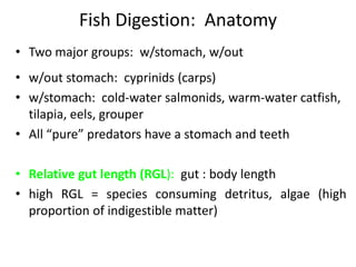
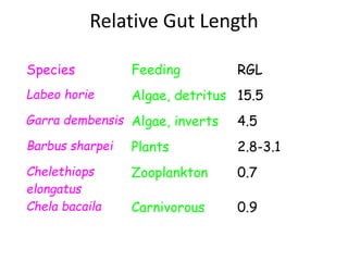
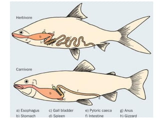
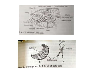
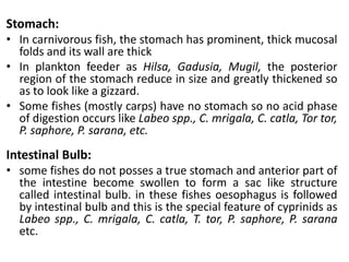
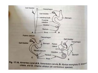
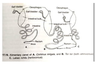
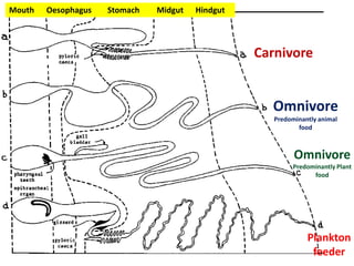
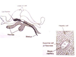
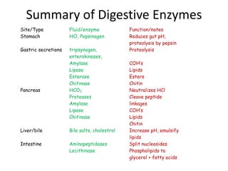
Digestive System
of Finfishes
• Two major groups: w/stomach, w/out
• w/out stomach: cyprinids (carps)
• w/stomach: cold-water salmonids, warm-water catfish,
tilapia, eels, grouper
• All “pure” predators have a stomach and teeth
• Relative gut length (RGL): gut : body length
• high RGL = species consuming detritus, algae (high
proportion of indigestible matter)
Fish Digestion: Anatomy
Relative Gut Length
Species Feeding RGL
Labeo horie Algae, detritus 15.5
Garra dembensis Algae, inverts 4.5
Barbus sharpei Plants 2.8-3.1
Chelethiops
elongatus
Zooplankton 0.7
Chela bacaila Carnivorous 0.9
Alimentary Canal
Fishes alimentary canal consist of mouth, oesophagus
(throat), and areas for the absorption of food
components (fore and midgut) and compaction of
indigestible waste material (hindgut)
The structure of alimentary canal varies in different
species of fishes, and is generally adapted in relation to
the food and feeding habits.
The variations are seen in the position of the mouth,
architecture of buccopharynx, relative length of the gut,
presence or absence of the stomach and pyloric caeca.
Gut and its Subdivisions
• The gut is a tubular structure beginning at the mouth and ending at the
anus. It is commonly divided into four parts.
• The most anterior part, the head gut is most often considered in terms of
its two components, the oral (buccal) and gill (branchial or pharyngeal)
cavities.
• The foregut begins at the posterior edge of the gills and includes
oesophagus, stomach and pylorus.
• The midgut includes the intestine posterior to the pylorus, often with no
distinct demarcation posteriorly between it and the hindgut. The midgut
is always the longest portion of the gut and may be coiled into
complicated loops when longer than the visceral cavity
• The beginning of the hindgut is marked by an increase in diameter of the
gut. The posterior end of the hindgut is the anus.
Alimentary Canal of Cartilaginous Fish (Scoliodon)
• It has a crescentic mouth on ventral side of the snout.
• The buccal cavity is lined by a thick and rough mucous
membrane. A large number of dermal denticles are
present in the mucous membrane, which become
enlarged to form the pointed teeth on the jaws.
• Teeth are arranged in several row, one behind the
other, of which only one row is functional at a time.
When teeth of the first row lost, other migrate to take
their place. Teeth of the Scoliodon are not used for
mastication.
• Tongue: Simple and non muscular without any gland.
• Pharynx: The buccal cavity leads into the pharynx,
which is perforated on each by the opening of gill
pouches. Mucous lining of the pharynx is also rough
due to the presence of dermal denticles in it.
• Oesophagus: pharynx opens into a short muscular
oesophagus whose internal lining is raised into
longitudinal folds.
• Stomach: oesophagus is followed by muscular stomach
which is J shaped with a long limb (cardiac) and a
shorter limb (pyloric). A small blind sac as an
outgrowth at the junction of the cardiac and pyloric
stomach is present. Pyloric stomach opens into
muscular chamber called Bursa entiana. The opening is
guarded by valve composed of circular muscle fiber.
• Intestine: Bursa entiana opens into the short
intestine which is wide tube open posteriorly
into rectum intestine have characteristic scroll
valve which has 2½ turns in anticlockwise. This
scroll valve check the rapid passage of food
through intestine and increase its absorptive
surface.
• Rectum: intestine is followed by rectum. One
small rectal gland also opens into the rectum.
Alimentary Canal of Scoliodon
Alimentary Canal of Teleost
• Alimentary canal of C. batrachus or M. seengala can be consider as a
typical example of teleost alimentary canal.
• Mouth: Lies at the anterior end of the snout and open behind into
the buccal cavity. Barbels surrounding the mouth are used for the
searching the food.
• Teeth: Numerous fine pointed teeth i.e. Maxillary, vomerine (behind
upper lip) and mandibular (behind lower lip) are present. Rest of the
buccophayrngeal cavity is lined with soft mucous membrane.
• Pharynx: The buccal cavity leads into the pharynx, which is
perforated ventro-laterally by means of oblique gill slits. Gill rakers
are present in one or two rows on each gill arch and are hard and
pointed structure. Gill rakers of the 1st arch are longest while 4th arch
are smallest in size. They prevent the escape of the food through gill
slits.
• Gullet: Pharyngeal cavity open into oesophagus by
the mean of circular aperture called gullet. A pair of
bony plates present on the dorsal side just anterior
to the gullet bears superior pharyngeal teeth.
Similarly a pair of bony plates borne by 4th gill arch
bear inferior pharyngeal teeth. Mucous lining the
posterior region of the bucco-pharynx is raised into
prominent folds near the gullet.
• Oesophagus: Short tube behind the gullet which
opens into the stomach.
• Stomach: Sac like structure with muscular walls. The
distal end of the stomach continues into the intestine
and the junction of the two is marked by a
constriction called the pylorus guarded by a muscular
sphincter. The mucous lining of the gut forms
prominent folds and thick in stomach
L. S. Head of Clarius batrachus
• Intestine: intestine is of moderate length having
1 or 2 coils only. Its wider proximal part and the
narrow distal part opens into a slightly wider
rectum. The mucous lining of the intestine
show a zig-zag pattern in proximal region and
reduce and indistinct at distal part.
• Rectum: Intestine is followed by rectum and
opens to the exterior by anus.
Alimentary Canal of C. batrachus
Anus
Histology of different
subdivisions of alimentary
canal
T. S. of Pharynx T. S. Of Oesophagus
T. S. of Stomach Part of gastric mucosa
Columnar
epithelium
T. S. of Intestine
Modification of
alimentary Canal
Modification of alimentary Canal
Fishes show great variation in the morphology of alimentary
canal or gut in all the sub division i.e. Fore-gut, mid-gut and
hind-gut.
Dentition:
• Carnivorous and predatory fishes have strong and pointed
teeth. eg: W. Attu, M. Seengala, C. Marulius, C. Punctatus,
C. Straitus, Notopterus chitala, Harpodon neherius etc. N.
Chitala and N. Notopterus posses teeth on the tongue also.
• Herbivorous species, teeth are completely absent from the
jaws and palate.
• In some omnivore like C. batrachus posses fine teeth on
jaws and palate which other like T. tor, P. sarana, C. reba, C.
carpio teeth are absent from the jaws and plate.
• In plankton feeder like H. Hilsa and Gadusia chapra havw
complete edentulous buccopharynx.
Buccopharynx:
• In carnivorous species there is no clear demarcation between
buccal cavity and pharyngeal cavity.
• In herbivore, there is clear demarcation between the buccal and
pharyngeal cavity.
Gill Rakers:
• In carnivorous and predatory fishes i.e. W. attu, M. seengala, N.
chitala, C. marulius, H. neherius, gill rakers are long, hard and
teeth like.
• In omnivorous species as the T. tor, P. sarana, the gill rakers are
short stumpy structure.
• In herbivorous fishes i.e. L. rohita, L. gonius gill rakers form a broad
sieve across the gill slits.
• In plankton feeder like C. catla, H. hilsa, G. chapra etc., gill rakers
are fairly long and thin and form a fine sieve to retain zoo and
phyto-planktons.
Taste buds and mucus secreting cells:
• In herbivore and omnivore fishes, a soft cushiony pad is
present in the roof of the buccal cavity which has large
number of papillae bearing taste buds and mucus secreting
cells.
• Large number of taste buds are present in C. catla, Puntius
spp., Tor spp., C. mrigala, Schizothorax,
• In carnivorous species like Channa and Mystus taste buds are
less or absent
Oesophagus:
• In herbivorous and some omnivorous like L. rohita, C. carpio,
C. mrigala, P. saphore, T. tor, L. calbasu, the oesophagus is
short and narrow.
• In carnivorous and predatory fishes i.e. W. attu, M.
seenghala, Channa spp. Oesophagus is much longer and
wider.
Stomach:
• In carnivorous fish, the stomach has prominent, thick mucosal
folds and its wall are thick
• In plankton feeder as Hilsa, Gadusia, Mugil, the posterior
region of the stomach reduce in size and greatly thickened so
as to look like a gizzard.
• Some fishes (mostly carps) have no stomach so no acid phase
of digestion occurs like Labeo spp., C. mrigala, C. catla, Tor tor,
P. saphore, P. sarana, etc.
Intestinal Bulb:
• some fishes do not posses a true stomach and anterior part of
the intestine become swollen to form a sac like structure
called intestinal bulb. in these fishes oesophagus is followed
by intestinal bulb and this is the special feature of cyprinids as
Labeo spp., C. mrigala, C. catla, T. tor, P. saphore, P. sarana
etc.
Intestine:
• The main variation or modification in intestine is the length
only.
• In most of the carnivore, length of intestine is short.
• In most of herbivore and plankton feeder, length of intestine is
very long
• In omnivore, intestine is of moderate length.
Pyloric Caeca:
• Several finger like outgrowth at anterior part of the intestine or
in the region of pylorus are called pyloric caeca.
• Pyloric caeca are present in species like Notopterus, Channa,
Hilsa, Harpodon, Mastacembelus.
• Pyloric caeca are absent in stomach less fish species like IMC.
• They are also absent in Wallago, Mystus.
• They vary in number and size.
Carnivore
Omnivore
Predominantly animal
food
Omnivore
Predominantly Plant
food
Plankton
feeder
Mouth Oesophagus Stomach Midgut Hindgut
Digestive Glands
& Digestion
Digestive glands
• Three main digestives glands are liver, pancreas
and gall bladder.
• Liver
• Pancreas
• Gall bladder
• Both organs produce digestive secretions
• liver produces bile but is also the primary organ for
synthesis, detoxification and storage of many nutrients
• pancreas is primary source of digestive enzymes in
most animals
• it also produces zymogens (precursors to enzymes)
Digestive Glands: liver & pancreas (fish)
 Channel catfish: have true stomach that secretes HCl and
pepsinogen (enzyme)
 Common carp: no stomach; however, “bulb” at anterior end of
digestive tract, bile and pancreatic secretions empty into intestine
posterior to cardiac sphincter, no secretion of gastrin (low pH)
 Tilapia: modified stomach, secretes HCl, well-defined pocket, pH
varies w/digestal flow, has pyloric sphincter
 Shrimp: cardiac/pyloric sections, gastric secretions, gastric mill,
straight shot to midgut
Digestive Gland Modification: stomach
 Channel catfish: length less than whole body, no large/small
version, slightly basic pH, digestive secretions, nutrient
absorption, many folds for absorption
 Common carp: digestive tract is 3x whole body length, similar in
activity to that of channel catfish
 Tilapia: tract is 6-8x that of body length, activities similar to that
of other species
 Shrimp: short midgut w/midgut gland used for
absorption/secretion/storage of nutrients, enzymes), slightly
basic, blind tubules
Digestive Gland Modification: Intestine
 Digestion: the preparation of food by the animal for
absorption
 involves the following processes:
 1) mechanical reduction of particle size;
 2) enzyme solubilization of organics;
 3) pH solubilization of inorganics;
 4) emulsification of fats
 Absorption: various processes that allow ions and
molecules to pass through membranes of the intestinal tract
into the blood, lymph, hemolymph, etc. to be metabolized
by the animal
Summary of Digestive Enzymes
Site/Type Fluid/enzyme Function/notes
Stomach HCl, Pepsinogen Reduces gut pH,
proteolysis by pepsin
Gastric secretions tripsynogen,
enterokinases,
Amylase
Lipase
Esterase
Chitinase
Proteolysis
COH’s
Lipids
Esters
Chitin
Pancreas HCO3
Proteases
Amylase
Lipase
Chitinase
Neutralizes HCl
Cleave peptide
linkages
COH’s
Lipids
Chitin
Liver/bile Bile salts, cholestrol Increase pH, emulsify
lipids
Intestine Aminopeptidases
Lecithinase
Split nucleosides
Phospholipids to
glycerol + fatty acids

Digestive-System-of-Finfishes.pdf

  • 1.
  • 2.
    • Two majorgroups: w/stomach, w/out • w/out stomach: cyprinids (carps) • w/stomach: cold-water salmonids, warm-water catfish, tilapia, eels, grouper • All “pure” predators have a stomach and teeth • Relative gut length (RGL): gut : body length • high RGL = species consuming detritus, algae (high proportion of indigestible matter) Fish Digestion: Anatomy
  • 3.
    Relative Gut Length SpeciesFeeding RGL Labeo horie Algae, detritus 15.5 Garra dembensis Algae, inverts 4.5 Barbus sharpei Plants 2.8-3.1 Chelethiops elongatus Zooplankton 0.7 Chela bacaila Carnivorous 0.9
  • 4.
    Alimentary Canal Fishes alimentarycanal consist of mouth, oesophagus (throat), and areas for the absorption of food components (fore and midgut) and compaction of indigestible waste material (hindgut) The structure of alimentary canal varies in different species of fishes, and is generally adapted in relation to the food and feeding habits. The variations are seen in the position of the mouth, architecture of buccopharynx, relative length of the gut, presence or absence of the stomach and pyloric caeca.
  • 6.
    Gut and itsSubdivisions • The gut is a tubular structure beginning at the mouth and ending at the anus. It is commonly divided into four parts. • The most anterior part, the head gut is most often considered in terms of its two components, the oral (buccal) and gill (branchial or pharyngeal) cavities. • The foregut begins at the posterior edge of the gills and includes oesophagus, stomach and pylorus. • The midgut includes the intestine posterior to the pylorus, often with no distinct demarcation posteriorly between it and the hindgut. The midgut is always the longest portion of the gut and may be coiled into complicated loops when longer than the visceral cavity • The beginning of the hindgut is marked by an increase in diameter of the gut. The posterior end of the hindgut is the anus.
  • 7.
    Alimentary Canal ofCartilaginous Fish (Scoliodon) • It has a crescentic mouth on ventral side of the snout. • The buccal cavity is lined by a thick and rough mucous membrane. A large number of dermal denticles are present in the mucous membrane, which become enlarged to form the pointed teeth on the jaws. • Teeth are arranged in several row, one behind the other, of which only one row is functional at a time. When teeth of the first row lost, other migrate to take their place. Teeth of the Scoliodon are not used for mastication. • Tongue: Simple and non muscular without any gland.
  • 8.
    • Pharynx: Thebuccal cavity leads into the pharynx, which is perforated on each by the opening of gill pouches. Mucous lining of the pharynx is also rough due to the presence of dermal denticles in it. • Oesophagus: pharynx opens into a short muscular oesophagus whose internal lining is raised into longitudinal folds. • Stomach: oesophagus is followed by muscular stomach which is J shaped with a long limb (cardiac) and a shorter limb (pyloric). A small blind sac as an outgrowth at the junction of the cardiac and pyloric stomach is present. Pyloric stomach opens into muscular chamber called Bursa entiana. The opening is guarded by valve composed of circular muscle fiber.
  • 9.
    • Intestine: Bursaentiana opens into the short intestine which is wide tube open posteriorly into rectum intestine have characteristic scroll valve which has 2½ turns in anticlockwise. This scroll valve check the rapid passage of food through intestine and increase its absorptive surface. • Rectum: intestine is followed by rectum. One small rectal gland also opens into the rectum.
  • 10.
  • 11.
    Alimentary Canal ofTeleost • Alimentary canal of C. batrachus or M. seengala can be consider as a typical example of teleost alimentary canal. • Mouth: Lies at the anterior end of the snout and open behind into the buccal cavity. Barbels surrounding the mouth are used for the searching the food. • Teeth: Numerous fine pointed teeth i.e. Maxillary, vomerine (behind upper lip) and mandibular (behind lower lip) are present. Rest of the buccophayrngeal cavity is lined with soft mucous membrane. • Pharynx: The buccal cavity leads into the pharynx, which is perforated ventro-laterally by means of oblique gill slits. Gill rakers are present in one or two rows on each gill arch and are hard and pointed structure. Gill rakers of the 1st arch are longest while 4th arch are smallest in size. They prevent the escape of the food through gill slits.
  • 12.
    • Gullet: Pharyngealcavity open into oesophagus by the mean of circular aperture called gullet. A pair of bony plates present on the dorsal side just anterior to the gullet bears superior pharyngeal teeth. Similarly a pair of bony plates borne by 4th gill arch bear inferior pharyngeal teeth. Mucous lining the posterior region of the bucco-pharynx is raised into prominent folds near the gullet. • Oesophagus: Short tube behind the gullet which opens into the stomach. • Stomach: Sac like structure with muscular walls. The distal end of the stomach continues into the intestine and the junction of the two is marked by a constriction called the pylorus guarded by a muscular sphincter. The mucous lining of the gut forms prominent folds and thick in stomach
  • 13.
    L. S. Headof Clarius batrachus
  • 14.
    • Intestine: intestineis of moderate length having 1 or 2 coils only. Its wider proximal part and the narrow distal part opens into a slightly wider rectum. The mucous lining of the intestine show a zig-zag pattern in proximal region and reduce and indistinct at distal part. • Rectum: Intestine is followed by rectum and opens to the exterior by anus.
  • 15.
    Alimentary Canal ofC. batrachus Anus
  • 16.
  • 17.
    T. S. ofPharynx T. S. Of Oesophagus
  • 18.
    T. S. ofStomach Part of gastric mucosa
  • 19.
  • 20.
  • 21.
    Modification of alimentaryCanal Fishes show great variation in the morphology of alimentary canal or gut in all the sub division i.e. Fore-gut, mid-gut and hind-gut. Dentition: • Carnivorous and predatory fishes have strong and pointed teeth. eg: W. Attu, M. Seengala, C. Marulius, C. Punctatus, C. Straitus, Notopterus chitala, Harpodon neherius etc. N. Chitala and N. Notopterus posses teeth on the tongue also. • Herbivorous species, teeth are completely absent from the jaws and palate. • In some omnivore like C. batrachus posses fine teeth on jaws and palate which other like T. tor, P. sarana, C. reba, C. carpio teeth are absent from the jaws and plate. • In plankton feeder like H. Hilsa and Gadusia chapra havw complete edentulous buccopharynx.
  • 22.
    Buccopharynx: • In carnivorousspecies there is no clear demarcation between buccal cavity and pharyngeal cavity. • In herbivore, there is clear demarcation between the buccal and pharyngeal cavity. Gill Rakers: • In carnivorous and predatory fishes i.e. W. attu, M. seengala, N. chitala, C. marulius, H. neherius, gill rakers are long, hard and teeth like. • In omnivorous species as the T. tor, P. sarana, the gill rakers are short stumpy structure. • In herbivorous fishes i.e. L. rohita, L. gonius gill rakers form a broad sieve across the gill slits. • In plankton feeder like C. catla, H. hilsa, G. chapra etc., gill rakers are fairly long and thin and form a fine sieve to retain zoo and phyto-planktons.
  • 23.
    Taste buds andmucus secreting cells: • In herbivore and omnivore fishes, a soft cushiony pad is present in the roof of the buccal cavity which has large number of papillae bearing taste buds and mucus secreting cells. • Large number of taste buds are present in C. catla, Puntius spp., Tor spp., C. mrigala, Schizothorax, • In carnivorous species like Channa and Mystus taste buds are less or absent Oesophagus: • In herbivorous and some omnivorous like L. rohita, C. carpio, C. mrigala, P. saphore, T. tor, L. calbasu, the oesophagus is short and narrow. • In carnivorous and predatory fishes i.e. W. attu, M. seenghala, Channa spp. Oesophagus is much longer and wider.
  • 26.
    Stomach: • In carnivorousfish, the stomach has prominent, thick mucosal folds and its wall are thick • In plankton feeder as Hilsa, Gadusia, Mugil, the posterior region of the stomach reduce in size and greatly thickened so as to look like a gizzard. • Some fishes (mostly carps) have no stomach so no acid phase of digestion occurs like Labeo spp., C. mrigala, C. catla, Tor tor, P. saphore, P. sarana, etc. Intestinal Bulb: • some fishes do not posses a true stomach and anterior part of the intestine become swollen to form a sac like structure called intestinal bulb. in these fishes oesophagus is followed by intestinal bulb and this is the special feature of cyprinids as Labeo spp., C. mrigala, C. catla, T. tor, P. saphore, P. sarana etc.
  • 29.
    Intestine: • The mainvariation or modification in intestine is the length only. • In most of the carnivore, length of intestine is short. • In most of herbivore and plankton feeder, length of intestine is very long • In omnivore, intestine is of moderate length. Pyloric Caeca: • Several finger like outgrowth at anterior part of the intestine or in the region of pylorus are called pyloric caeca. • Pyloric caeca are present in species like Notopterus, Channa, Hilsa, Harpodon, Mastacembelus. • Pyloric caeca are absent in stomach less fish species like IMC. • They are also absent in Wallago, Mystus. • They vary in number and size.
  • 30.
  • 31.
  • 32.
    Digestive glands • Threemain digestives glands are liver, pancreas and gall bladder. • Liver • Pancreas • Gall bladder
  • 33.
    • Both organsproduce digestive secretions • liver produces bile but is also the primary organ for synthesis, detoxification and storage of many nutrients • pancreas is primary source of digestive enzymes in most animals • it also produces zymogens (precursors to enzymes) Digestive Glands: liver & pancreas (fish)
  • 35.
     Channel catfish:have true stomach that secretes HCl and pepsinogen (enzyme)  Common carp: no stomach; however, “bulb” at anterior end of digestive tract, bile and pancreatic secretions empty into intestine posterior to cardiac sphincter, no secretion of gastrin (low pH)  Tilapia: modified stomach, secretes HCl, well-defined pocket, pH varies w/digestal flow, has pyloric sphincter  Shrimp: cardiac/pyloric sections, gastric secretions, gastric mill, straight shot to midgut Digestive Gland Modification: stomach
  • 36.
     Channel catfish:length less than whole body, no large/small version, slightly basic pH, digestive secretions, nutrient absorption, many folds for absorption  Common carp: digestive tract is 3x whole body length, similar in activity to that of channel catfish  Tilapia: tract is 6-8x that of body length, activities similar to that of other species  Shrimp: short midgut w/midgut gland used for absorption/secretion/storage of nutrients, enzymes), slightly basic, blind tubules Digestive Gland Modification: Intestine
  • 37.
     Digestion: thepreparation of food by the animal for absorption  involves the following processes:  1) mechanical reduction of particle size;  2) enzyme solubilization of organics;  3) pH solubilization of inorganics;  4) emulsification of fats  Absorption: various processes that allow ions and molecules to pass through membranes of the intestinal tract into the blood, lymph, hemolymph, etc. to be metabolized by the animal
  • 38.
    Summary of DigestiveEnzymes Site/Type Fluid/enzyme Function/notes Stomach HCl, Pepsinogen Reduces gut pH, proteolysis by pepsin Gastric secretions tripsynogen, enterokinases, Amylase Lipase Esterase Chitinase Proteolysis COH’s Lipids Esters Chitin Pancreas HCO3 Proteases Amylase Lipase Chitinase Neutralizes HCl Cleave peptide linkages COH’s Lipids Chitin Liver/bile Bile salts, cholestrol Increase pH, emulsify lipids Intestine Aminopeptidases Lecithinase Split nucleosides Phospholipids to glycerol + fatty acids