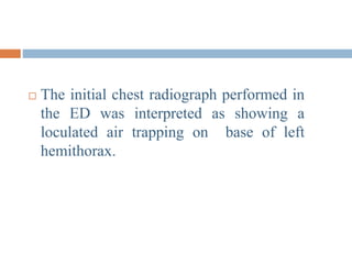
Download to read offline
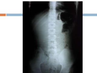
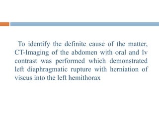

A 16-year-old male presented with abdominal pain, vomiting, and constipation for 3 days. Imaging showed a left diaphragmatic rupture with herniation of abdominal organs into the left hemithorax, likely resulting from a previous stab wound injury 2 months prior that was repaired but resulted in a diaphragmatic defect. The patient underwent surgery to repair the diaphragmatic rupture and his recovery was uneventful.


![management of Chest_trauma for nursing [1].ppt](https://cdn.slidesharecdn.com/ss_thumbnails/malamulochesttrauma1-241127110255-71befbaa-thumbnail.jpg?width=640&height=640&fit=bounds)
![Chest_trauma types and management[1].ppt](https://cdn.slidesharecdn.com/ss_thumbnails/malamulochesttrauma1-241126062258-4b388e87-thumbnail.jpg?width=640&height=640&fit=bounds)


