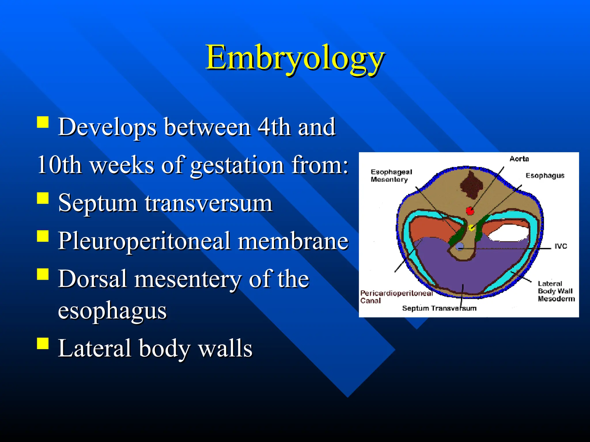
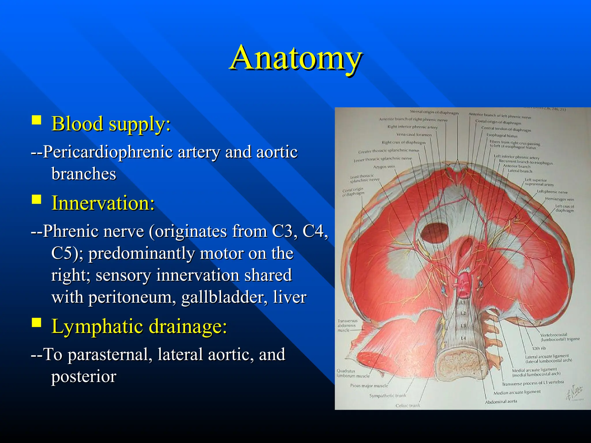
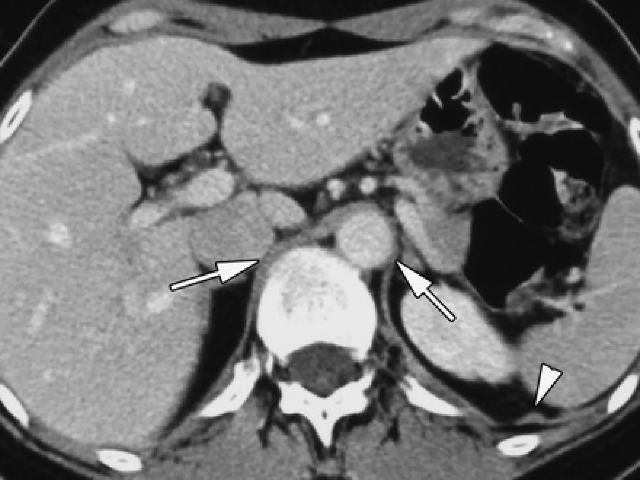
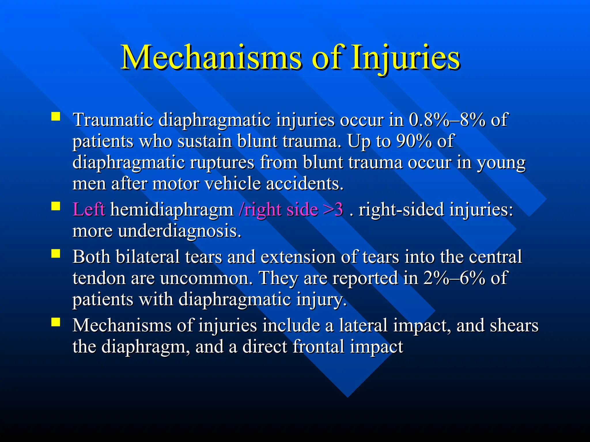
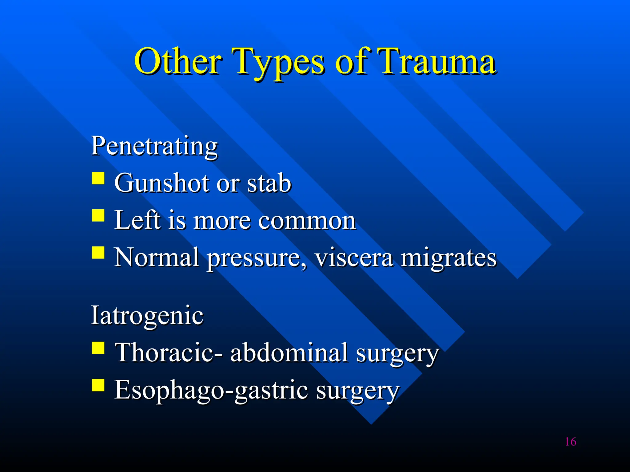
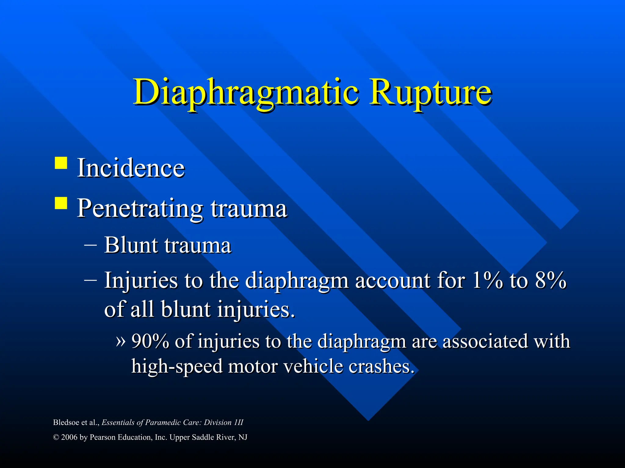
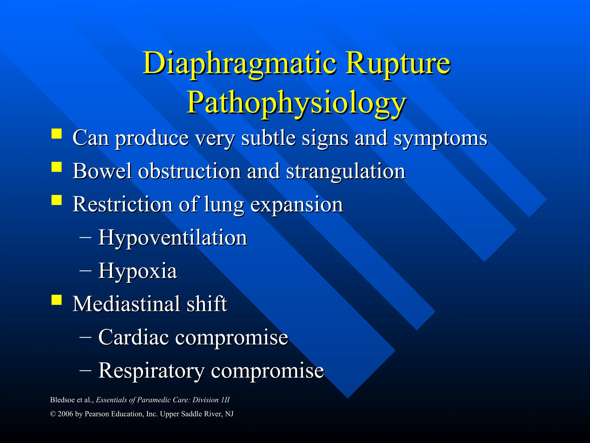
Diaphragmatic Rupture
HAMED RASHAD
HAMED RASHAD
Professor of Surgery -Egypt
Embryology
Embryology
 Develops between 4th and
Develops between 4th and
10th weeks of gestation from:
 Septum transversum
Septum transversum
 Pleuroperitoneal membrane
Pleuroperitoneal membrane
 Dorsal mesentery of the
Dorsal mesentery of the
esophagus
esophagus
 Lateral body walls
Lateral body walls
Image from
Image from
laparoscopy shows the intrathoracic herniation of
the stomach and the diaphragmatic tear.
Anatomy
Anatomy
Lumbar part
Lumbar part: Crura arises from the anterior surface of the
: Crura arises from the anterior surface of the
lumbar vertebrae; other fibers originate from lumbocostal
arches; insertion at the central tendon
Costal part:
Costal part: Originates from the six caudal ribs, and inserts in the
Originates from the six caudal ribs, and inserts in the
central tendon. Lumbocostal trigone (Bochdalek’s gap)
Tendinous part
Tendinous part Location: Depends on the extent of inhalation or
Location: Depends on the extent of inhalation or
exhalation, age, sex, momentary posture, extent that intestines
are filled, and general body structure At rest, the right dome is at
ICS 4 and left dome is approximately 1 to 2 cm lower; at
maximal inhalation at ICS 6 on right and ICS 7 on left
Anatomy
Anatomy
 Central tendon
Central tendon
 Crus
Crus
 Hiatus*3
Hiatus*3
 Celiac trunk
Celiac trunk
 Inf. Phrenic n.
Inf. Phrenic n.
Anatomy
Anatomy
 Blood supply:
Blood supply:
--Pericardiophrenic artery and aortic
branches
branches
 Innervation:
Innervation:
--Phrenic nerve (originates from C3, C4,
C5); predominantly motor on the
right; sensory innervation shared
with peritoneum, gallbladder, liver
 Lymphatic drainage:
Lymphatic drainage:
--To parasternal, lateral aortic, and
posterior
posterior
Unilateral Diaphragmatic Elevation
1.Subpulmonic pleural effusion
dome of pseudodiaphragm migrates toward the costophrenic
dome of pseudodiaphragm migrates toward the costophrenic angle and
angle and
flattens
flattens
2.Altered pulmonary volume
(a)Atelectasis
(a)Atelectasis
associated pulmonary density
(b)Postoperative lobectomy / pneumonectomy
rib defects, metallic sutures
(c)Hypoplastic lung
(c)Hypoplastic lung
small hemithorax (more often on the right), crowding of ribs,
small hemithorax (more often on the right), crowding of ribs, mediastinal
mediastinal
shift, absent / small pulmonary artery, frequently
shift, absent / small pulmonary artery, frequently associated with dextrocardia +
associated with dextrocardia +
anomalous pulmonary venous
anomalous pulmonary venous return
return
3.Phrenic nerve paralysis
(a)Primary lung tumor
(b)Malignant mediastinal tumor
(c)Iatrogenic
(c)Iatrogenic
(d)Idiopathic
(d)Idiopathic
paradoxic motion on fluoroscopy (patient in lateral position
paradoxic motion on fluoroscopy (patient in lateral position sniffing)
sniffing)
4.Abdominal disease
4.Abdominal disease
(a)Subphrenic abscess: history of surgery, accompanied by pleural
(a)Subphrenic abscess: history of surgery, accompanied by pleural effusion
effusion
(b)Distended stomach / colon
(c)Interposition of colon
(d)Liver mass (tumor, echinococcal cyst, abscess)
5.Diaphragmatic hernia
6.Eventration of diaphragm
7.Traumatic rupture of diaphragm
Associated with rib fractures, pulmonary contusion, hemothorax
8.Diaphragmatic tumor
Mesothelioma, fibroma, lipoma, lymphoma, metastases
12
Diaphragmatic Rupture
 Present in 1~6% major thoracic trauma
Present in 1~6% major thoracic trauma
 Penetrating, blunt, iatrogenic
Penetrating, blunt, iatrogenic
 Mostly in blunt trauma (traffic accident or
Mostly in blunt trauma (traffic accident or
a fall from height)
a fall from height)
Diaphragmatic Injury
 Incidence -0.8%-1.6% in BTA
Incidence -0.8%-1.6% in BTA
 High index of suspicion required , may be missed.
High index of suspicion required , may be missed.
 40 to 50% are diagnosed immediately
40 to 50% are diagnosed immediately
 Presentation may be delayed
Presentation may be delayed
 Imaging
Imaging
Nasogastric tube seen in the thorax
Abdominal contents in the thorax
Elevated hemidiaphragm (>4 cm Lt vs Rt)
Distortion of diaphragmatic margin.
 Lt- 69% , Rt -24% B/L- 15%
Lt- 69% , Rt -24% B/L- 15%
Mechanisms of Injuries
 Traumatic diaphragmatic injuries occur in 0.8%–8% of
Traumatic diaphragmatic injuries occur in 0.8%–8% of
patients who sustain blunt trauma. Up to 90% of
diaphragmatic ruptures from blunt trauma occur in young
men after motor vehicle accidents.
 Left
Left hemidiaphragm
hemidiaphragm /right side >3
/right side >3 . right-sided injuries:
. right-sided injuries:
more underdiagnosis.
 Both bilateral tears and extension of tears into the central
Both bilateral tears and extension of tears into the central
tendon are uncommon. They are reported in 2%–6% of
patients with diaphragmatic injury.
 Mechanisms of injuries include a lateral impact, and shears
Mechanisms of injuries include a lateral impact, and shears
the diaphragm, and a direct frontal impact
15
Diaphramatic Rupture
Diaphramatic Rupture in Blunt
in Blunt
Trauma
Trauma
 Only 10% cases diagnosed in acute phase
Only 10% cases diagnosed in acute phase
 Right : 15~24%; left : 70%~80%; bilateral: 5~8%
Right : 15~24%; left : 70%~80%; bilateral: 5~8%
( Liver had a protective effect)
∵
( Liver had a protective effect)
∵
 Raised pressure in abdomen
Raised pressure in abdomen
 tear of diaphragm (A
tear of diaphragm (A
new defect in health diaphragm or weakened impaired
segment of closed diaphragm)
segment of closed diaphragm) 
 abdominal organs
abdominal organs
through the defect
through the defect
– Stomach (31.8%), colon (27.2%), liver, spleen, kidney,
Stomach (31.8%), colon (27.2%), liver, spleen, kidney,
small intestine
small intestine
 Complication: hernia with gastro-intestinal
Complication: hernia with gastro-intestinal
obstruction, incarceration or perforation
16
Other Types of Trauma
Penetrating
Penetrating
 Gunshot or stab
Gunshot or stab
 Left is more common
Left is more common
 Normal pressure, viscera migrates
Normal pressure, viscera migrates
Iatrogenic
Iatrogenic
 Thoracic- abdominal surgery
Thoracic- abdominal surgery
 Esophago-gastric surgery
Esophago-gastric surgery
Diaphragmatic rupture
 Most ruptures are longer than 10
Most ruptures are longer than 10
cm and occur at the posterolateral
aspect of the hemidiaphragm
between the lumbar and intercostal
attachments and spread in a radial
direction
direction
 Penetrating injuries such as
Penetrating injuries such as
gunshot wounds or stab injuries are
more random
more random
Sites of injuries. Drawing shows radial (A),
transverse (B), and central (C) ruptures and a
peripheral detachment (D). Radial tears appear
to be the most frequently found injury at
surgery, whereas peripheral detachments are the
least frequent.
Bledsoe et al., Essentials of Paramedic Care: Division 1II
© 2006 by Pearson Education, Inc. Upper Saddle River, NJ
Diaphragmatic Rupture
 Incidence
Incidence
 Penetrating trauma
Penetrating trauma
– Blunt trauma
Blunt trauma
– Injuries to the diaphragm account for 1% to 8%
Injuries to the diaphragm account for 1% to 8%
of all blunt injuries.
» 90% of injuries to the diaphragm are associated with
90% of injuries to the diaphragm are associated with
high-speed motor vehicle crashes.
Natural history
Natural history
 Diaphragmatic hernias are not believed to close spontaneously1
Diaphragmatic hernias are not believed to close spontaneously1
 A pressure gradient exists between the thoracic and abdominal cavities
A pressure gradient exists between the thoracic and abdominal cavities
 The diaphragm is thin and in constant motion
The diaphragm is thin and in constant motion
 In animal models, however, some spontaneous healing is noted,
In animal models, however, some spontaneous healing is noted,
particularly when injury is “protected”:
 56 rats with 5-mm trocar injuries to diaphragm. At 150 days 100% of
56 rats with 5-mm trocar injuries to diaphragm. At 150 days 100% of
right-sided and 83% of left-sided punctures had healed2
 8 pigs with 1.5- to 2-cm lacerations to each diaphragm. At 6 weeks, 15/16
8 pigs with 1.5- to 2-cm lacerations to each diaphragm. At 6 weeks, 15/16
lacerations had healed3
 1 Ali J, Qi W. The effects of positive airway pressure and intra-abdominal pressure in diaphragmatic rupture.
1 Ali J, Qi W. The effects of positive airway pressure and intra-abdominal pressure in diaphragmatic rupture.
 World J Surg 1992;16:1120-5
World J Surg 1992;16:1120-5
 2 Gianni JA, Saad JR, Rasslan S, Lanceloti C. The natural course of penetratic diaphragmatic injury: an
2 Gianni JA, Saad JR, Rasslan S, Lanceloti C. The natural course of penetratic diaphragmatic injury: an
 experimental study (Abst). J Trauma 2002;53:194
experimental study (Abst). J Trauma 2002;53:194
 3 Shatney CH, Sensaki K, Morgan L. The natural history of stab wounds of the diaphragm: implications for a new
3 Shatney CH, Sensaki K, Morgan L. The natural history of stab wounds of the diaphragm: implications for a new
 management scheme for patients with penetrating thoracoabdominal trauma
management scheme for patients with penetrating thoracoabdominal trauma
Associated Injuries
Associated Injuries
 Common associated injuries include pelvic fractures
Common associated injuries include pelvic fractures
(40%–55%), splenic injuries (60%), and renal injuries
 There is also a high frequency of liver injuries, which are
There is also a high frequency of liver injuries, which are
more frequently associated with right than with left
diaphragmatic tears. Indeed, liver injuries are seen in 93%
of patients with right diaphragmatic injuries and 24% of
patients with left-sided lesions
 Thoracic injuries such as pneumohemothoraces and rib
Thoracic injuries such as pneumohemothoraces and rib
fractures are seen in 90% of patients. Aortic thoracic
injuries are reported in 5% of patients
Bledsoe et al., Essentials of Paramedic Care: Division 1II
© 2006 by Pearson Education, Inc. Upper Saddle River, NJ
Diaphragmatic Rupture
 Rupture can allow intra-abdominal organs
Rupture can allow intra-abdominal organs
to enter the thoracic cavity, which may
cause the following:
– Compression of the lung with reduced
Compression of the lung with reduced
ventilation
ventilation
– Decreased venous return
Decreased venous return
– Decreased cardiac output
Decreased cardiac output
– Shock
Shock
Bledsoe et al., Essentials of Paramedic Care: Division 1II
© 2006 by Pearson Education, Inc. Upper Saddle River, NJ
Diaphragmatic Rupture
Pathophysiology
Pathophysiology
 Can produce very subtle signs and symptoms
Can produce very subtle signs and symptoms
 Bowel obstruction and strangulation
Bowel obstruction and strangulation
 Restriction of lung expansion
Restriction of lung expansion
– Hypoventilation
Hypoventilation
– Hypoxia
Hypoxia
 Mediastinal shift
Mediastinal shift
– Cardiac compromise
Cardiac compromise
– Respiratory compromise
Respiratory compromise
23
Clinical Symptoms -- 3 Phases
1.
1. Acute phase
Acute phase
– Absent
Absent
– Dyspnea, chest pain, abdominal pain and vomiting
Dyspnea, chest pain, abdominal pain and vomiting
– PE: breathing sound
PE: breathing sound 
, bowel sounds in thorax
, bowel sounds in thorax
– Cardiac and respiratory insufficiency in massive herniation
Cardiac and respiratory insufficiency in massive herniation
Bledsoe et al., Essentials of Paramedic Care: Division 1II
© 2006 by Pearson Education, Inc. Upper Saddle River, NJ
Findings
Findings
 Tachypnea
Tachypnea
 Tachycardia
Tachycardia
 Respiratory distress
Respiratory distress
 Dullness to percussion
Dullness to percussion
 Scaphoid abdomen (hollow or empty appearance)
Scaphoid abdomen (hollow or empty appearance)
– If a large quantity of the abdominal contents are displaced into the
If a large quantity of the abdominal contents are displaced into the
chest
chest
 Bowel sounds in the affected hemithorax
Bowel sounds in the affected hemithorax
 Decreased breath sounds on the affected side
Decreased breath sounds on the affected side
 Possible chest or abdominal pain
Possible chest or abdominal pain
25
Clinical Symptoms -- 3 Phases
2.
2.Latent phase
Latent phase
–
– Symptom free – or just mild dyspnea or cardiac arrythmia
Symptom free – or just mild dyspnea or cardiac arrythmia
3. Late/ Obstructive phase
– Mortality : 25-66 %
Mortality : 25-66 %
– Mediastinal shift with cardiorespiratory compromise or
Mediastinal shift with cardiorespiratory compromise or
intestinal infarction with perforation
26
Diagnosis
Diagnosis
 High suspicious
High suspicious
 Proper studies:
Proper studies:
Chest X ray or abdominal X ray,
abdominal echo, barium studies, CT
and MRI
and MRI
 30~40% of patient had a true pre-
30~40% of patient had a true pre-
operative diagnosis
operative diagnosis
27
Image Study
Image Study
 CXR
CXR
– As first-line, only 20~34% positive findings
As first-line, only 20~34% positive findings
– Visceral gas in thorax
Visceral gas in thorax
– Strangulated bowel
Strangulated bowel 
 intrathoracic air-fluid levels
intrathoracic air-fluid levels
– Perforated bowel
Perforated bowel 
 pneumothorax
pneumothorax
 Fluoroscopy :
Fluoroscopy :
– Evaluation of integrity and motion of diaphragm
Evaluation of integrity and motion of diaphragm
 Barium studies
Barium studies
– A complement to diagnosis
A complement to diagnosis
 Abdominal echo
Abdominal echo
– Diaphragmatic position and integrity
Diaphragmatic position and integrity
– Pleural-peritoneal fluid
Pleural-peritoneal fluid
– Abdominal organ injuries
Abdominal organ injuries
29
Image Study
Image Study
 CT: useful and reliable
CT: useful and reliable
– Intrathoracic herniation of abdominal contents, and
Intrathoracic herniation of abdominal contents, and
waist-like constriction of abdominal viscera
– Diaphragmatic avulsion
Diaphragmatic avulsion
– Diaphragmatic discontinuity
Diaphragmatic discontinuity
* Helical CT: 78% left-sided; 50% right-sided injuries
 MRI: for delayed TDR
MRI: for delayed TDR
– T1-weighted sagittal view
T1-weighted sagittal view
 Laparpscopy and thoracoscopy
Laparpscopy and thoracoscopy
– For diagnosis and repair
For diagnosis and repair
30
31
Various Signs of Diaphragmatic Rupture
---CXR
---CXR
 initial radiographs allow diagnosis of 27%–60% of left-
initial radiographs allow diagnosis of 27%–60% of left-
sided injuries but only 17% of right-sided injuries
 Specific diagnostic findings:
Specific diagnostic findings:
(a)
(a) intrathoracic herniation of a hollow viscus (stomach,
intrathoracic herniation of a hollow viscus (stomach,
colon, small bowel) with or without focal constriction of
the viscus at the site of the tear
the viscus at the site of the tear (collar sign
(collar sign)
)
(b)
(b) visualization of a nasogastric tube above the
visualization of a nasogastric tube above the
hemidiaphragm on the left side
 Findings suggestive of hemidiaphragmatic rupture include
Findings suggestive of hemidiaphragmatic rupture include
elevation of the hemidiaphragm, distortion or obliteration
of the outline of the hemidiaphragm, and contralateral shift
of the mediastinum
of the mediastinum
Left diaphragmatic tear in a 24-
year-old woman who was injured
in a motor vehicle accident. Initial
chest radiograph shows
intrathoracic herniation of the
stomach (S), a pleural effusion, a
Left diaphragmatic tear in a
48-year-old man after a motor
vehicle accident. Initial chest
radiograph shows a gas-filled
viscus above the left
hemidiaphragm that
Various Signs of Diaphragmatic
Rupture ---CT
Rupture ---CT
 Conventional CT has a variable sensitivity of 14%–61% and
specificity of 76%–99% in the diagnosis of diaphragmatic
rupture
 Helical CT has proved to be more valuable in the detection
of diaphragmatic injuries with a sensitivity of 71% (78% for
left-sided injuries and 50% for right-sided injuries), a
specificity of 100%, and an accuracy of 88% for left-sided
injuries and 70% for right-sided injuries.
 Moreover, because of the high frequency of associated
injuries with blunt diaphragmatic tears, most
hemodynamically stable patients with suspected
diaphragmatic injuries require an admission CT examination
to evaluate the extent and anatomic sites of coexisting
thoracoabdominal injuries to guide clinical management.
 Left diaphragmatic tear in a 65-year-old patient after blunt trauma. CT scan
Left diaphragmatic tear in a 65-year-old patient after blunt trauma. CT scan
obtained at the level of the hepatic hilum shows a defect in the continuity of
the anterolateral left hemidiaphragm (arrows).
 CT scan of the midthoracic region shows intrathoracic herniation of the
CT scan of the midthoracic region shows intrathoracic herniation of the
stomach
stomach
Coronal reformatted image clearly shows a
Coronal reformatted image clearly shows a waistlike constriction
waistlike constriction of the liver
of the liver
(arrowheads).
(arrowheads).
Coronal contrast material-enhanced fat-suppressed fast gradient-echo MR image
shows a high position of the liver in the thoracic cavity. The
shows a high position of the liver in the thoracic cavity. The constricting rim of the
constricting rim of the
diaphragm
diaphragm is seen as a low-signal-intensity structure around the herniated liver
is seen as a low-signal-intensity structure around the herniated liver
(arrowheads).
(arrowheads).
1: Coronal reformatted image shows elevation and focal
constriction of the liver.
2: Sagittal single-shot fast spin-echo MR image clearly
shows the posterior diaphragm (arrow), which is outlined
by hemoperitoneum and pleural effusion.
3: Coronal contrast-enhanced fast gradient-echo MR
image clearly shows waistlike constriction of the liver at
the level of the diaphragmatic tear.
32-year-old man with left-sided diaphragmatic rupture. Axial
CT scan shows discontinuity of left hemidiaphragm (arrows
indicate extent of diaphragmatic tear) with gastric and left renal
herniation. Stomach lies dependent on left posterior ribs, which
is positive "dependent viscera" sign.
Diagnosis
Diagnosis
Laparoscopy
Laparoscopy
Safe and effective with a specificity of 100% in some
Safe and effective with a specificity of 100% in some
series3
series3
Can be therapeutic as well as diagnostic
Can be therapeutic as well as diagnostic
Complications are minor and include atelectasis,
Complications are minor and include atelectasis,
pneumothorax,and transient hypoxia or hemodynamic
instability during insufflation of abdomen
Expensive: addition to protocol increases costs by
Expensive: addition to protocol increases costs by
$1000 per patient
$1000 per patient
Bledsoe et al., Essentials of Paramedic Care: Division 1II
© 2006 by Pearson Education, Inc. Upper Saddle River, NJ
Diaphragmatic Rupture Management
 Airway and ventilation
Airway and ventilation
– High-concentration oxygen
High-concentration oxygen
– Positive-pressure ventilation if necessary
Positive-pressure ventilation if necessary
– Caution: positive pressure may worsen the injury
Caution: positive pressure may worsen the injury
 Circulation—IV access
Circulation—IV access
 Nonpharmacological—do not place patient in
Nonpharmacological—do not place patient in
Trendelenburg position
 Transport considerations
Transport considerations
– Appropriate mode
Appropriate mode
– Appropriate facility
Appropriate facility
Management
Management
 Acute injuries are best approached through
Acute injuries are best approached through
laparotomy
laparotomy
- Single layer closure with
nonabsorbable suture
 Chronic diaphragmatic hernias are best
Chronic diaphragmatic hernias are best
repaired through the chest
Diaphragmatic injury
Diaphragm Rupture /Hernia
S Lal,
S Lal, Y Kailasia, S Chouhan, APS Gaharwar, GP Shrivastava. Delayed presentation of
Y Kailasia, S Chouhan, APS Gaharwar, GP Shrivastava. Delayed presentation of
post traumatic diaphragmatic hernia. JSCR 2011. 7:6
46
Operation
Operation
 Operation time: according to the
Operation time: according to the
patient’s status
patient’s status
 Method: surgical repair
Method: surgical repair
– Primary closure
Primary closure
– If defect > 10cm
If defect > 10cm 
 synthetic grafts
synthetic grafts
 Post-operative mortality: 21%
Post-operative mortality: 21%
Eventration
Eventration
 a diaphragmatic eventration can mimic a rupture.
a diaphragmatic eventration can mimic a rupture.
Coronal and sagittal reformation images are helpful
to avoid false-positive diagnosis of diaphragmatic
injury and to identify the site of injury
Diaphragm eventration
 Incomplete muscularization of the diaphragm.
 This very common abnormality consists of a thin membranous sheet
replacing the diaphragmatic muscle.
 Usually it is partial, involving one half to one third of the hemidiaphragm,
frequently the anteromedial portion of the right hemidiaphragm.
 The lack of muscle is manifested on the chest radiograph by an elevation
of the affected portion of the diaphragm, usually depicted as a smooth
hump on the contour of the hemidiaphragm.
 When the entire hemidiaphragm is involved, it appears elevated and on
fluoroscopy, there is poor, or absent, paradoxical movement.
 In many cases, the distinction between this condition and acquired
paralysis of the phrenic nerve is impossible
diaphragmatic eventration
 upward displacement of abdominal contents
upward displacement of abdominal contents
secondary to a congenitally thin hypoplastic
diaphragm
diaphragm
 location:
location:
– anteromedial on right
anteromedial on right
– total involvement on the left
total involvement on the left
– R:L = 5:1
R:L = 5:1
 findings:
findings:
 small diaphragmatic excursions
small diaphragmatic excursions
 often lobulated contour
often lobulated contour
Diaphragmatic paralysis
 may be unilateral or bilateral. The most common cause of
unilateral paralysis of the diaphragm is involvement of the
phrenic nerve by a tumour. But there are many other
causes. It occasionally occurs as a complication of various
neurological diseases. Also injury to the phrenic nerve as a
result of trauma to the thoracic cage or cervical spine and
pressure upon the phrenic nerve from a substernal thyroid
or aortic aneurysm can cause paralysis. Infectious processes
involving the lung, pleura and/or mediastinum may result
in temporary or permanent diaphragmatic paralysis. Finally
diaphragmatic paralysis can be idiopathic.
Diaphragmatic paralysis
 The radiological evaluation of diaphragmatic paralysis
requires chest radiographs, adequate fluoroscopic tests and
clinical information. In some cases chest ultrasonography can
also be useful for studying and monitoring diaphragmatic
movement.
 Four signs indicate diaphragmatic paralysis:
– elevation of the diaphragm above the normal range;
– diminished, absent or paradoxical movement on inspiration;
– mediastinal shift on inspiration; and
– paradoxical movement during sniffing (Fig.1).
Diaphragmatic paralysis
 All these signs need not be present simultaneously but paradoxical
movement during sniffing is generally considered as the sine qua non
of diaphragmatic paralysis. However, these signs should be interpreted
with care since false positive and false negative results occur and other
diseases such as lung fibrosis, atelectasis, (hydro)pneumothorax,
bronchial stenosis, pneumonia, emphysema and diaphragmatic injury
can cause abnormal diaphragmatic position and motion. In addition,
the sniff test can be normal in apparently normal patients. Though in
these circumstances the paradoxal motion is small (< 2 cm) and
predominantly seen in the anterior part of the diaphragm. The
radiological diagnosis of bilateral diaphragmatic paralysis can be
difficult and the fluoroscopic study of bilateral diaphragmatic
paralysis requires simultaneous evaluation of diaphragmatic and chest
wall movement.
 Isolated elevation of the diaphragm in a 59-year-old man after blunt trauma.
Isolated elevation of the diaphragm in a 59-year-old man after blunt trauma. (a, b)
(a, b) CT scan
CT scan (a)
(a) and
and
sagittal reformatted image
sagittal reformatted image (b)
(b) show an isolated elevation of the diaphragm (arrow) without
show an isolated elevation of the diaphragm (arrow) without
discontinuity. Note the right-sided rib fracture on the scan
 Sagittal single-shot fast spin-echo
Sagittal single-shot fast spin-echo (c)
(c) and contrast-enhanced fat-suppressed fast gradient-echo
and contrast-enhanced fat-suppressed fast gradient-echo (d)
(d)
MR images show the diaphragm (arrow) as a thin hypointense band. Fat suppression and contrast
enhancement
enhancement (d)
(d) are used for better demonstration of the diaphragm and for differentiation
are used for better demonstration of the diaphragm and for differentiation
between a pleural effusion and a pulmonary contusion or atelectasis (arrowhead).
 Direct discontinuity
Direct discontinuity of the hemidiaphragm:
of the hemidiaphragm:
sensitivity 73%, specificity 90%.
 Intrathoracic herniation of abdominal contents:
Intrathoracic herniation of abdominal contents:
sensitivity 55%, specificity 100%.
 The
The collar sign
collar sign:
:
sensitivity 36% with conventional CT
63% with helical CT
63% with helical CT
– On the right side, the collar sign can appear as a focal
On the right side, the collar sign can appear as a focal
indentation of the liver, a subtle sign easily overlooked
 The
The dependent viscera sign
dependent viscera sign:
:
sensitivity: 100%: left-sided
83%: right-sided
83%: right-sided
Findings suggestive of
hemidiaphragmatic tears
56
Conclusion
Conclusion
 1~6% major thoracic trauma
1~6% major thoracic trauma
 25-66 % mortality in late phase
25-66 % mortality in late phase
 Difficulty in diagnosis, overlooked in acute
Difficulty in diagnosis, overlooked in acute
phase
phase
 High suspicious & PE & image studies:
High suspicious & PE & image studies:
– CXR is the first-line image study
CXR is the first-line image study
– Abdominal echo is another choice
Abdominal echo is another choice
– CT is superior in quality and tolerability
CT is superior in quality and tolerability
 Surgical repair with primary suture or grafts
Surgical repair with primary suture or grafts
Conclusions
Conclusions
 Diaphragmatic injury after penetrating thoracoabdominal
Diaphragmatic injury after penetrating thoracoabdominal
trauma can be clinically silent. However, the results of a
missed injury can be catastrophic. Clinical suspicion must be
high.
high.
 Penetrating injuries with a
Penetrating injuries with a trajectory
trajectory below the mammary line
below the mammary line
and above the costal margin – particularly on the left – should
arouse suspicion of a diaphragmatic injury
 DPL is superior to imaging modalities in diagnosing
DPL is superior to imaging modalities in diagnosing
diaphragmatic trauma is nonetheless imperfect
 Laparoscopy should be considered whenever diaphragmatic
Laparoscopy should be considered whenever diaphragmatic
injury is suspected, even if the remainder of the workup is
negative
negative
58
Refrences
Diaphragm rupture
Diaphragm rupture
Clinical radiology.61(6):467-77, 2006 Jun.
Ultrasound detection of right-sided diaphragmatic injury; the
“liver sliding” sign
American Journal of Emergency Medicine (2006) 24, 251–257
Traumatic rupture of the diaphragm: experience with 65
patients
patients
Injury, Int. J. Care Injured 34 (2003) 169–172
Diagnosis of Blunt Rupture of the Right Hemidiaphragm by
Technetium Scan
Technetium Scan
The American Surgeon [Am Surg], 1999 Aug; Vol. 65 (8), pp. 761-5
THANK YOU
THANK YOU

Diaphragmatic rupture & injuries how to repair the lect.ppt

  • 1.
    Diaphragmatic Rupture Diaphragmatic Rupture HAMEDRASHAD HAMED RASHAD Professor of Surgery -Egypt Professor of Surgery -Egypt
  • 2.
    Embryology Embryology  Develops between4th and Develops between 4th and 10th weeks of gestation from: 10th weeks of gestation from:  Septum transversum Septum transversum  Pleuroperitoneal membrane Pleuroperitoneal membrane  Dorsal mesentery of the Dorsal mesentery of the esophagus esophagus  Lateral body walls Lateral body walls
  • 3.
    Image from Image from laparoscopyshows the intrathoracic herniation of laparoscopy shows the intrathoracic herniation of the stomach and the diaphragmatic tear. the stomach and the diaphragmatic tear.
  • 5.
    Anatomy Anatomy Lumbar part Lumbar part:Crura arises from the anterior surface of the : Crura arises from the anterior surface of the lumbar vertebrae; other fibers originate from lumbocostal lumbar vertebrae; other fibers originate from lumbocostal arches; insertion at the central tendon arches; insertion at the central tendon Costal part: Costal part: Originates from the six caudal ribs, and inserts in the Originates from the six caudal ribs, and inserts in the central tendon. Lumbocostal trigone (Bochdalek’s gap) central tendon. Lumbocostal trigone (Bochdalek’s gap) Tendinous part Tendinous part Location: Depends on the extent of inhalation or Location: Depends on the extent of inhalation or exhalation, age, sex, momentary posture, extent that intestines exhalation, age, sex, momentary posture, extent that intestines are filled, and general body structure At rest, the right dome is at are filled, and general body structure At rest, the right dome is at ICS 4 and left dome is approximately 1 to 2 cm lower; at ICS 4 and left dome is approximately 1 to 2 cm lower; at maximal inhalation at ICS 6 on right and ICS 7 on left maximal inhalation at ICS 6 on right and ICS 7 on left
  • 6.
    Anatomy Anatomy  Central tendon Centraltendon  Crus Crus  Hiatus*3 Hiatus*3  Celiac trunk Celiac trunk  Inf. Phrenic n. Inf. Phrenic n.
  • 7.
    Anatomy Anatomy  Blood supply: Bloodsupply: --Pericardiophrenic artery and aortic --Pericardiophrenic artery and aortic branches branches  Innervation: Innervation: --Phrenic nerve (originates from C3, C4, --Phrenic nerve (originates from C3, C4, C5); predominantly motor on the C5); predominantly motor on the right; sensory innervation shared right; sensory innervation shared with peritoneum, gallbladder, liver with peritoneum, gallbladder, liver  Lymphatic drainage: Lymphatic drainage: --To parasternal, lateral aortic, and --To parasternal, lateral aortic, and posterior posterior
  • 10.
    Unilateral Diaphragmatic Elevation UnilateralDiaphragmatic Elevation 1.Subpulmonic pleural effusion 1.Subpulmonic pleural effusion dome of pseudodiaphragm migrates toward the costophrenic dome of pseudodiaphragm migrates toward the costophrenic angle and angle and flattens flattens 2.Altered pulmonary volume 2.Altered pulmonary volume (a)Atelectasis (a)Atelectasis associated pulmonary density associated pulmonary density (b)Postoperative lobectomy / pneumonectomy (b)Postoperative lobectomy / pneumonectomy rib defects, metallic sutures rib defects, metallic sutures (c)Hypoplastic lung (c)Hypoplastic lung small hemithorax (more often on the right), crowding of ribs, small hemithorax (more often on the right), crowding of ribs, mediastinal mediastinal shift, absent / small pulmonary artery, frequently shift, absent / small pulmonary artery, frequently associated with dextrocardia + associated with dextrocardia + anomalous pulmonary venous anomalous pulmonary venous return return
  • 11.
    3.Phrenic nerve paralysis 3.Phrenicnerve paralysis (a)Primary lung tumor (a)Primary lung tumor (b)Malignant mediastinal tumor (b)Malignant mediastinal tumor (c)Iatrogenic (c)Iatrogenic (d)Idiopathic (d)Idiopathic paradoxic motion on fluoroscopy (patient in lateral position paradoxic motion on fluoroscopy (patient in lateral position sniffing) sniffing) 4.Abdominal disease 4.Abdominal disease (a)Subphrenic abscess: history of surgery, accompanied by pleural (a)Subphrenic abscess: history of surgery, accompanied by pleural effusion effusion (b)Distended stomach / colon (b)Distended stomach / colon (c)Interposition of colon (c)Interposition of colon (d)Liver mass (tumor, echinococcal cyst, abscess) (d)Liver mass (tumor, echinococcal cyst, abscess) 5.Diaphragmatic hernia 5.Diaphragmatic hernia 6.Eventration of diaphragm 6.Eventration of diaphragm 7.Traumatic rupture of diaphragm 7.Traumatic rupture of diaphragm Associated with rib fractures, pulmonary contusion, hemothorax Associated with rib fractures, pulmonary contusion, hemothorax 8.Diaphragmatic tumor 8.Diaphragmatic tumor Mesothelioma, fibroma, lipoma, lymphoma, metastases Mesothelioma, fibroma, lipoma, lymphoma, metastases
  • 12.
    12 Diaphragmatic Rupture Diaphragmatic Rupture Present in 1~6% major thoracic trauma Present in 1~6% major thoracic trauma  Penetrating, blunt, iatrogenic Penetrating, blunt, iatrogenic  Mostly in blunt trauma (traffic accident or Mostly in blunt trauma (traffic accident or a fall from height) a fall from height)
  • 13.
    Diaphragmatic Injury Diaphragmatic Injury Incidence -0.8%-1.6% in BTA Incidence -0.8%-1.6% in BTA  High index of suspicion required , may be missed. High index of suspicion required , may be missed.  40 to 50% are diagnosed immediately 40 to 50% are diagnosed immediately  Presentation may be delayed Presentation may be delayed  Imaging Imaging Nasogastric tube seen in the thorax Nasogastric tube seen in the thorax Abdominal contents in the thorax Abdominal contents in the thorax Elevated hemidiaphragm (>4 cm Lt vs Rt) Elevated hemidiaphragm (>4 cm Lt vs Rt) Distortion of diaphragmatic margin. Distortion of diaphragmatic margin.  Lt- 69% , Rt -24% B/L- 15% Lt- 69% , Rt -24% B/L- 15%
  • 14.
    Mechanisms of Injuries Mechanismsof Injuries  Traumatic diaphragmatic injuries occur in 0.8%–8% of Traumatic diaphragmatic injuries occur in 0.8%–8% of patients who sustain blunt trauma. Up to 90% of patients who sustain blunt trauma. Up to 90% of diaphragmatic ruptures from blunt trauma occur in young diaphragmatic ruptures from blunt trauma occur in young men after motor vehicle accidents. men after motor vehicle accidents.  Left Left hemidiaphragm hemidiaphragm /right side >3 /right side >3 . right-sided injuries: . right-sided injuries: more underdiagnosis. more underdiagnosis.  Both bilateral tears and extension of tears into the central Both bilateral tears and extension of tears into the central tendon are uncommon. They are reported in 2%–6% of tendon are uncommon. They are reported in 2%–6% of patients with diaphragmatic injury. patients with diaphragmatic injury.  Mechanisms of injuries include a lateral impact, and shears Mechanisms of injuries include a lateral impact, and shears the diaphragm, and a direct frontal impact the diaphragm, and a direct frontal impact
  • 15.
    15 Diaphramatic Rupture Diaphramatic Rupturein Blunt in Blunt Trauma Trauma  Only 10% cases diagnosed in acute phase Only 10% cases diagnosed in acute phase  Right : 15~24%; left : 70%~80%; bilateral: 5~8% Right : 15~24%; left : 70%~80%; bilateral: 5~8% ( Liver had a protective effect) ∵ ( Liver had a protective effect) ∵  Raised pressure in abdomen Raised pressure in abdomen  tear of diaphragm (A tear of diaphragm (A new defect in health diaphragm or weakened impaired new defect in health diaphragm or weakened impaired segment of closed diaphragm) segment of closed diaphragm)   abdominal organs abdominal organs through the defect through the defect – Stomach (31.8%), colon (27.2%), liver, spleen, kidney, Stomach (31.8%), colon (27.2%), liver, spleen, kidney, small intestine small intestine  Complication: hernia with gastro-intestinal Complication: hernia with gastro-intestinal obstruction, incarceration or perforation obstruction, incarceration or perforation
  • 16.
    16 Other Types ofTrauma Other Types of Trauma Penetrating Penetrating  Gunshot or stab Gunshot or stab  Left is more common Left is more common  Normal pressure, viscera migrates Normal pressure, viscera migrates Iatrogenic Iatrogenic  Thoracic- abdominal surgery Thoracic- abdominal surgery  Esophago-gastric surgery Esophago-gastric surgery
  • 17.
    Diaphragmatic rupture Diaphragmatic rupture Most ruptures are longer than 10 Most ruptures are longer than 10 cm and occur at the posterolateral cm and occur at the posterolateral aspect of the hemidiaphragm aspect of the hemidiaphragm between the lumbar and intercostal between the lumbar and intercostal attachments and spread in a radial attachments and spread in a radial direction direction  Penetrating injuries such as Penetrating injuries such as gunshot wounds or stab injuries are gunshot wounds or stab injuries are more random more random Sites of injuries. Drawing shows radial (A), transverse (B), and central (C) ruptures and a peripheral detachment (D). Radial tears appear to be the most frequently found injury at surgery, whereas peripheral detachments are the least frequent.
  • 18.
    Bledsoe et al.,Essentials of Paramedic Care: Division 1II © 2006 by Pearson Education, Inc. Upper Saddle River, NJ Diaphragmatic Rupture Diaphragmatic Rupture  Incidence Incidence  Penetrating trauma Penetrating trauma – Blunt trauma Blunt trauma – Injuries to the diaphragm account for 1% to 8% Injuries to the diaphragm account for 1% to 8% of all blunt injuries. of all blunt injuries. » 90% of injuries to the diaphragm are associated with 90% of injuries to the diaphragm are associated with high-speed motor vehicle crashes. high-speed motor vehicle crashes.
  • 19.
    Natural history Natural history Diaphragmatic hernias are not believed to close spontaneously1 Diaphragmatic hernias are not believed to close spontaneously1  A pressure gradient exists between the thoracic and abdominal cavities A pressure gradient exists between the thoracic and abdominal cavities  The diaphragm is thin and in constant motion The diaphragm is thin and in constant motion  In animal models, however, some spontaneous healing is noted, In animal models, however, some spontaneous healing is noted, particularly when injury is “protected”: particularly when injury is “protected”:  56 rats with 5-mm trocar injuries to diaphragm. At 150 days 100% of 56 rats with 5-mm trocar injuries to diaphragm. At 150 days 100% of right-sided and 83% of left-sided punctures had healed2 right-sided and 83% of left-sided punctures had healed2  8 pigs with 1.5- to 2-cm lacerations to each diaphragm. At 6 weeks, 15/16 8 pigs with 1.5- to 2-cm lacerations to each diaphragm. At 6 weeks, 15/16 lacerations had healed3 lacerations had healed3  1 Ali J, Qi W. The effects of positive airway pressure and intra-abdominal pressure in diaphragmatic rupture. 1 Ali J, Qi W. The effects of positive airway pressure and intra-abdominal pressure in diaphragmatic rupture.  World J Surg 1992;16:1120-5 World J Surg 1992;16:1120-5  2 Gianni JA, Saad JR, Rasslan S, Lanceloti C. The natural course of penetratic diaphragmatic injury: an 2 Gianni JA, Saad JR, Rasslan S, Lanceloti C. The natural course of penetratic diaphragmatic injury: an  experimental study (Abst). J Trauma 2002;53:194 experimental study (Abst). J Trauma 2002;53:194  3 Shatney CH, Sensaki K, Morgan L. The natural history of stab wounds of the diaphragm: implications for a new 3 Shatney CH, Sensaki K, Morgan L. The natural history of stab wounds of the diaphragm: implications for a new  management scheme for patients with penetrating thoracoabdominal trauma management scheme for patients with penetrating thoracoabdominal trauma
  • 20.
    Associated Injuries Associated Injuries Common associated injuries include pelvic fractures Common associated injuries include pelvic fractures (40%–55%), splenic injuries (60%), and renal injuries (40%–55%), splenic injuries (60%), and renal injuries  There is also a high frequency of liver injuries, which are There is also a high frequency of liver injuries, which are more frequently associated with right than with left more frequently associated with right than with left diaphragmatic tears. Indeed, liver injuries are seen in 93% diaphragmatic tears. Indeed, liver injuries are seen in 93% of patients with right diaphragmatic injuries and 24% of of patients with right diaphragmatic injuries and 24% of patients with left-sided lesions patients with left-sided lesions  Thoracic injuries such as pneumohemothoraces and rib Thoracic injuries such as pneumohemothoraces and rib fractures are seen in 90% of patients. Aortic thoracic fractures are seen in 90% of patients. Aortic thoracic injuries are reported in 5% of patients injuries are reported in 5% of patients
  • 21.
    Bledsoe et al.,Essentials of Paramedic Care: Division 1II © 2006 by Pearson Education, Inc. Upper Saddle River, NJ Diaphragmatic Rupture Diaphragmatic Rupture  Rupture can allow intra-abdominal organs Rupture can allow intra-abdominal organs to enter the thoracic cavity, which may to enter the thoracic cavity, which may cause the following: cause the following: – Compression of the lung with reduced Compression of the lung with reduced ventilation ventilation – Decreased venous return Decreased venous return – Decreased cardiac output Decreased cardiac output – Shock Shock
  • 22.
    Bledsoe et al.,Essentials of Paramedic Care: Division 1II © 2006 by Pearson Education, Inc. Upper Saddle River, NJ Diaphragmatic Rupture Diaphragmatic Rupture Pathophysiology Pathophysiology  Can produce very subtle signs and symptoms Can produce very subtle signs and symptoms  Bowel obstruction and strangulation Bowel obstruction and strangulation  Restriction of lung expansion Restriction of lung expansion – Hypoventilation Hypoventilation – Hypoxia Hypoxia  Mediastinal shift Mediastinal shift – Cardiac compromise Cardiac compromise – Respiratory compromise Respiratory compromise
  • 23.
    23 Clinical Symptoms --3 Phases Clinical Symptoms -- 3 Phases 1. 1. Acute phase Acute phase – Absent Absent – Dyspnea, chest pain, abdominal pain and vomiting Dyspnea, chest pain, abdominal pain and vomiting – PE: breathing sound PE: breathing sound  , bowel sounds in thorax , bowel sounds in thorax – Cardiac and respiratory insufficiency in massive herniation Cardiac and respiratory insufficiency in massive herniation
  • 24.
    Bledsoe et al.,Essentials of Paramedic Care: Division 1II © 2006 by Pearson Education, Inc. Upper Saddle River, NJ Findings Findings  Tachypnea Tachypnea  Tachycardia Tachycardia  Respiratory distress Respiratory distress  Dullness to percussion Dullness to percussion  Scaphoid abdomen (hollow or empty appearance) Scaphoid abdomen (hollow or empty appearance) – If a large quantity of the abdominal contents are displaced into the If a large quantity of the abdominal contents are displaced into the chest chest  Bowel sounds in the affected hemithorax Bowel sounds in the affected hemithorax  Decreased breath sounds on the affected side Decreased breath sounds on the affected side  Possible chest or abdominal pain Possible chest or abdominal pain
  • 25.
    25 Clinical Symptoms --3 Phases Clinical Symptoms -- 3 Phases 2. 2.Latent phase Latent phase – – Symptom free – or just mild dyspnea or cardiac arrythmia Symptom free – or just mild dyspnea or cardiac arrythmia 3. Late/ Obstructive phase 3. Late/ Obstructive phase – Mortality : 25-66 % Mortality : 25-66 % – Mediastinal shift with cardiorespiratory compromise or Mediastinal shift with cardiorespiratory compromise or intestinal infarction with perforation intestinal infarction with perforation
  • 26.
    26 Diagnosis Diagnosis  High suspicious Highsuspicious  Proper studies: Proper studies: Chest X ray or abdominal X ray, Chest X ray or abdominal X ray, abdominal echo, barium studies, CT abdominal echo, barium studies, CT and MRI and MRI  30~40% of patient had a true pre- 30~40% of patient had a true pre- operative diagnosis operative diagnosis
  • 27.
    27 Image Study Image Study CXR CXR – As first-line, only 20~34% positive findings As first-line, only 20~34% positive findings – Visceral gas in thorax Visceral gas in thorax – Strangulated bowel Strangulated bowel   intrathoracic air-fluid levels intrathoracic air-fluid levels – Perforated bowel Perforated bowel   pneumothorax pneumothorax  Fluoroscopy : Fluoroscopy : – Evaluation of integrity and motion of diaphragm Evaluation of integrity and motion of diaphragm  Barium studies Barium studies – A complement to diagnosis A complement to diagnosis  Abdominal echo Abdominal echo – Diaphragmatic position and integrity Diaphragmatic position and integrity – Pleural-peritoneal fluid Pleural-peritoneal fluid – Abdominal organ injuries Abdominal organ injuries
  • 29.
    29 Image Study Image Study CT: useful and reliable CT: useful and reliable – Intrathoracic herniation of abdominal contents, and Intrathoracic herniation of abdominal contents, and waist-like constriction of abdominal viscera waist-like constriction of abdominal viscera – Diaphragmatic avulsion Diaphragmatic avulsion – Diaphragmatic discontinuity Diaphragmatic discontinuity * Helical CT: 78% left-sided; 50% right-sided injuries * Helical CT: 78% left-sided; 50% right-sided injuries  MRI: for delayed TDR MRI: for delayed TDR – T1-weighted sagittal view T1-weighted sagittal view  Laparpscopy and thoracoscopy Laparpscopy and thoracoscopy – For diagnosis and repair For diagnosis and repair
  • 30.
  • 31.
  • 32.
    Various Signs ofDiaphragmatic Rupture Various Signs of Diaphragmatic Rupture ---CXR ---CXR  initial radiographs allow diagnosis of 27%–60% of left- initial radiographs allow diagnosis of 27%–60% of left- sided injuries but only 17% of right-sided injuries sided injuries but only 17% of right-sided injuries  Specific diagnostic findings: Specific diagnostic findings: (a) (a) intrathoracic herniation of a hollow viscus (stomach, intrathoracic herniation of a hollow viscus (stomach, colon, small bowel) with or without focal constriction of colon, small bowel) with or without focal constriction of the viscus at the site of the tear the viscus at the site of the tear (collar sign (collar sign) ) (b) (b) visualization of a nasogastric tube above the visualization of a nasogastric tube above the hemidiaphragm on the left side hemidiaphragm on the left side  Findings suggestive of hemidiaphragmatic rupture include Findings suggestive of hemidiaphragmatic rupture include elevation of the hemidiaphragm, distortion or obliteration elevation of the hemidiaphragm, distortion or obliteration of the outline of the hemidiaphragm, and contralateral shift of the outline of the hemidiaphragm, and contralateral shift of the mediastinum of the mediastinum
  • 33.
    Left diaphragmatic tearin a 24- year-old woman who was injured in a motor vehicle accident. Initial chest radiograph shows intrathoracic herniation of the stomach (S), a pleural effusion, a Left diaphragmatic tear in a 48-year-old man after a motor vehicle accident. Initial chest radiograph shows a gas-filled viscus above the left hemidiaphragm that
  • 34.
    Various Signs ofDiaphragmatic Various Signs of Diaphragmatic Rupture ---CT Rupture ---CT  Conventional CT has a variable sensitivity of 14%–61% and specificity of 76%–99% in the diagnosis of diaphragmatic rupture  Helical CT has proved to be more valuable in the detection of diaphragmatic injuries with a sensitivity of 71% (78% for left-sided injuries and 50% for right-sided injuries), a specificity of 100%, and an accuracy of 88% for left-sided injuries and 70% for right-sided injuries.  Moreover, because of the high frequency of associated injuries with blunt diaphragmatic tears, most hemodynamically stable patients with suspected diaphragmatic injuries require an admission CT examination to evaluate the extent and anatomic sites of coexisting thoracoabdominal injuries to guide clinical management.
  • 35.
     Left diaphragmatictear in a 65-year-old patient after blunt trauma. CT scan Left diaphragmatic tear in a 65-year-old patient after blunt trauma. CT scan obtained at the level of the hepatic hilum shows a defect in the continuity of obtained at the level of the hepatic hilum shows a defect in the continuity of the anterolateral left hemidiaphragm (arrows). the anterolateral left hemidiaphragm (arrows).
  • 36.
     CT scanof the midthoracic region shows intrathoracic herniation of the CT scan of the midthoracic region shows intrathoracic herniation of the stomach stomach
  • 38.
    Coronal reformatted imageclearly shows a Coronal reformatted image clearly shows a waistlike constriction waistlike constriction of the liver of the liver (arrowheads). (arrowheads). Coronal contrast material-enhanced fat-suppressed fast gradient-echo MR image Coronal contrast material-enhanced fat-suppressed fast gradient-echo MR image shows a high position of the liver in the thoracic cavity. The shows a high position of the liver in the thoracic cavity. The constricting rim of the constricting rim of the diaphragm diaphragm is seen as a low-signal-intensity structure around the herniated liver is seen as a low-signal-intensity structure around the herniated liver (arrowheads). (arrowheads).
  • 39.
    1: Coronal reformattedimage shows elevation and focal constriction of the liver. 2: Sagittal single-shot fast spin-echo MR image clearly shows the posterior diaphragm (arrow), which is outlined by hemoperitoneum and pleural effusion. 3: Coronal contrast-enhanced fast gradient-echo MR image clearly shows waistlike constriction of the liver at the level of the diaphragmatic tear.
  • 40.
    32-year-old man withleft-sided diaphragmatic rupture. Axial CT scan shows discontinuity of left hemidiaphragm (arrows indicate extent of diaphragmatic tear) with gastric and left renal herniation. Stomach lies dependent on left posterior ribs, which is positive "dependent viscera" sign.
  • 41.
    Diagnosis Diagnosis Laparoscopy Laparoscopy Safe and effectivewith a specificity of 100% in some Safe and effective with a specificity of 100% in some series3 series3 Can be therapeutic as well as diagnostic Can be therapeutic as well as diagnostic Complications are minor and include atelectasis, Complications are minor and include atelectasis, pneumothorax,and transient hypoxia or hemodynamic pneumothorax,and transient hypoxia or hemodynamic instability during insufflation of abdomen instability during insufflation of abdomen Expensive: addition to protocol increases costs by Expensive: addition to protocol increases costs by $1000 per patient $1000 per patient
  • 42.
    Bledsoe et al.,Essentials of Paramedic Care: Division 1II © 2006 by Pearson Education, Inc. Upper Saddle River, NJ Diaphragmatic Rupture Management Diaphragmatic Rupture Management  Airway and ventilation Airway and ventilation – High-concentration oxygen High-concentration oxygen – Positive-pressure ventilation if necessary Positive-pressure ventilation if necessary – Caution: positive pressure may worsen the injury Caution: positive pressure may worsen the injury  Circulation—IV access Circulation—IV access  Nonpharmacological—do not place patient in Nonpharmacological—do not place patient in Trendelenburg position Trendelenburg position  Transport considerations Transport considerations – Appropriate mode Appropriate mode – Appropriate facility Appropriate facility
  • 43.
    Management Management  Acute injuriesare best approached through Acute injuries are best approached through laparotomy laparotomy - Single layer closure with - Single layer closure with nonabsorbable suture nonabsorbable suture  Chronic diaphragmatic hernias are best Chronic diaphragmatic hernias are best repaired through the chest repaired through the chest
  • 44.
  • 45.
    Diaphragm Rupture /Hernia DiaphragmRupture /Hernia S Lal, S Lal, Y Kailasia, S Chouhan, APS Gaharwar, GP Shrivastava. Delayed presentation of Y Kailasia, S Chouhan, APS Gaharwar, GP Shrivastava. Delayed presentation of post traumatic diaphragmatic hernia. JSCR 2011. 7:6 post traumatic diaphragmatic hernia. JSCR 2011. 7:6
  • 46.
    46 Operation Operation  Operation time:according to the Operation time: according to the patient’s status patient’s status  Method: surgical repair Method: surgical repair – Primary closure Primary closure – If defect > 10cm If defect > 10cm   synthetic grafts synthetic grafts  Post-operative mortality: 21% Post-operative mortality: 21%
  • 47.
    Eventration Eventration  a diaphragmaticeventration can mimic a rupture. a diaphragmatic eventration can mimic a rupture. Coronal and sagittal reformation images are helpful Coronal and sagittal reformation images are helpful to avoid false-positive diagnosis of diaphragmatic to avoid false-positive diagnosis of diaphragmatic injury and to identify the site of injury injury and to identify the site of injury
  • 48.
    Diaphragm eventration  Incompletemuscularization of the diaphragm.  This very common abnormality consists of a thin membranous sheet replacing the diaphragmatic muscle.  Usually it is partial, involving one half to one third of the hemidiaphragm, frequently the anteromedial portion of the right hemidiaphragm.  The lack of muscle is manifested on the chest radiograph by an elevation of the affected portion of the diaphragm, usually depicted as a smooth hump on the contour of the hemidiaphragm.  When the entire hemidiaphragm is involved, it appears elevated and on fluoroscopy, there is poor, or absent, paradoxical movement.  In many cases, the distinction between this condition and acquired paralysis of the phrenic nerve is impossible
  • 49.
    diaphragmatic eventration diaphragmatic eventration upward displacement of abdominal contents upward displacement of abdominal contents secondary to a congenitally thin hypoplastic secondary to a congenitally thin hypoplastic diaphragm diaphragm  location: location: – anteromedial on right anteromedial on right – total involvement on the left total involvement on the left – R:L = 5:1 R:L = 5:1  findings: findings:  small diaphragmatic excursions small diaphragmatic excursions  often lobulated contour often lobulated contour
  • 50.
    Diaphragmatic paralysis Diaphragmatic paralysis may be unilateral or bilateral. The most common cause of unilateral paralysis of the diaphragm is involvement of the phrenic nerve by a tumour. But there are many other causes. It occasionally occurs as a complication of various neurological diseases. Also injury to the phrenic nerve as a result of trauma to the thoracic cage or cervical spine and pressure upon the phrenic nerve from a substernal thyroid or aortic aneurysm can cause paralysis. Infectious processes involving the lung, pleura and/or mediastinum may result in temporary or permanent diaphragmatic paralysis. Finally diaphragmatic paralysis can be idiopathic.
  • 51.
    Diaphragmatic paralysis Diaphragmatic paralysis The radiological evaluation of diaphragmatic paralysis requires chest radiographs, adequate fluoroscopic tests and clinical information. In some cases chest ultrasonography can also be useful for studying and monitoring diaphragmatic movement.  Four signs indicate diaphragmatic paralysis: – elevation of the diaphragm above the normal range; – diminished, absent or paradoxical movement on inspiration; – mediastinal shift on inspiration; and – paradoxical movement during sniffing (Fig.1).
  • 52.
    Diaphragmatic paralysis Diaphragmatic paralysis All these signs need not be present simultaneously but paradoxical movement during sniffing is generally considered as the sine qua non of diaphragmatic paralysis. However, these signs should be interpreted with care since false positive and false negative results occur and other diseases such as lung fibrosis, atelectasis, (hydro)pneumothorax, bronchial stenosis, pneumonia, emphysema and diaphragmatic injury can cause abnormal diaphragmatic position and motion. In addition, the sniff test can be normal in apparently normal patients. Though in these circumstances the paradoxal motion is small (< 2 cm) and predominantly seen in the anterior part of the diaphragm. The radiological diagnosis of bilateral diaphragmatic paralysis can be difficult and the fluoroscopic study of bilateral diaphragmatic paralysis requires simultaneous evaluation of diaphragmatic and chest wall movement.
  • 53.
     Isolated elevationof the diaphragm in a 59-year-old man after blunt trauma. Isolated elevation of the diaphragm in a 59-year-old man after blunt trauma. (a, b) (a, b) CT scan CT scan (a) (a) and and sagittal reformatted image sagittal reformatted image (b) (b) show an isolated elevation of the diaphragm (arrow) without show an isolated elevation of the diaphragm (arrow) without discontinuity. Note the right-sided rib fracture on the scan discontinuity. Note the right-sided rib fracture on the scan
  • 54.
     Sagittal single-shotfast spin-echo Sagittal single-shot fast spin-echo (c) (c) and contrast-enhanced fat-suppressed fast gradient-echo and contrast-enhanced fat-suppressed fast gradient-echo (d) (d) MR images show the diaphragm (arrow) as a thin hypointense band. Fat suppression and contrast MR images show the diaphragm (arrow) as a thin hypointense band. Fat suppression and contrast enhancement enhancement (d) (d) are used for better demonstration of the diaphragm and for differentiation are used for better demonstration of the diaphragm and for differentiation between a pleural effusion and a pulmonary contusion or atelectasis (arrowhead). between a pleural effusion and a pulmonary contusion or atelectasis (arrowhead).
  • 55.
     Direct discontinuity Directdiscontinuity of the hemidiaphragm: of the hemidiaphragm: sensitivity 73%, specificity 90%. sensitivity 73%, specificity 90%.  Intrathoracic herniation of abdominal contents: Intrathoracic herniation of abdominal contents: sensitivity 55%, specificity 100%. sensitivity 55%, specificity 100%.  The The collar sign collar sign: : sensitivity 36% with conventional CT sensitivity 36% with conventional CT 63% with helical CT 63% with helical CT – On the right side, the collar sign can appear as a focal On the right side, the collar sign can appear as a focal indentation of the liver, a subtle sign easily overlooked indentation of the liver, a subtle sign easily overlooked  The The dependent viscera sign dependent viscera sign: : sensitivity: 100%: left-sided sensitivity: 100%: left-sided 83%: right-sided 83%: right-sided Findings suggestive of Findings suggestive of hemidiaphragmatic tears hemidiaphragmatic tears
  • 56.
    56 Conclusion Conclusion  1~6% majorthoracic trauma 1~6% major thoracic trauma  25-66 % mortality in late phase 25-66 % mortality in late phase  Difficulty in diagnosis, overlooked in acute Difficulty in diagnosis, overlooked in acute phase phase  High suspicious & PE & image studies: High suspicious & PE & image studies: – CXR is the first-line image study CXR is the first-line image study – Abdominal echo is another choice Abdominal echo is another choice – CT is superior in quality and tolerability CT is superior in quality and tolerability  Surgical repair with primary suture or grafts Surgical repair with primary suture or grafts
  • 57.
    Conclusions Conclusions  Diaphragmatic injuryafter penetrating thoracoabdominal Diaphragmatic injury after penetrating thoracoabdominal trauma can be clinically silent. However, the results of a trauma can be clinically silent. However, the results of a missed injury can be catastrophic. Clinical suspicion must be missed injury can be catastrophic. Clinical suspicion must be high. high.  Penetrating injuries with a Penetrating injuries with a trajectory trajectory below the mammary line below the mammary line and above the costal margin – particularly on the left – should and above the costal margin – particularly on the left – should arouse suspicion of a diaphragmatic injury arouse suspicion of a diaphragmatic injury  DPL is superior to imaging modalities in diagnosing DPL is superior to imaging modalities in diagnosing diaphragmatic trauma is nonetheless imperfect diaphragmatic trauma is nonetheless imperfect  Laparoscopy should be considered whenever diaphragmatic Laparoscopy should be considered whenever diaphragmatic injury is suspected, even if the remainder of the workup is injury is suspected, even if the remainder of the workup is negative negative
  • 58.
    58 Refrences Diaphragm rupture Diaphragm rupture Clinicalradiology.61(6):467-77, 2006 Jun. Clinical radiology.61(6):467-77, 2006 Jun. Ultrasound detection of right-sided diaphragmatic injury; the Ultrasound detection of right-sided diaphragmatic injury; the “liver sliding” sign “liver sliding” sign American Journal of Emergency Medicine (2006) 24, 251–257 American Journal of Emergency Medicine (2006) 24, 251–257 Traumatic rupture of the diaphragm: experience with 65 Traumatic rupture of the diaphragm: experience with 65 patients patients Injury, Int. J. Care Injured 34 (2003) 169–172 Injury, Int. J. Care Injured 34 (2003) 169–172 Diagnosis of Blunt Rupture of the Right Hemidiaphragm by Diagnosis of Blunt Rupture of the Right Hemidiaphragm by Technetium Scan Technetium Scan The American Surgeon [Am Surg], 1999 Aug; Vol. 65 (8), pp. 761-5 The American Surgeon [Am Surg], 1999 Aug; Vol. 65 (8), pp. 761-5
  • 59.