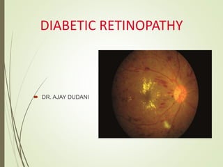
Diabetic retinopathy is damage to the blood vessels of the retina due to diabetes. It can progress to proliferative diabetic retinopathy where new abnormal blood vessels grow on the retina or optic disc which can lead to vision loss. A clinical trial studied whether initial treatment of proliferative diabetic retinopathy with injections of the anti-VEGF drug ranibizumab followed by deferred pan-retinal photocoagulation if needed could provide visual outcomes equal to or better than prompt pan-retinal photocoagulation. At two years, vision was maintained or improved in the ranibizumab group while remaining unchanged in the laser group, suggesting anti-VEGF therapy may delay or reduce the need for laser treatment
updating in diabetic macular edema including old and new approach era, including DRCR protocol
how to approach, how to treat, when to surgery
plus knownledge about anti-VEGF therapy up to date
Novel Development in treatment of Diabetic Macular Edema, by Dr. Fritz Allen, presented at VO, Lecture Series 11, Feb 20, 2011
COPE Course ID: 30657-PS
Lecture on Management of Cataract Surgery and Diabetes Mellitus. 2010 World Congress, American Society of Cataract & Refractive Surgery. Boston, MA 2010
Retinal vein occlusion (RVO) is an obstruction of the retinal venous system by thrombus formation and may involve the central, hemi-central or branch retinal vein.
The most common aetiological factor is compression by adjacent atherosclerotic retinal arteries.
Other possible causes are external compression or disease of the vein wall e.g. vasculitis.
In this case-based presentation, Dr. Lori Myers unscrambles the alphabet soup of Diabetic Retinopathy, providing clear explanations and outstanding images to describe the diagnosis, risk stratification, and treatment of diabetic retinopathy.
Use of digital retinal camera to detect prevalence and severity of diabetic ...Riyad Banayot
ABStrAct
BAckGround: The purpose of this study was to determine the prevalence of diabetic retinopathy among Palestinian
refugees serviced by the Diabetic Retinopathy Screening Program in the Occupied Palestinian Territories (DRS-
-OPT).
MAterIAl And MethodS: This is a retrospective study of retinal images of 1891 diabetic patients in 15 urban
UNRWA clinics participating in the DRS-OPT program in Palestine over 12 months. A nonmydriatic Canon CR-2
fundus retinal camera was used to capture two 450 non-stereo fundus images for each eye. Qualified graders (nurses)
performed the grading based on the DRS-OPT grading system.
reSultS: Out of the 1891 diabetic patients screened, 1694 had at least one gradable eye. 16% of patients had
diabetic retinopathy (5.7% had mild nonproliferative diabetic retinopathy, 4.3% had moderate nonproliferative
diabetic retinopathy, 1.1% had severe, moderate nonproliferative diabetic retinopathy, and 1.7% had proliferative
diabetic retinopathy. Maculopathy without retinopathy amounted to 3%. Other findings included the identification
of blinding diseases such as age-related macular degeneration and optic disc glaucomatous cupping.
concluSIonS: The retinopathy screening program using a nonmydriatic fundus camera identified diabetic retino-
pathy in 16% of diabetic Palestinian refugees. A total of 72% of these patients were diabetics with nonproliferative
retinopathy. This program can be used to prevent progression by facilitating the education of patients and early
intervention.
An Amalgamation-Based System for Micro aneurysm Detection and Diabetic Retino...IJMER
We propose an ensemble-based framework to improve microaneurysm detection. Unlike
the well-known approach of considering the output of multiple classifiers, we propose a combination of
internal components of microaneurysm detectors, namely preprocessing methods and candidate
extractors. We have evaluated our approach for microaneurysm detection in an online competition,
where this algorithm is currently ranked as first, and also on two other databases.
Similar to Diabetic retinopathy for GENERAL OPHTHALMOLOGIST (20)
Factory Supply Best Quality Pmk Oil CAS 28578–16–7 PMK Powder in Stockrebeccabio
Factory Supply Best Quality Pmk Oil CAS 28578–16–7 PMK Powder in Stock
Telegram: bmksupplier
signal: +85264872720
threema: TUD4A6YC
You can contact me on Telegram or Threema
Communicate promptly and reply
Free of customs clearance, Double Clearance 100% pass delivery to USA, Canada, Spain, Germany, Netherland, Poland, Italy, Sweden, UK, Czech Republic, Australia, Mexico, Russia, Ukraine, Kazakhstan.Door to door service
Hot Selling Organic intermediates
Couples presenting to the infertility clinic- Do they really have infertility...Sujoy Dasgupta
Dr Sujoy Dasgupta presented the study on "Couples presenting to the infertility clinic- Do they really have infertility? – The unexplored stories of non-consummation" in the 13th Congress of the Asia Pacific Initiative on Reproduction (ASPIRE 2024) at Manila on 24 May, 2024.
Tom Selleck Health: A Comprehensive Look at the Iconic Actor’s Wellness Journeygreendigital
Tom Selleck, an enduring figure in Hollywood. has captivated audiences for decades with his rugged charm, iconic moustache. and memorable roles in television and film. From his breakout role as Thomas Magnum in Magnum P.I. to his current portrayal of Frank Reagan in Blue Bloods. Selleck's career has spanned over 50 years. But beyond his professional achievements. fans have often been curious about Tom Selleck Health. especially as he has aged in the public eye.
Follow us on: Pinterest
Introduction
Many have been interested in Tom Selleck health. not only because of his enduring presence on screen but also because of the challenges. and lifestyle choices he has faced and made over the years. This article delves into the various aspects of Tom Selleck health. exploring his fitness regimen, diet, mental health. and the challenges he has encountered as he ages. We'll look at how he maintains his well-being. the health issues he has faced, and his approach to ageing .
Early Life and Career
Childhood and Athletic Beginnings
Tom Selleck was born on January 29, 1945, in Detroit, Michigan, and grew up in Sherman Oaks, California. From an early age, he was involved in sports, particularly basketball. which played a significant role in his physical development. His athletic pursuits continued into college. where he attended the University of Southern California (USC) on a basketball scholarship. This early involvement in sports laid a strong foundation for his physical health and disciplined lifestyle.
Transition to Acting
Selleck's transition from an athlete to an actor came with its physical demands. His first significant role in "Magnum P.I." required him to perform various stunts and maintain a fit appearance. This role, which he played from 1980 to 1988. necessitated a rigorous fitness routine to meet the show's demands. setting the stage for his long-term commitment to health and wellness.
Fitness Regimen
Workout Routine
Tom Selleck health and fitness regimen has evolved. adapting to his changing roles and age. During his "Magnum, P.I." days. Selleck's workouts were intense and focused on building and maintaining muscle mass. His routine included weightlifting, cardiovascular exercises. and specific training for the stunts he performed on the show.
Selleck adjusted his fitness routine as he aged to suit his body's needs. Today, his workouts focus on maintaining flexibility, strength, and cardiovascular health. He incorporates low-impact exercises such as swimming, walking, and light weightlifting. This balanced approach helps him stay fit without putting undue strain on his joints and muscles.
Importance of Flexibility and Mobility
In recent years, Selleck has emphasized the importance of flexibility and mobility in his fitness regimen. Understanding the natural decline in muscle mass and joint flexibility with age. he includes stretching and yoga in his routine. These practices help prevent injuries, improve posture, and maintain mobilit
Explore natural remedies for syphilis treatment in Singapore. Discover alternative therapies, herbal remedies, and lifestyle changes that may complement conventional treatments. Learn about holistic approaches to managing syphilis symptoms and supporting overall health.
These lecture slides, by Dr Sidra Arshad, offer a quick overview of physiological basis of a normal electrocardiogram.
Learning objectives:
1. Define an electrocardiogram (ECG) and electrocardiography
2. Describe how dipoles generated by the heart produce the waveforms of the ECG
3. Describe the components of a normal electrocardiogram of a typical bipolar leads (limb II)
4. Differentiate between intervals and segments
5. Enlist some common indications for obtaining an ECG
Study Resources:
1. Chapter 11, Guyton and Hall Textbook of Medical Physiology, 14th edition
2. Chapter 9, Human Physiology - From Cells to Systems, Lauralee Sherwood, 9th edition
3. Chapter 29, Ganong’s Review of Medical Physiology, 26th edition
4. Electrocardiogram, StatPearls - https://www.ncbi.nlm.nih.gov/books/NBK549803/
5. ECG in Medical Practice by ABM Abdullah, 4th edition
6. ECG Basics, http://www.nataliescasebook.com/tag/e-c-g-basics
Title: Sense of Smell
Presenter: Dr. Faiza, Assistant Professor of Physiology
Qualifications:
MBBS (Best Graduate, AIMC Lahore)
FCPS Physiology
ICMT, CHPE, DHPE (STMU)
MPH (GC University, Faisalabad)
MBA (Virtual University of Pakistan)
Learning Objectives:
Describe the primary categories of smells and the concept of odor blindness.
Explain the structure and location of the olfactory membrane and mucosa, including the types and roles of cells involved in olfaction.
Describe the pathway and mechanisms of olfactory signal transmission from the olfactory receptors to the brain.
Illustrate the biochemical cascade triggered by odorant binding to olfactory receptors, including the role of G-proteins and second messengers in generating an action potential.
Identify different types of olfactory disorders such as anosmia, hyposmia, hyperosmia, and dysosmia, including their potential causes.
Key Topics:
Olfactory Genes:
3% of the human genome accounts for olfactory genes.
400 genes for odorant receptors.
Olfactory Membrane:
Located in the superior part of the nasal cavity.
Medially: Folds downward along the superior septum.
Laterally: Folds over the superior turbinate and upper surface of the middle turbinate.
Total surface area: 5-10 square centimeters.
Olfactory Mucosa:
Olfactory Cells: Bipolar nerve cells derived from the CNS (100 million), with 4-25 olfactory cilia per cell.
Sustentacular Cells: Produce mucus and maintain ionic and molecular environment.
Basal Cells: Replace worn-out olfactory cells with an average lifespan of 1-2 months.
Bowman’s Gland: Secretes mucus.
Stimulation of Olfactory Cells:
Odorant dissolves in mucus and attaches to receptors on olfactory cilia.
Involves a cascade effect through G-proteins and second messengers, leading to depolarization and action potential generation in the olfactory nerve.
Quality of a Good Odorant:
Small (3-20 Carbon atoms), volatile, water-soluble, and lipid-soluble.
Facilitated by odorant-binding proteins in mucus.
Membrane Potential and Action Potential:
Resting membrane potential: -55mV.
Action potential frequency in the olfactory nerve increases with odorant strength.
Adaptation Towards the Sense of Smell:
Rapid adaptation within the first second, with further slow adaptation.
Psychological adaptation greater than receptor adaptation, involving feedback inhibition from the central nervous system.
Primary Sensations of Smell:
Camphoraceous, Musky, Floral, Pepperminty, Ethereal, Pungent, Putrid.
Odor Detection Threshold:
Examples: Hydrogen sulfide (0.0005 ppm), Methyl-mercaptan (0.002 ppm).
Some toxic substances are odorless at lethal concentrations.
Characteristics of Smell:
Odor blindness for single substances due to lack of appropriate receptor protein.
Behavioral and emotional influences of smell.
Transmission of Olfactory Signals:
From olfactory cells to glomeruli in the olfactory bulb, involving lateral inhibition.
Primitive, less old, and new olfactory systems with different path
Anti ulcer drugs and their Advance pharmacology ||
Anti-ulcer drugs are medications used to prevent and treat ulcers in the stomach and upper part of the small intestine (duodenal ulcers). These ulcers are often caused by an imbalance between stomach acid and the mucosal lining, which protects the stomach lining.
||Scope: Overview of various classes of anti-ulcer drugs, their mechanisms of action, indications, side effects, and clinical considerations.
Title: Sense of Taste
Presenter: Dr. Faiza, Assistant Professor of Physiology
Qualifications:
MBBS (Best Graduate, AIMC Lahore)
FCPS Physiology
ICMT, CHPE, DHPE (STMU)
MPH (GC University, Faisalabad)
MBA (Virtual University of Pakistan)
Learning Objectives:
Describe the structure and function of taste buds.
Describe the relationship between the taste threshold and taste index of common substances.
Explain the chemical basis and signal transduction of taste perception for each type of primary taste sensation.
Recognize different abnormalities of taste perception and their causes.
Key Topics:
Significance of Taste Sensation:
Differentiation between pleasant and harmful food
Influence on behavior
Selection of food based on metabolic needs
Receptors of Taste:
Taste buds on the tongue
Influence of sense of smell, texture of food, and pain stimulation (e.g., by pepper)
Primary and Secondary Taste Sensations:
Primary taste sensations: Sweet, Sour, Salty, Bitter, Umami
Chemical basis and signal transduction mechanisms for each taste
Taste Threshold and Index:
Taste threshold values for Sweet (sucrose), Salty (NaCl), Sour (HCl), and Bitter (Quinine)
Taste index relationship: Inversely proportional to taste threshold
Taste Blindness:
Inability to taste certain substances, particularly thiourea compounds
Example: Phenylthiocarbamide
Structure and Function of Taste Buds:
Composition: Epithelial cells, Sustentacular/Supporting cells, Taste cells, Basal cells
Features: Taste pores, Taste hairs/microvilli, and Taste nerve fibers
Location of Taste Buds:
Found in papillae of the tongue (Fungiform, Circumvallate, Foliate)
Also present on the palate, tonsillar pillars, epiglottis, and proximal esophagus
Mechanism of Taste Stimulation:
Interaction of taste substances with receptors on microvilli
Signal transduction pathways for Umami, Sweet, Bitter, Sour, and Salty tastes
Taste Sensitivity and Adaptation:
Decrease in sensitivity with age
Rapid adaptation of taste sensation
Role of Saliva in Taste:
Dissolution of tastants to reach receptors
Washing away the stimulus
Taste Preferences and Aversions:
Mechanisms behind taste preference and aversion
Influence of receptors and neural pathways
Impact of Sensory Nerve Damage:
Degeneration of taste buds if the sensory nerve fiber is cut
Abnormalities of Taste Detection:
Conditions: Ageusia, Hypogeusia, Dysgeusia (parageusia)
Causes: Nerve damage, neurological disorders, infections, poor oral hygiene, adverse drug effects, deficiencies, aging, tobacco use, altered neurotransmitter levels
Neurotransmitters and Taste Threshold:
Effects of serotonin (5-HT) and norepinephrine (NE) on taste sensitivity
Supertasters:
25% of the population with heightened sensitivity to taste, especially bitterness
Increased number of fungiform papillae
ARTIFICIAL INTELLIGENCE IN HEALTHCARE.pdfAnujkumaranit
Artificial intelligence (AI) refers to the simulation of human intelligence processes by machines, especially computer systems. It encompasses tasks such as learning, reasoning, problem-solving, perception, and language understanding. AI technologies are revolutionizing various fields, from healthcare to finance, by enabling machines to perform tasks that typically require human intelligence.
- Video recording of this lecture in English language: https://youtu.be/lK81BzxMqdo
- Video recording of this lecture in Arabic language: https://youtu.be/Ve4P0COk9OI
- Link to download the book free: https://nephrotube.blogspot.com/p/nephrotube-nephrology-books.html
- Link to NephroTube website: www.NephroTube.com
- Link to NephroTube social media accounts: https://nephrotube.blogspot.com/p/join-nephrotube-on-social-media.html
Report Back from SGO 2024: What’s the Latest in Cervical Cancer?bkling
Are you curious about what’s new in cervical cancer research or unsure what the findings mean? Join Dr. Emily Ko, a gynecologic oncologist at Penn Medicine, to learn about the latest updates from the Society of Gynecologic Oncology (SGO) 2024 Annual Meeting on Women’s Cancer. Dr. Ko will discuss what the research presented at the conference means for you and answer your questions about the new developments.
TEST BANK for Operations Management, 14th Edition by William J. Stevenson, Ve...kevinkariuki227
TEST BANK for Operations Management, 14th Edition by William J. Stevenson, Verified Chapters 1 - 19, Complete Newest Version.pdf
TEST BANK for Operations Management, 14th Edition by William J. Stevenson, Verified Chapters 1 - 19, Complete Newest Version.pdf
2. Diabetic Retinopathy
Damage to the blood vessels in the retina due to diabetes.
https://www.contouravisionindia.com/single-post/What-causes-diabetic-retinopathy as visited on 13th Jan 2020
Biyani RS, Patre BM. Algorithms for red lesion detection in Diabetic Retinopathy: A review. Biomedicine &
3. Projections of DR :How big is the
problem going to be ?
https://www.nei.nih.gov/learn-about-eye-health/resources-for-health-educators/eye-health-data-and-
Between 2010 and 2050, It is
expected that the number of
people with the most common
eye diseases to double, and it will
rise from 7.7 Mn to 14.6 Mn
4. Global Burden of Diabetic
Retinopathy (DR)
35 studies = 22,896 patients 21
Among those with diabetes:
• 34.6% with any DR
• 6.95% with proliferative DR
• 6.81% with diabetic macular
edema
• 10.2% with vision-threatening DR
Among those with diabetes, increased
risk of diabetic retinopathy:
• Longer duration of diabetes
• Poorer glycemic control
• Poorer blood pressure control
• Poorer control of blood cholesterol levels
https://www.nei.nih.gov/learn-about-eye-health/resources-for-health-educators/eye-health-data-and-
DR
35%
PDR
7%
DME
7%
Vision
Threatnein
g DR
10%
6. Diabetic Eye Disease
Key Points
k best
• Treatme
before v
nts exist but wo
ision is lost
RECOMMENDED EYE EXAMINATION SCHEDULE
Diabetes type Recommended time
of first examination
Recommended
follow-up*
Type 1 3-5 years after diagnosis Yearly
Type 2 At time of diagnosis Yearly
Prior to pregnancy
(type 1 or type 2)
Prior to conception and
early in the first trimester
No retinopathy to mild-
moderate NPDR -
every 3-12 months
rSevere NPDR or worse -
every 1-3 months
*Abnormal findings may dictate more frequent follow-up examinations
7. • Microangiopathy due to hyperglycemia
• Endothelial barrier decompensation leads to serum leakage and
retinal edema
• Later stages, VEGF
produced by ischemic
retina causes
neovascularisation
PATHOGENESIS
8. Diabetic Retinopathy
Five pathologic processes:
• Formation of micro aneurysms (outpouchings of the small
vessels)
• Excessive vascular permeability (leakage)
• Vascular occlusions (closure of blood vessels)
• Proliferation of new vessels (± hemorrhage)
• Contraction of new blood vessels: Scarring, retinal
detachment
https://www.nei.nih.gov/learn-about-eye-health/resources-for-health-educators/eye-health-data-and-
9. NON-PROLIFERATIVE DIABETIC RETINOPATHY (NPDR)
NO DR Review in 12 months
VERY MILD
Microaneurysms only
Review most patients in 12 months
MILD
Any or all of:
microaneurysms, retinal hemorrhages, exudates, cotton
wool spots
Review range 6-12 months, depending on
severity of signs, stability, systemic factors,
and patient’s personal circumstances
MODERATE
Severe retinal haemorrhages in 1-3 quadrants or mild IRMA
Significant venous beading in no more than 1 quadrant
Cotton wool spots
Review in approximately 6 months
(PDR in up to 26%, high-risk PDR in up to 8%
within a year)
SEVERE
The 4-2-1 rule-
Severe retinal haemorrhages in all 4 quadrants
Significant venous beading in ≥2 quadrants
Moderate IRMA in ≥1 quadrants
Review in 4 months
(PDR in up to 50%, high-risk PDR in up to 15%
within a year)
VERY SEVERE
≥2 of the criteria for severe
Review in 2-3 months
(High-risk PDR in up to 45% within a year)
ABBREVIATED EARLY TREATMENT DIABETIC RETINOPATHY STUDY (ETDRS) CLASSIFICATION
CATEGORY MANAGEMENT
10. CATEGORY MANAGEMENT
PROLIFERATIVE DIABETIC RETINOPATHY (PDR)
MILD-MODERATE
New vessels on the disc (NVD) < 1/3 disc area
New vessels elsewhere (NVE) < 1/2 disc area
Treatment considered according to severity of signs,
stability, systemic factors, and patient’s personal
circumstances
If not treated, review in up to 2 months
HIGH-RISK
NVD > 1/3 disc area
Any NVD with vitreous or preretinal
hemorrhage
NVE >1/2 disc area with vitreous or preretinal
hemorrhage
Laser photocoagulation
Intravitreal anti-VEGF agents
Intravitreal triamcinolone
Pars plana vitrectomy
Lipid lowering drugs
ADVANCED DIABETIC EYE DISEASE
Preretinal (retrohyaloid) and/or intragel
hemorrhage
Tractional retinal detachment
Tractional retinoschisis
Rubeosis iridis (iris neovascularisation)
Pars plana vitrectomy
PDR – formation of new vessels or fibrovascular tissue on the optic disc or inner
retina
11. OTHER CATEGORIES
• BACKGROUND DIABETIC RETINOPATHY (BDR)
It’s the earliest phase of DR.
Characterised by microaneurysms, dot and blot hemorrhages and exudates.
• DIABETIC MACULOPATHY
Refers to presence of any retinopathy at the macula.
• PRE-PROLIFERATIVE DIABETIC RETINOPATHY (PPDR)
Cotton wool spots, venous changes, IRMA and deep retinal hemorrhages.
• DIABETIC PAPILLOPATHY
It is a form of optic neuropathy seen in young type I diabetics. It is unrelated to
glycemic control or any other known feature of diabetes.
12. APPROXIMATE EQUIVALENCE OF
THE CLASSIFICATION
SYSTEMS
ETDRS NSC SDRGS AAO RCOphth
10 - None R0 - None R0 - None No apparent retinopathy None
20 - Microaneurysms only R1 - Background R1 - BDR Mild NPDR Low risk
35 - Mild NPDR Moderate NPDR
43 - Moderate NPDR R2 - Pre-
proliferative
R2 Moderate BDR High risk
47 - Moderately severe
NPDR
53 A-D - Severe NPDR R3 - Severe BDR Severe NPDR
53 E - Very severe NPDR
61 - Mild PDR
65 - Moderate PDR
R3 - Proliferative R4 - PDR PDR PDR
71, 75 - High risk PDR
81, 85 - Advanced PDR
18. DIABETIC MACULAR OEDEMA
• Most common cause of visual impairment in diabetic retinopathy
• According to the Wisconsin Epidemiologic Study of Diabetic
Retinopathy (WESDR), the prevalence rate of macular oedema is 10 %
in the diabetic population.
• Best detected by slit-lamp biomicroscopy and stereoscopic fundus
photography.
19. Retinal thickening
within 500 µm of
centre of macula
Exudates within
500 µm of centre
of macula, if
associated with
retinal thickening
Retinal thickening one disc area (1500 µm)
or larger, any part for which is within
one disc diameter of centre of macula
CLINICALLY SIGNIFICANT MACULAR EDEMA (CSME)
As defined by ETDRS-
20. OCT
A. Diffuse edema
B. Cystoid macular edema
C. Serous retinal detachment
D. Posterior hyaloid traction
22. Diabetic Retinopathy National Institutes
of Health-supported Clinical Trials
https://www.nei.nih.gov/learn-about-eye-health/resources-for-health-educators/eye-health-data-and-
23. DIABETIC RETINOPATHY STUDY
(DRS)
ELIGIBILITY CRITERIA
1. Visual acuity ≥ 20/100 (6/36) in each eye
2. PDR in at least one eye or severe NPDR in both
3. Both eyes suitable for photocoagulation
STUDY DESIGN
One eye of each patient was assigned randomly to photocoagulation. The
other eye was assigned to follow-up without photocoagulation
CONCLUSIONS
1. Photocoagulation reduced the risk of severe visual loss by 50 % or more
2. Modest risks of decrease in visual acuity and visual field
3. Treatment benefit outweighs risks for eye with high-risk PDR
24. EARLY TREATMENT
DIABETIC RETINOPATHY
STUDY (ETDRS)
ELIGIBILITY CRITERIA
1. Visual acuity ≥ 20/40 (6/12) {20/400 (60/120) if reduction caused by macular
oedema}
2. Mild NPDR to non-high risk PDR, with or without macular oedema
3. Both eyes suitable for photocoagulation
STUDY DESIGN
1. One eye of each patient was assigned randomly to early photocoagulation and
the to deferral (careful follow-up and photocoagulation if high risk PDR
develops).
2. Patients assigned randomly to aspirin or placebo.
25. CONCLUSIONS
1. Focal photocoagulation
- reduced the risk of moderate visual loss by 50 % or more
- increased the chance of a small improvement in visual acuity
- reduced retinal thickening
2. Early scatter photocoagulation
- small reduction in risk of severe vision loss
- not indicated in mild to moderate retinopathy
- most effective in type 2 diabetes mellitus
3. Aspirin did not alter the progression of diabetic retinopathy
26. DIABETIC RETINOPATHY
VITRECTOMY STUDY
(DRVS)
ELIGIBILITY CRITERIA
1. Visual acuity ≤ 5/200 (5/60)
2. Vitreous hemorrhage consistent with visual acuity, duration 1-6 months
3. Macula attached
STUDY DESIGN
Eligible eye or eyes assigned randomly to early vitrectomy or conventional
management (vitrectomy if center of macula detaches or if vitreous hemorrhage
persists for 1 year, photocoagulation as needed and as possible)
CONCLUSIONS
Chances of recovery of VA ≥ 10/20 (3/6) increased by early vitrectomy, at least
in patients with type I diabetes, who were younger and had more severe PDR
27. GROUP NR – Very Severe PDR with Useful Vision
MAJOR ELIGIBILITY CRITERIA
1. Visual acuity ≥ 10/200 (3/60)
2. Center of macula attached
3. Extensive, active, neovascular, or fibrovascular proliferations
MAJOR DESIGN FEATURES
Same as Group H (except conventional management included vitrectomy
after a 6 months waiting period in eyes that developed severe VH)
MAJOR CONCLUSIONS
Chances of of VA ≥ 10/20 (3/6) increased by early vitrectomy, at least for eyes
with severe new vessels.
Early vitrectomy for eyes with recent severe VH and VA < 5/200 (5/60) was
beneficial, especially for patients with type I DM. Furthermore, the chances of
achieving VA of 10/20 (3/6) or better increased when early vitrectomy was
performed in eyes with severe new vessels, again especially for patients with
type I DM.
28. Treatments for Diabetic
Retinopathy
Standard therapies:
• Laser photocoagulation
• Surgical intervention (vitrectomy)
• Medical therapies delivered into
the eye (intravitreal injections*)
• Systemic medical therapies
involving blood sugar, blood
pressure, and cholesterol control
* Note : Some drugs are used as an off label indication for the
management of DR
Berco E, Rappoport D, Pollack A, Kleinmann G, Greenwald Y. Management of Diabetic Retinopathy and Other Ocular Complications in Type 1
29. Background :
Laser and supplemental therapy for
management of DR
Protocol S:
This study showed that ranibizumab injections are effective in treating proliferative
diabetic retinopathy.
At two years, vision of the ranibizumab group on average improved by half a line on
an eye chart.
Vision of the laser group remained unchanged
• Current treatment for PDR is pan-retinal photocoagulation (PRP)
Inherently destructive
Adverse effects on visual function
• Some eyes with PDR+DME now receive anti-VEGF as standard care for DME
• Would initial treatment of PDR with intravitreal anti-VEGF delay or prevent need for
PRP?
30. STUDY OBJECTIVE AND
TREATMENT GROUPS
Prompt
PRP
To determine if visual acuity outcomes at 2 years
in eyes with PDR (with or without concurrent
DME) that receive anti-VEGF therapy with
deferred PRP are non-inferior to those in eyes
that receive prompt PRP therapy.
0.5mg
ranibizuma
b with
deferred
PRP
(Note: Study ranibizumab may be given as needed for DME using Protocol I
retreatment as guidelines.)
Bressler NM, Beck RW, Ferris III FL. Panretinal photocoagulation for proliferative diabetic retinopathy. New England Journal of Medicine.
2011 Oct 20;365(16):1520-6.
31. Endpoints for measuring the
outcome
Primary End Point
Is visual acuity using ranibizumab for PDR not worse than treatment with
PRP at 2 years?
Non-inferiority margin of 5 letters
Secondary End Point
Are there potential benefits of ranibizumab on:
Vision throughout follow-up (area under the curve)
Peripheral vision
Macular edema
Incidence of vitrectomy
32. Follow-up Schedule
Baseline to
1 Year
PRP group: Visits every 16
weeks*
Ranibizumab group: Visits
every 4 weeks to assess for
PDR treatment
Both groups simultaneously
evaluated for DME treatment
1 Year to
2Years
PRP group: Visits every 16
weeks*
Ranibizumab group: Visits
every 4wk to 16wk to assess
for PDR treatment
Interval is extended if
injections for PDR and DME
deferred (“Defer and Extend”)
Bressler NM, Beck RW, Ferris III FL. Panretinal photocoagulation for proliferative diabetic retinopathy. New England Journal of Medicine.
2011 Oct 20;365(16):1520-6.
*Eyes with DME could be seen more frequently for DME
treatment as needed.
33. Ranibizu
mab
Group
N = 191
N = 160
(84%)
N = 88%
N = 22
(18, 24)
Randomization
Participants:
N = 304
Eyes: N = 394
PRP Group
N = 203
N =
168 (83%)
N =
86%
2-
Years
2-Years
Excludi
ng
Death
Baseli
ne
N =
16 (9, 22)
Median
(Quartiles) No.
Visits over 2
years
Bressler NM, Beck RW, Ferris III FL. Panretinal photocoagulation for proliferative diabetic retinopathy. New England Journal of Medicine.
2011 Oct 20;365(16):1520-6.
34. Baseline Characteristics
Ranibizu
mab
Group
(N = 191)
PRP
Group
(N = 203)
Age (yrs) – Median 52 51
Women 43% 45%
Race
White 52% 50%
Type 2 diabetes 73% 76%
Duration of Diabetes
(yrs)
18 17
Median HbA1c (%) 8.6 8.9
35. Ocular Baseline Characteristics
Ranibizu
mab
Group
(N = 189)
PRP
Group
(N = 199)
Diabetic Retinopathy Severity by Reading Center
NPDR† 10% 13%
Mild to moderate
PDR
52% 49%
High risk PDR to
advanced PDR
38% 37%
† There were 46 eyes (12%) for which NV was not identified by the reading center on the submitted color images or
quality precluded identification. In 29 of these cases (63%), subsequent review of additional images (e.g. FA) confirmed
NV, leaving 17 (4%) of 394 subjects with no photographic documentation of PDR.
36. Ocular Baseline Characteristics
Ranibizum
ab
Group
(N = 189)
PRP
Group
(N = 201)
Mean OCT CST* (µm) 262 249
< 250 µm 66% 67%
250 to 349 µm 19% 26%
≥ 350 µm 15% 7%
Presence of central-
involved DME with VA
loss**
22% 23%
*OCT values are Stratus equivalents
**Eyes with visual acuity letter score ≤ 78 (20/32 or worse) AND OCT CST ≥ machine and gender
Required ranibizumab at baseline
38. PRP Group
Overall
(N =
203)
Completed initial full
PRP
98%
Performed in 1
Sitting
54%
Baseline PRP
Overall
(N = 203)
Eyes given additional PRP
(after completing initial full
PRP)
45%
Distribution of timing to additional PRP
From completion of initial full
PRP:
median time to additional
PRP
~7 months
Additional PRP
39. Ranibizumab Group
# of Ranibizumab Injections
Eyes With
Baseline DME
(N = 36)
Eyes Without
Baseline DME
(N = 133)
Prior to 1-year Visit (Max possible = 13)
Median 9 7
Mean 8.9 6.9
Prior to 2-year visit (Max possible= 26)
Median 14 10
Mean 13.3 10.1
Note: 97% of protocol-required injections for PDR were given
Overall
N = 191
Received PRP* before 2
years
12 (6%)
Received PRP for PDR
*1 met failure criteria, 1 with Protocol Chair approval,
1 without Chair approval, 8 during vitrectomy (e.g., via
endolaser), and 1 by non-study physician
40. Mean Change in Visual Acuity
Outlying values were truncated to 3 SD from the mean
-5
0
5
10
15
0 16 32 52 68 84 104
Mean
Visual
Acuity
Change
(Letter
Score)
Visit Week
Ranibizumab Group PRP Group
N =
N =
203
+
2.8
+ 0.2
N =
168
N =
160
2-Year Adjusted
Mean Difference:
+2.2 letters
95% Confidence
Interval: (-0.5, +5.0)
(Meets pre-specified
non-inferiority
criterion: lower
bounds of the 95%
CI of -0.5 letters was
greater than the non-
inferiority limit of -
5.0 letters)
41. -4
1
6
11
0 16 32 52 68 84 104
Mean
Visual
Acuity
Change
(Letter
Score)
Visit Week
With “Baseline DME”
Ranibizumab Group PRP Group
+
+7.
-5
0
5
10
0 16 32 52 68 84 104
Visit Week
Without “Baseline
DME”
-
0.5
+1.
8
N = 42 N = 33N =
147
N = 46 N = 37N =
155
N =
130
N =
126
*Outlying values were truncated to 3 SD from the
an Change in Visual Acuity
Stratified by Baseline DMEMe
42. Discussion
DRCR.net Protocol S (PRP vs. Ranibizumab
for PDR):
Treatment with 0.5-mg ranibizumab met primary non-inferiority outcome for
VA being no worse than PRP
Summary of Ranibizumab group results vs. PRP:
Mean change in VA from baseline to 2-years with ranibizumab no worse
than with PRP
Superior mean visual acuity over course of 2-years (area under the curve
analysis)
Superior mean visual field outcomes
Decreased occurrence of vitrectomies
Decreased development of central involved DME
PRP rarely given for failure or futility of ranibizumab
43. Discussion
No systemic safety concerns with ranibizumab
identified among pre-specified major safety outcomes
Increased frequency of adverse events defined by
cardiac, endocrine, respiratory, infections/infestations,
skin and surgical conditions MedDRA systems in
ranibizumab groups could be real, due to chance, or
due to ascertainment bias (more visits in ranibizumab
group)
Interpretation of systemic safety difficult since large
proportion of PRP group received ranibizumab per
protocol for DME
44. SYSTEMIC MANAGEMENT
• Glycemic control – Insulin, OHG
• Blood pressure control – Anti-hypertensive medications
• Cholesterol control – Statins, Fibrates
• Support renal function – ACEI, ARB
• Lifestyle modification – Smoking and alcohol cessation, exercise ,weight
control
45. DIABETES CONTROL &
COMPLICATION TRIAL (DCCT)
STUDY GROUP
Intensive management of blood glucose (multiple daily
insulin injection) vs conventional management
CONCLUSIONS
intensive control reduced the risk of developing retinopathy by
76% and slowed progression of retinopathy by 54%.
Intensive control reduced the risk of clinical neuropathy by
60% and albuminuria (Nephropathy) by 54%
46. UNITED KINGDOM
PROSPECTIVE DIABETES
STUDY (UKPDS)
Patients were assigned to a conventional policy starting
with diet or to an intensive policy starting with a sulfonyl
urea or insulin. If overweight and in the intensive group,
patients were assigned to start treatment with metformin
Patients were randomly assigned to tight control of BP
(ACE inhibitor or beta blocker) or to less tight control
47. RESULTS
Intensive control of blood glucose level slowed progression
of retinopathy and reduced other microvascular
complications
Intensive control of BP slowed progression of diabetic
reinopathy and reduced other microvascular &
macrovascular complications
48. PROMINENT-Eye Ancillary Study
To assess whether treatment with pemafibrate (0.2 mg
orally BID) compared with placebo reduces the hazard rate
of diabetic retinopathy worsening in adults with type 2
diabetes and diabetic retinopathy without
neovascularization in at least one eye who are participating
in the parent PROMINENT trial.
FIELD (Fenofibrate Intervetion and Event Lowering in
Diabetes) and The Action to Control Cardiovascular Risk in
Diabetes (ACCORD)-eye study, have demonstrated
clinically important reduction in progression of retinopathy in
patients with diabetes assigned to fibrate compared with
placebo.
50. 1. ARGON LASER (514.5 nm)
• All eyes with CSMO should be considered for laser photocoagulation
irrespective of the level of visual acuity.
• Reduces the risk of visual loss by 50%.
• Two types- focal and grid
i. Focal treatment - Burns are applied to microaneurysms and microvascular
lesions in the centre of rings of exudates located 500-3000 µm from the
centre of the macula.
51. ii. Grid treatment – Burns are applied
to areas of diffuse retinal thickening
more than 500 µm from the centre
of the macula and 500 µm from the
temporal margin of the optic disc.
Spot size - 50-100 µm
Exposure time – 0.1-0.5sec
• 70% of eyes achieve stable visual acuity after laser photocoagulation
• 15% show improvement
• 15% subsequently deteriorate.
53. LASER SETTINGS
• SPOT SIZE depends on the contact lens used.
With Goldmann lens spot size is set at 200-500 µm, but with a
panfundoscopic-type lens it is set at 100-300 µm because of induced
magnification.
Other lenses used are – Volk, Mainster, Rodenstock
• DURATION OF BURN - 0.05-0.1 sec
• POWER - 250-570 mW, sufficient to produce only a light intensity burn
causing stimulation of the retinal pigment epithelium. The end point is a
whitening or darkening of the microaneurysms.
54. ASSESSMENT AFTER LASER PHOTOCOAGULATION
GOOD INVOLUTION POOR INVOLUTION
• Regression of neovascularization leaving
‘ghost’ vessels or fibrous tissue
• Decrease in venous changes
• Absorption of hemorrhages
• Disc pallor
• Persistent neovascularization
• Hemorrhage
58. BEVACIZUMAB
Recombinant humanized monoclonal antibody
Inhibits VEGF A & blocks angiogenesis
Approved for use in metastatic colon cancer, certain lung
cancer, renal and ovarian cancers
Not yet approved by FDA, off label use in ophthalmology
Dose: 1.25 mg in 0.05 ml