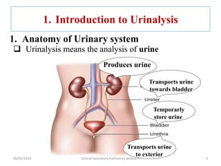
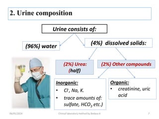
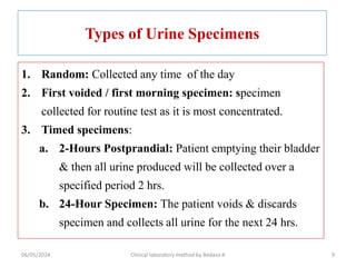
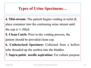
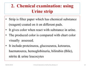
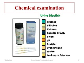
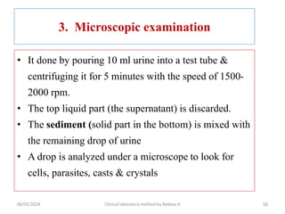
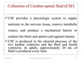
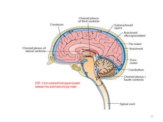
CHAPTER 6
Basic Urinalysis
06/05/2024 Clinical laboratory method by Bedasa A 1
Chapter objective
At the end of this chapter, students are expected
to:
To state different urine specimen type
Describe about urine formation and its
composition
Explain macroscopic, chemical and
microscopic examination on urine
06/05/2024 Clinical laboratory method by Bedasa A 2
1. Introduction to Urinalysis
 Urinalysis means the analysis of urine
3
Produces urine
Transports urine
towards bladder
Temporarly
store urine
Transports urine
to exterior
1. Anatomy of Urinary system
06/05/2024 Clinical laboratory method by Bedasa A
Significance of Urinalysis
1. Provides information about kidneys
2. Provides information about metabolic
processes
3. To test urinary tract infections (UTIs)
06/05/2024 Clinical laboratory method by Bedasa A 4
Introduction to Urinalysis……..
2. Physiology of Urinary System:
1. Excretion & Elimination of wastes products
(urea, creatinine, uric acid) through urine
2. Homeostatic regulation of water-salt & acid-
base Balance
3. Endocrine function: Hormones production
06/05/2024 Clinical laboratory method by Bedasa A 5
2. Urine
1. Urine Formation: there are four processes
I. Filtration: small molecules move from the
capillaries into the bowman's capsule
II. Re-absorption: molecules are reabsorbed from the
nephron into the capillary blood. e.g. Glucose
III. Secretion: substances are actively removed from
blood & added to tubular fluid. e.g. H+,
IV. Excretion: removal of waste products through urine
6
06/05/2024 Clinical laboratory method by Bedasa A
Urine consists of:
(96%) water
Inorganic:
• Cl-, Na, K.
• trace amounts of:
sulfate, HCO3 etc.)
(4%) dissolved solids:
(2%) Urea:
(half)
(2%) Other compounds
Organic:
• creatinine, uric
acid
7
06/05/2024 Clinical laboratory method by Bedasa A
Factors Affecting Urine Composition
1. Dietary intake
2. Physical activity
3. Body metabolism
4. Endocrine function
8
06/05/2024 Clinical laboratory method by Bedasa A
Types of Urine Specimens
1. Random: Collected any time of the day
2. First voided / first morning specimen: specimen
collected for routine test as it is most concentrated.
3. Timed specimens:
a. 2-Hours Postprandial: Patient emptying their bladder
& then all urine produced will be collected over a
specified period 2 hrs.
b. 24-Hour Specimen: The patient voids & discards
specimen and collects all urine for the next 24 hrs.
9
06/05/2024 Clinical laboratory method by Bedasa A
Types of Urine Specimens…
4. Mid-stream: The patient begins voiding in toilet &
place container into the continuing urine stream until
the cup is ½ filled.
5. Clean Catch: Prior to the voiding process, the
patient should be provided clean cup.
6. Catheterized Specimen: Collected from a hollow
tube threaded up the urethra into the bladder.
7. Supra-pubic needle aspiration: For culture purpose
10
06/05/2024 Clinical laboratory method by Bedasa A
Handling & Preserving Urine
Specimens
• When urine placed at room temperature
– Bacteria multiply
– Glucose decreases
– Casts and cellular elements decompose
• So, examine within 2 hrs of collection OR use of
preservatives like
A. Physical methods (Refrigeration @ 4-6° C for up to 8
hrs)
B. Chemical methods (boric acid, thymole, toluene,
formalin, chloroform, HCL, …)
11
06/05/2024 Clinical laboratory method by Bedasa A
Types of Urine examination = Urinalysis
1. Macroscopic examination
2. Chemical examination
3. Microscopic examination
12
06/05/2024 Clinical laboratory method by Bedasa A
1. Macroscopic/Physical/ Exam
• Physical examination of urine, usually give hint for the
subsequent urinalysis
• Physical properties include….
 Volume
 Color
 Transparency
 Odor
 Foam
06/05/2024 Clinical laboratory method by Bedasa A 13
2. Chemical examination: using
Urine strip
• Strip is filter paper which has chemical substance
(reagent) coated on it on different pads.
• It gives color when react with substance in urine.
• The produced color is compared with chart color
visually assessed.
• It include proteinurea, glucoseurea, ketourea,
haematourea, hemoglobinuria, bilirubin (Bile),
nitrite & urine leucocytes
06/05/2024 Clinical laboratory method by Bedasa A 14
Chemical examination
Glucose
Bilirubin
Ketones
Specific Gravity
Blood
pH
Protein
Urobilinogen
Nitrite
Leukocyte Esterase
Urine Dipstick
06/05/2024 Clinical laboratory method by Bedasa A 15
3. Microscopic examination
• It done by pouring 10 ml urine into a test tube &
centrifuging it for 5 minutes with the speed of 1500-
2000 rpm.
• The top liquid part (the supernatant) is discarded.
• The sediment (solid part in the bottom) is mixed with
the remaining drop of urine
• A drop is analyzed under a microscope to look for
cells, parasites, casts & crystals
16
06/05/2024 Clinical laboratory method by Bedasa A
Classification of Urinary Sediments
• Urine sediments can grossly be
categorized into two based on the
substances they are composed of.
1. Organized
2. Non-organized sediments
06/05/2024 Clinical laboratory method by Bedasa A 17
06/05/2024 Clinical laboratory method by Bedasa A 18
Microscopic Examination: Abnormal
Findings
Per High Power Field (HPF) (400x)
– > 3 erythrocytes
– > 5 leukocytes
– > 2 renal tubular cells
– > 10 bacteria
Per Low Power Field (LPF) (200x)
– > 3 hyaline casts or > 1 granular cast
– > 10 squamous cells (indicative of contaminated
specimen)
– Any other cast (RBCs, WBCs)
Presence of:
– Fungal hyphae or yeast, parasite, viral inclusions
– Pathological crystals (cystine, leucine, tyrosine)
– Large number of uric acid or calcium oxalate crystals
06/05/2024 Clinical laboratory method by Bedasa A 19
20
06/05/2024 Clinical laboratory method by Bedasa A
Collection of Cerebro-spinal fluid (CSF)
CSF provides a physiologic system to supply
nutrients to the nervous tissue, remove metabolic
wastes, and produce a mechanical barrier to
cushion the brain and spinal cord against trauma.
CSF is produced in the choroid plexuses of the
two lumbar ventricles and the third and fourth
ventricles. In adults, approximately 20 mL of
fluid is produced every hour.
06/05/2024 Clinical laboratory method by Bedasa A 21
22
CSF: in b/n subarachnoid space located
between the arachnoid and pia mater
CSF…
• Its volume is 90 to 150 mL in adults and 10 to 60 mL in neonates.
• The circulating fluid is reabsorbed back into the blood capillaries in
the arachnoid granulations/villae at a rate equal to its production
• Collection by lumbar puncture done by experienced medical
personnel
• About 1-2ml of CSF is collected for examination
– lumbar puncture is made from the space between the 3rd to 5th
lumbar vertebrae under sterile conditions.
06/05/2024 Clinical laboratory method by Bedasa A 23
Collecting CSF specimen
Location of CSF
Can be Collected in three sequentially
labeled tubes
 Tube 1-Chemical and immunologic tests
 Tube 2-Microbiology
 Tube 3-Hematology (gross examination,
total WBC & Diff)
 This is the least likely to contain cells
introduced by the puncture procedure
24
25
06/05/2024 Clinical laboratory method by Bedasa A

Chapter 6 urbedndenernernrnrnrnrnine.ppt

  • 1.
    CHAPTER 6 Basic Urinalysis 06/05/2024Clinical laboratory method by Bedasa A 1
  • 2.
    Chapter objective At theend of this chapter, students are expected to: To state different urine specimen type Describe about urine formation and its composition Explain macroscopic, chemical and microscopic examination on urine 06/05/2024 Clinical laboratory method by Bedasa A 2
  • 3.
    1. Introduction toUrinalysis  Urinalysis means the analysis of urine 3 Produces urine Transports urine towards bladder Temporarly store urine Transports urine to exterior 1. Anatomy of Urinary system 06/05/2024 Clinical laboratory method by Bedasa A
  • 4.
    Significance of Urinalysis 1.Provides information about kidneys 2. Provides information about metabolic processes 3. To test urinary tract infections (UTIs) 06/05/2024 Clinical laboratory method by Bedasa A 4
  • 5.
    Introduction to Urinalysis…….. 2.Physiology of Urinary System: 1. Excretion & Elimination of wastes products (urea, creatinine, uric acid) through urine 2. Homeostatic regulation of water-salt & acid- base Balance 3. Endocrine function: Hormones production 06/05/2024 Clinical laboratory method by Bedasa A 5
  • 6.
    2. Urine 1. UrineFormation: there are four processes I. Filtration: small molecules move from the capillaries into the bowman's capsule II. Re-absorption: molecules are reabsorbed from the nephron into the capillary blood. e.g. Glucose III. Secretion: substances are actively removed from blood & added to tubular fluid. e.g. H+, IV. Excretion: removal of waste products through urine 6 06/05/2024 Clinical laboratory method by Bedasa A
  • 7.
    Urine consists of: (96%)water Inorganic: • Cl-, Na, K. • trace amounts of: sulfate, HCO3 etc.) (4%) dissolved solids: (2%) Urea: (half) (2%) Other compounds Organic: • creatinine, uric acid 7 06/05/2024 Clinical laboratory method by Bedasa A
  • 8.
    Factors Affecting UrineComposition 1. Dietary intake 2. Physical activity 3. Body metabolism 4. Endocrine function 8 06/05/2024 Clinical laboratory method by Bedasa A
  • 9.
    Types of UrineSpecimens 1. Random: Collected any time of the day 2. First voided / first morning specimen: specimen collected for routine test as it is most concentrated. 3. Timed specimens: a. 2-Hours Postprandial: Patient emptying their bladder & then all urine produced will be collected over a specified period 2 hrs. b. 24-Hour Specimen: The patient voids & discards specimen and collects all urine for the next 24 hrs. 9 06/05/2024 Clinical laboratory method by Bedasa A
  • 10.
    Types of UrineSpecimens… 4. Mid-stream: The patient begins voiding in toilet & place container into the continuing urine stream until the cup is ½ filled. 5. Clean Catch: Prior to the voiding process, the patient should be provided clean cup. 6. Catheterized Specimen: Collected from a hollow tube threaded up the urethra into the bladder. 7. Supra-pubic needle aspiration: For culture purpose 10 06/05/2024 Clinical laboratory method by Bedasa A
  • 11.
    Handling & PreservingUrine Specimens • When urine placed at room temperature – Bacteria multiply – Glucose decreases – Casts and cellular elements decompose • So, examine within 2 hrs of collection OR use of preservatives like A. Physical methods (Refrigeration @ 4-6° C for up to 8 hrs) B. Chemical methods (boric acid, thymole, toluene, formalin, chloroform, HCL, …) 11 06/05/2024 Clinical laboratory method by Bedasa A
  • 12.
    Types of Urineexamination = Urinalysis 1. Macroscopic examination 2. Chemical examination 3. Microscopic examination 12 06/05/2024 Clinical laboratory method by Bedasa A
  • 13.
    1. Macroscopic/Physical/ Exam •Physical examination of urine, usually give hint for the subsequent urinalysis • Physical properties include….  Volume  Color  Transparency  Odor  Foam 06/05/2024 Clinical laboratory method by Bedasa A 13
  • 14.
    2. Chemical examination:using Urine strip • Strip is filter paper which has chemical substance (reagent) coated on it on different pads. • It gives color when react with substance in urine. • The produced color is compared with chart color visually assessed. • It include proteinurea, glucoseurea, ketourea, haematourea, hemoglobinuria, bilirubin (Bile), nitrite & urine leucocytes 06/05/2024 Clinical laboratory method by Bedasa A 14
  • 15.
    Chemical examination Glucose Bilirubin Ketones Specific Gravity Blood pH Protein Urobilinogen Nitrite LeukocyteEsterase Urine Dipstick 06/05/2024 Clinical laboratory method by Bedasa A 15
  • 16.
    3. Microscopic examination •It done by pouring 10 ml urine into a test tube & centrifuging it for 5 minutes with the speed of 1500- 2000 rpm. • The top liquid part (the supernatant) is discarded. • The sediment (solid part in the bottom) is mixed with the remaining drop of urine • A drop is analyzed under a microscope to look for cells, parasites, casts & crystals 16 06/05/2024 Clinical laboratory method by Bedasa A
  • 17.
    Classification of UrinarySediments • Urine sediments can grossly be categorized into two based on the substances they are composed of. 1. Organized 2. Non-organized sediments 06/05/2024 Clinical laboratory method by Bedasa A 17
  • 18.
    06/05/2024 Clinical laboratorymethod by Bedasa A 18
  • 19.
    Microscopic Examination: Abnormal Findings PerHigh Power Field (HPF) (400x) – > 3 erythrocytes – > 5 leukocytes – > 2 renal tubular cells – > 10 bacteria Per Low Power Field (LPF) (200x) – > 3 hyaline casts or > 1 granular cast – > 10 squamous cells (indicative of contaminated specimen) – Any other cast (RBCs, WBCs) Presence of: – Fungal hyphae or yeast, parasite, viral inclusions – Pathological crystals (cystine, leucine, tyrosine) – Large number of uric acid or calcium oxalate crystals 06/05/2024 Clinical laboratory method by Bedasa A 19
  • 20.
  • 21.
    Collection of Cerebro-spinalfluid (CSF) CSF provides a physiologic system to supply nutrients to the nervous tissue, remove metabolic wastes, and produce a mechanical barrier to cushion the brain and spinal cord against trauma. CSF is produced in the choroid plexuses of the two lumbar ventricles and the third and fourth ventricles. In adults, approximately 20 mL of fluid is produced every hour. 06/05/2024 Clinical laboratory method by Bedasa A 21
  • 22.
    22 CSF: in b/nsubarachnoid space located between the arachnoid and pia mater
  • 23.
    CSF… • Its volumeis 90 to 150 mL in adults and 10 to 60 mL in neonates. • The circulating fluid is reabsorbed back into the blood capillaries in the arachnoid granulations/villae at a rate equal to its production • Collection by lumbar puncture done by experienced medical personnel • About 1-2ml of CSF is collected for examination – lumbar puncture is made from the space between the 3rd to 5th lumbar vertebrae under sterile conditions. 06/05/2024 Clinical laboratory method by Bedasa A 23
  • 24.
    Collecting CSF specimen Locationof CSF Can be Collected in three sequentially labeled tubes  Tube 1-Chemical and immunologic tests  Tube 2-Microbiology  Tube 3-Hematology (gross examination, total WBC & Diff)  This is the least likely to contain cells introduced by the puncture procedure 24
  • 25.