2. 6/22/2022 Regulation of Cardiac Output by YeS 2
Regulation of Cardiac Output I
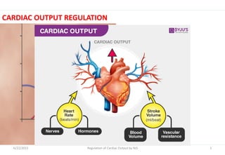
o Cardiac output (CO)
Volume of blood pumped by each ventricle per minute
CO (L/min) = HR (beasts/min) x SV (L/beat)
o Venous return
Is equally important with CO
Is the quantity of blood flowing from the veins into the right atrium each
minute.
3. 6/22/2022 Regulation of Cardiac Output by YeS 3
Regulation of Cardiac Output I
Stroke Volume (SV):
o Volume ejected during each beat , depends on venous return, can be
equal to venous return
Heart Rate (HR): number of beats per minute
o HR=72 beats/min , SV=0.07L/min (70ml)
CO (at rest) = 72 x 0.07 = 5-6 L/min
o CO increases when needed (e. g. exercise)
o Therefore, HR or SV or both can increase
4. 6/22/2022 Regulation of Cardiac Output by YeS 4
Regulation of Cardiac Output I
o The CO of the left ventricle equals the rate of blood flow through the
systemic circuit
o The right ventricle equals the rate of blood flow through the pulmonary
circuit.
o The left and right sides of the heart must have the same CO, or else
blood volume would shift from the pulmonary circuit to the systemic
circuit, or vice versa.
o Because the HR and the CO are the same for the right and left sides of
the heart, both ventricles must also have the same average SV.
5. 6/22/2022 Regulation of Cardiac Output by YeS 5
Regulation of Cardiac Output
I
Normal values for CO at rest and during activity
o CO varies widely with the level of activity of the body.
o Factors directly affect CO:
1. The basic level of body metabolism
2. Exercising
3. Age
4. The size of the body
o For young healthy men, resting CO averages about 5 - 6 L/min.
o For women, this value is about 4 - 5 L/min.
o With ↑ age, body activity and mass of some tissues (e.g. skeletal muscle)
diminish →↓CO.
6. 6/22/2022 Regulation of Cardiac Output by YeS 6
Regulation of Cardiac Output I
o Cardiac Reserve: the difference
b/n resting CO and maximum
volume of blood the heart is
capable of pumping per minute.
o CO during maximal exercise - CO
at rest,
o E.g. 35L - 5L=30L
7. 6/22/2022 Regulation of Cardiac Output by YeS 7
Regulation of Cardiac Output I
Cardiac Index (CI)
o Adequacy of CO considered in relation to tissue metabolic demand,
which varies by body size and activity
o Thus, CO expressed relative to body surface area is CI
CI= CO/BSA
o The average person who weighs 70 kg has a BSA of about 1.7 m2, which
means that the normal average CI for adults is about 3 L/min/m2 of
BSA; 5 L/min ÷1.7 =3 L/m2
8. 6/22/2022 Regulation of Cardiac Output by YeS 8
Regulation of Cardiac Output I
Effect of Age on CO
o The CI rises rapidly to a level
greater than 4 L/min/m2 at age 10
years and declines to about 2.4
L/min/m2 at age 80 years.
o The declining CI is indicative of
declining activity and/or declining
muscle mass with age.
CI for a person CO per m2 of surface
area at different ages
9. 6/22/2022 Regulation of Cardiac Output by YeS 9
Regulation of Cardiac Output I
Ejection fraction (EF)
o Measurement of ventricular performance
o Is fraction of EDV ejected from ventricles
EF= SV/EDV = 70/130 = 54%
A healthy man has EF of 50% or more
o Is primary clinical index of contractility
10. 6/22/2022 Regulation of Cardiac Output by YeS 10
Regulation of Cardiac Output I
Regulation of CO
i. Intrinsic regulation:
Aortic pressure
(afterload)
Venous filling pressure
(preload)
ii. Extrinsic regulation:
ANS effects
Hormonal effects
13. 6/22/2022 Regulation of Cardiac Output by YeS 13
Regulation of Cardiac Output I
I. Intrinsic Regulation of CO
o Intrinsic auto-regulation relates to myocardial length tension
relationship
o Demonstrated by:
Frank-Starling’s law of heart
According to this law, the contractile force of the heart is
proportional to the initial length of the muscle fibers
(EDV) within physiological limit.
14. 6/22/2022 Regulation of Cardiac Output by YeS 14
Regulation of Cardiac Output I
I. Intrinsic Regulation of CO…
o With stretch in cardiac muscle fibers → ↑tension and
contractility to a maximum and then decline as the stretch
becomes more extreme
o Intra-ventricular systolic pressure (IVSP)↓ and intra ventricular
diastolic pressure (IVDP) ↑ reaching theoretically a meeting
point.
o This is heterometric (change in size) auto-regulation, a pre-load
(EDV) phenomenon.
16. 6/22/2022 Regulation of Cardiac Output by YeS 16
Regulation of Cardiac Output I
Frank-Starling Law (Autoregulation)
o Volume regulation:
More filling EDVDistension SV (because of
force of contraction)
o Pressure regulation
Within limit, changes in arterial pressure has no effect on CO
Achieved in 2 phases:
a) diastolic aortic pressure intraventricular pressure
↓SV
b) thus, ESV , venous return further adds EDV SV
17. 6/22/2022 Regulation of Cardiac Output by YeS 17
Regulation of Cardiac Output I
A Starling curve showing how stroke volume changes in response to changes in EDV
18. 6/22/2022 Regulation of Cardiac Output by YeS 18
Regulation of Cardiac Output I
Factors shifting the Frank-
Starling’s curve of the
Heart
The Frank-Starling’s curve
of the heart may be
shifted to the left by
positive inotropism and to
the right by negative
inotropism.
19. 6/22/2022 Regulation of Cardiac Output by YeS 19
Regulation of Cardiac Output I
Stretching the heart causes an ↑
HR
o Stretch of the SA node has a
direct effect on the rhythmicity of
the node to ↑ HR as much as 10%
to 15%.
o The stretched RA → initiates
Bainbridge reflex →
↑sympathetic stimulation to
heart→↑HR.
20. 6/22/2022 Regulation of Cardiac Output by YeS 20
Regulation of Cardiac Output I
II. Extrinsic Regulation of CO
o Involves general factors, neural and hormonal factors.
1. General Factors
Change in CO Condition (factors)
CO Anxiety and excitement
Eating
Exercise
High environment TO
Pregnancy
CO Sitting or standing position
Arrhythmias
Heart disease
21. 6/22/2022 Regulation of Cardiac Output by YeS 21
Regulation of Cardiac Output I
Regulation of HR
1. Nervous factors: autonomic stimulation
2. Physical factors: temperature
3. Mechanical factors: right atrial distension
4. Chemical factors: catecholamines
22. 6/22/2022 Regulation of Cardiac Output by YeS 22
Regulation of Cardiac Output I
1. Neural control of HR and CO
Sympathetic neurons
o Sympathetic neurons release NE, which binds to β1 adrenergic receptors on
the SA nodal cells
o Activates the cAMP second messenger system; augments the opening of
funny channels and T-type Ca2+
o The net result is an ↑slope of the spontaneous depolarization and a ↓the
level of repolarization → such that threshold for an AP is reached more
quickly.
o ↑The frequency of APs → ↑HR ↑CO
24. 6/22/2022 Regulation of Cardiac Output by YeS 24
Regulation of Cardiac Output I
Sympathetic:
o Heart:
NE binds to adrenoceptors in the heart
Affinity : β1 > > β2 and α1
Produce positive inotropy, chronotropy, and dromotropy
o Blood vessels:
α1-adrenoceptors –vasoconstriction
β2 adrenoceptors- vasodilation
25. 6/22/2022 Regulation of Cardiac Output by YeS 25
Regulation of Cardiac Output I
o Sympathetic neurons → to the AV node and conduction system
→ influence the speed with which APs are conducted.
o ↑sympathetic activity → APs move faster
o Which ↓ the delay of impulse conduction
o Shortens the time it takes for APs to travel through the
ventricles.
o Ventricular contraction starts sooner after atrial contraction
and proceeds more quickly → ↓ the duration of systole.
26. 6/22/2022 Regulation of Cardiac Output by YeS 26
Regulation of Cardiac Output I
Sympathetic Stimulation
Inotropic effect
↑Chronotropic effect
Conduction velocity
Pacemaker activity
Coronary blood flow (β2)
↑HR
↑CO
27. 6/22/2022 Regulation of Cardiac Output by YeS 27
Regulation of Cardiac Output I
Parasympathetic neurons
o ↑ parasympathetic activity to the SA node
o ↓ the frequency of APs in the pacemaker cells
o By release ACh, which binds to M1 cholinergic receptors on the SA
nodal cells
o Binding augments the opening of K+ channels and suppresses the
opening of funny channels and T-type Ca++ channels.
o The net result is a ↓ the slope of the spontaneous depolarization
o Hyperpolarization of the membrane potential such that the threshold
for an AP is reached more slowly.
29. 6/22/2022 Regulation of Cardiac Output by YeS 29
Regulation of Cardiac Output I
Parasympathetic neurons…
o The frequency of APs↓ →↓HR → ↓CO
o They also influence impulse conduction through the AV node and the
rest of the conduction system.
o The speed of impulse conduction ↓, which increases the delay of
conduction between the atria and the ventricles
o Lengthens the time required for impulses to travel through the
ventricles.
o As a result, the duration of systole↑→↓HR and CO.
o Decrease Inotropy??, chrontropy and dromotropy
30. 6/22/2022 Regulation of Cardiac Output by YeS 30
Regulation of Cardiac Output I
Vagal stimulation.
i) Mild……slowing of HR, atrial contraction, conduction velocity
ii) Moderate…further slowing
iii) Strong …. Complete cessation of heart beat for a few seconds
↓
Vagal escape
31. 6/22/2022 Regulation of Cardiac Output by YeS 31
Regulation of Cardiac Output I
2. Hormonal control of HR and CO
o Epinephrine (E): Phosphorylation of Ca++ channels
o ↑Ca++ into cells by cAMP dependent protein kinase A (cAMP-
PKA)……..
o The effects of E, which is secreted by the adrenal medulla in response
to increased sympathetic activity, are similar to those exerted by
sympathetic neural activity
o E → ↑AP frequency at the SA node →↑HR and velocity of AP
conduction→↑CO
32. 6/22/2022 Regulation of Cardiac Output by YeS 32
Regulation of Cardiac Output I
Other hormones and drugs that affect HR and CO:
Cathecolamines (E and NE)
Digitalis
Angiotensin II
T3/T4
Insulin
Glucagon
o These hormones primarily increase the force of myocardial
contraction→↑CO
33. 6/22/2022 Regulation of Cardiac Output by YeS 33
Regulation of Cardiac Output I
3. Metabolic agents with negative effect on the CO
Hyperkalemia: -ve inotropic effect low amplitude AP—weak contraction
Hypercalcemia: +ve inotropic stronger systole & incomplete diastole.
Hypernatremia: -ve inotropic effect; stimulate Na+-Ca++ exchanger;
Ca++ out of cardiac myocyte, cytosolic Ca++ level decreases
Acidosis: -ve Inotropic effect, due to depression of affinity of troponin C to Ca++
Alkalosis: +inotropic effect due to increase affinity of troponin C to
Ca++
34. 6/22/2022 Regulation of Cardiac Output by YeS 34
Regulation of Cardiac Output I
Changes in stroke volume (SV) affect CO
o Like HR, SV can vary from moment to moment and depends on several
factors.
o Primary factors that affect SV are:
i. Ventricular contractility: a measure of the ventricles’ capacity for
generating force.
ii. End-diastolic volume (EDV)
iii. Aortic pressure (afterload): the pressure that the ventricles have
to work against as they pump blood out of the heart.
36. 6/22/2022 Regulation of Cardiac Output by YeS 36
Regulation of Cardiac Output I
i. Ventricular contractility
o Is a change in the force of ventricular contraction at any given
EDV.
o Any factor that causes the ventricles to contract with more
force will tend to make SV larger→ ↑CO.
o This is true regardless of whether the change in contractile
force is due to a change in contractility or a change in EDV.
37. 6/22/2022 Regulation of Cardiac Output by YeS 37
Regulation of Cardiac Output I
Sympathetic nervous control of ventricular contractility
o Autonomic control of SV is exerted almost entirely by the SNS
o No parasympathetic influence on ventricular contractility because of
the sparse distribution in the ventricular myocardium.
o Some of sympathetic neurons project to the atria and influence the
force of atrial contraction.
o ↑Increased sympathetic activity →↑force contraction → which raises
atrial pressure and increases the volume of blood the atria pump into
the ventricles.
38. 6/22/2022 Regulation of Cardiac Output by YeS 38
Regulation of Cardiac Output I
Sympathetic nervous control…
o Sympathetic neurons project to the ventricular myocardium
where they exert a direct influence on myocardial
contractility.
o APs in these neurons trigger the release of NE, which binds to
β1 adrenergic receptors on the contractile cells.
o ↑sympathetic activity→↑ ventricular contractility →↑ SV;
CO.
39. 6/22/2022 Regulation of Cardiac Output by YeS 39
Regulation of Cardiac Output I
Effects of sympathetic activity on ventricular contractility
41. 6/22/2022 Regulation of Cardiac Output by YeS 41
Regulation of Cardiac Output I
Changes in ventricular contractility induced by sympathetic activity
42. 6/22/2022 Regulation of Cardiac Output by YeS 42
Regulation of Cardiac Output I
ii. The influence of EDV on SV and CO
o An ↑ in EDV occurs, the force of ventricular contraction rises,
producing an ↑ in SV and CO.
o Conversely, if the EDV ↓, the force of ventricular contraction
declines, producing a ↓ in SV and CO.
o ↑ in EDV cause muscle fibers in the ventricular
myocardium to lengthen.
o Such stretching of the muscle fibers causes an ↑ in the force of
contraction by two mechanisms.
43. 6/22/2022 Regulation of Cardiac Output by YeS 43
Regulation of Cardiac Output I
i. Increasing the length of the muscle by increasing the EDV stretches
the muscle fibers closer to their optimal length for contraction, so
that they contract with greater force.
ii. Stretching of the muscle fibers induces an increase in the affinity of
troponin for calcium.
o As a consequence, binding between troponin and Ca++ is increased,
which increases the number of cross bridges that are activated with
each contraction.
44. 6/22/2022 Regulation of Cardiac Output by YeS 44
Regulation of Cardiac Output I
o Changes in either sympathetic
activity or EDV affect the force of
ventricular contraction
o It is possible to alter SV either by
changing sympathetic activity
without changing EDV or by
changing EDV without changing
sympathetic activity.
o As a result, SV at any given EDV
increases, reflecting the fact that
ventricular contractility has
increased.
A family of Starling curves, which shows
the influence of sympathetic input on
ventricular contractility
45. 6/22/2022 Regulation of Cardiac Output by YeS 45
Regulation of Cardiac Output I
Factors affecting EDV; is primarily determined by:
i. End diastolic pressure (preload):
Ventricular end-diastolic pressure is called preload because it
places tension (or load) on the myocardium before it begins to
contract.
When a ventricle fills with blood during diastole, the process is
similar to what happens when you blow up a balloon with air:
As pressure inside rises, the balloon expands.
Therefore, the final volume of a given balloon is determined by
the final pressure of the air inside it.
46. 6/22/2022 Regulation of Cardiac Output by YeS 46
Regulation of Cardiac Output I
Factors affecting EDV…
o Likewise, the EDV of a ventricle is determined by the pressure of the
blood inside it at the end of diastole.
o As preload increases, EDV increases, and SV increases according to
Starling’s law.
o Preload is determined by a number of factors:
i. Filling time, which depends on HR
ii. Atrial pressure, which is determined by venous return and the force
of atrial contraction.
47. 6/22/2022 Regulation of Cardiac Output by YeS 47
Regulation of Cardiac Output I
INCREASED PRELOAD (FEELING)
48. 6/22/2022 Regulation of Cardiac Output by YeS 48
Regulation of Cardiac Output I
Factors affecting EDV…
o As HR ↓ → filling time ↑ → because diastole ↑ in duration.
o At a HR = 60 beats/min → diastole is approximately 0.6
second long
o When the HR = 180 beats/min → diastole decreases to 0.1
second.
o Because more time is allowed for the entry of blood into the
ventricles when the HR is lower, a ↓HR (increase in filling
time) tends to ↑both preload and EDV.
49. 6/22/2022 Regulation of Cardiac Output by YeS 49
Regulation of Cardiac Output I
o Atrial pressure, rises in response to ↑ in venous return.
o The most important factor influencing venous return is central venous
pressure (the pressure of blood contained in the large veins that lead into
the heart).
o CVP is affected: changes in blood volume, muscular activity, and even
posture (as when a person stands up or lies down).
o As CVP↑ → venous return ↑ (the increased pressure forces more blood
to flow into the atria).
o This effect raises atrial pressure, which leads to an increase in preload and
EDV, which produces an increase in SV.
50. 6/22/2022 Regulation of Cardiac Output by YeS 50
Regulation of Cardiac Output I
The influence of afterload on SV
o However, SV depends not only on how much force the ventricular
muscle develops, but also on how large a force it has to work
against.
o Consider a person attempting to push a wagon up a slope:
o The speed of the wagon depends not only on how much force the
person exerts, but also on how much the wagon weighs.
o When the heart ejects blood, the ventricular muscle works
against arterial pressure.
51. 6/22/2022 Regulation of Cardiac Output by YeS 51
Regulation of Cardiac Output I
o An ↑ in arterial pressure tend to cause SV to decrease.
o Because arterial pressure places a load on the myocardium
after contraction starts, it is called afterload.
o For the left ventricle, afterload is determined by the pressure
in the aorta during the ejection period.
o Generally speaking, afterload increases as mean arterial
pressure rises→↓SV, CO.