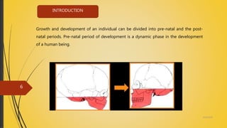
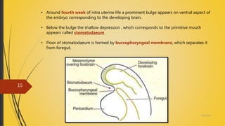
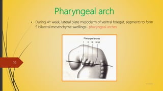
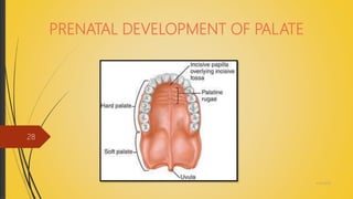
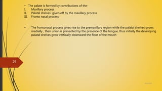
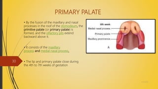
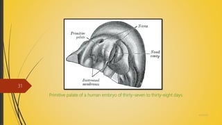
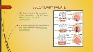
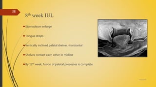
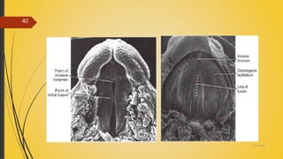
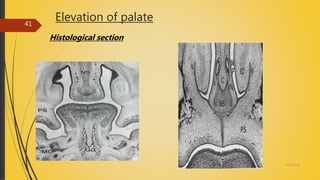
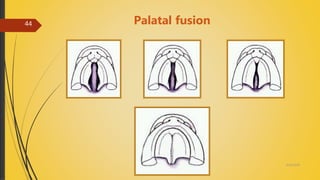
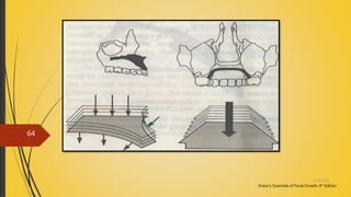
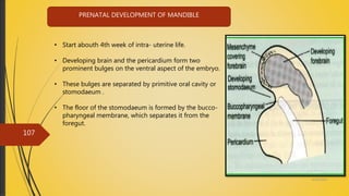
DR. SWATI SAHU
MDS FELLOW
DEPT. OF ORAL & MAXILLOFACIAL SURGERY
4/14/2018
1
4/14/2018
2
MAXILLA
4/14/2018
3
Introduction
Growth
Development
Prenatal growth & development of maxilla
 Prenatal growth & development of palate
Postnatal growth & development of maxilla
CONTENTS
4/14/2018
4
Development of Tongue
• Formation of Anterior 2/3rd of the Tongue
• Formation of Posterior 1/3rd of the Tongue
• Developmental defect of Tongue
Developmental defect of maxilla
4/14/2018
5
Growth and development of an individual can be divided into pre-natal and the post-
natal periods. Pre-natal period of development is a dynamic phase in the development
of a human being.
INTRODUCTION
4/14/2018
6
• Stewart – It may be defined as a developmental increase in mass. In other
words it is a process that leads to an increase in the physical size of cells, tissues,
organs or organisms as a whole.
• Proffit – an increase in size or number.
• Pinkham – an increase, expansion or extension of a given tissue.
GROWTH
4/14/2018
7
DEVELOPMENT
• Moyers – all the naturally occurring unidirectional changes in the life of an
individual from its existence as a single cell to its elaboration as a multifunctional unit
terminating in death.
• Todd – development means progress towards maturity.
• Proffit – development is in complexity.
4/14/2018
8
 Indicator of general health.
 Identify unusual growth patterns at an early stage.
 Etiology and development of malocclusion.
 Identify abnormal occlusion – early stage.
 Poorly timed eruptions - malocclusion
 Growth - effects stability of occlusion.
 Use of growth spurts.
IMPORTANCE OF GROWTH
4/14/2018
9
 Growth and development of an individual can be divided into PRENATAL & the
POSTNATAL periods.
 The pre-natal period of development is a dynamic phase in the development of a
human being.
 During this period, the height increases by almost 5000 times as compared to only a
threefold increase during the post-natal period.
 The pre-natal life can be arbitrarily divided into three periods.
1. Period of the Ovum
2. Period of the Embryo
3. Period of the Fetus
4/14/2018
10
 This period extends for a period of approximately two weeks from the time of
fertilization.
 During this period the cleavage of the ovum and the attachment of the ovum to
the intra-uterine wall occurs.
PERIOD OF OVUM -
4/14/2018
11
PERIOD OF EMBRYO -
 This period extends from the fourteenth day to the fifty sixth day of intra-uterine life.
 During this period the major part of the development of the facial & the cranial
region occurs.
4/14/2018
12
PERIOD OF FETUS -
 This phase extends between the fifty sixth day of intra-uterine life till birth.
 In this period ,accelerated growth of the cranio-facial structures occurs
resulting in an increase in their size.
 In addition, a change in proportion between the various structures also occurs
4/14/2018
13
PRENATAL DEVELOPMENT OF MAXILLA
4/14/2018
14
• Around fourth week of intra uterine life a prominent bulge appears on ventral aspect of
the embryo corresponding to the developing brain.
• Below the bulge the shallow depression , which corresponds to the primitive mouth
appears called stomatodaeum .
• Floor of stomatodaeum is formed by buccopharyngeal membrane, which separates it
from foregut.
4/14/2018
15
Pharyngeal arch
• During 4th week, lateral plate mesoderm of ventral foregut, segments to form
5 bilateral mesenchyme swellings– pharyngeal arches
4/14/2018
16
Each PHARYNGEAL ARCH consists of
4/14/2018
17
1. A central cartilage rod that form
the skeleton of the arch.
1. A muscular component termed as bronchomere
2. A vascular component.
3. A neural element.
4/14/2018
18
• Mesoderm covering the developing forebrain proliferates, and forms a downward
projection that overlaps the upper part of the stomatodeum this downward projection
is called the frontonasal process
• The first branchial arch is called the mandibular arch and plays an important role in the
development of the naso maxillary region.
• The stomodeum is overlapped superiorly by frontonasal process & laterally by
mandibular arches of both sides.
4/14/2018
19
4/14/2018
20
• The ectoderm overlying the Fronto-nasal Process shows bilateral localized thickenings
above the stomatodeum. These are called the Nasal Placodes.
• These Placodes soon sink and form the Nasal Pits.
• Formation of nasal pits divides frontonasal process into 2 parts :
- The medial nasal process
- The lateral nasal process
• The mandibular arch gives off a bud from its
dorsal end called the maxillary process which
grows ventro-medio-cranial to the mandibular
process.
4/14/2018
21
4/14/2018
22 • At this stage the stomodeum is overlapped from above by the frontal process, below by
the mandibular process & on either side by the maxillary process.
• The two mandibular processes grow medially & fuse to form the lower lip & the lower
jaw.
• Maxillary process undergoes growth, frontonasal process becomes narrower so that 2 nasal
pits come closer.
• Line of fusion of maxillary process & the medial nasal process corresponds to the
nasolacrimal duct.
4/14/2018
23
4/14/2018
24
• Each maxillary process now grows medially and fuses , first with the lateral nasal
process and then with the medial nasal process .
• Medial and lateral process also fuse with each other in this way the nasal pit(external
nares) are cut off from the stomatodeum .
• As the Maxillary Process undergoes growth the Fronto-nasal process becomes narrow
so that the two Nasal Pits come closer.
• Mesodermal basis of the median part of the lip(called philtrum)is formed from the
frontonasal process.
DEVELOPMENT OF NASAL CAVITY
• Nasal cavity are formed by extension of nasal pits .
• Soon the medial and lateral processes fuse , and form a partition between the pit and the
stomatodeum this is called primitive palate and is derived from the frontonasal process.
• The nasal pits now deepen to form the nasal sac .
4/14/2018
25
4/14/2018
26
• The dorsal part of this sac is , at first , separated from the stomatodeum by thin
membrane called the bucconasal membrane or nasal fin this soon breaks down.
• Nasal sac now has a ventral orifice that opens on the face (anterior or external nares) and
dorsal orifice that open into stomatodeum (primitive posterior nasal aperture) .
• Frontonasal process becomes progressively narrower .
• This narrowing of the frontonasal process and the enlargement of the nasal cavities
themselves being closer together the intervening tissue becomes much thinned to form the
nasal septum ventrally attached to below the primitive palate and dorsally bucconasal
membrane.
4/14/2018
27
PRENATAL DEVELOPMENT OF PALATE
4/14/2018
28
• The palate is formed by contributions of the-
I. Maxillary process
II. Palatal shelves given off by the maxillary process
III. Fronto nasal process
• The frontonasal process gives rise to the premaxillary region while the palatal shelves grows
medially , their union is prevented by the presence of the tongue, thus initially the developing
palatal shelves grow vertically downward the floor of the mouth
4/14/2018
29
PRIMARY PALATE
• By the fusion of the maxillary and nasal
processes in the roof of the stomodeum, the
primitive palate (or primary palate) is
formed, and the olfactory pits extend
backward above it.
• It consists of the maxillary
process and medial nasal process.
• The lip and primary palate close during
the 4th to 7th weeks of gestation
4/14/2018
30
Primitive palate of a human embryo of thirty-seven to thirty-eight days.
4/14/2018
31
SECONDARY PALATE
• The development of the secondary
palate commences in the sixth week
of human embryological
development.
• It is characterised by the formation of
two palatal shelves on the maxillary
prominences
4/14/2018
32
• As the palatal shelves grow medially their, their union is prevented by the presence of
tongue
• Initially the developing palatal shelves grow vertically toward the floor of mouth
4/14/2018
33
• During 7th week of intrauterine life, a transformation in the position of the palatine shelf
occurs
• They change from a vertical to a horizontal position
4/14/2018
34
Various reasons are given to explain how this transformation occurs. They are:
• Alteration in biochemical and physical consistency of the connective tissue of the
palatal shelves
• Alteration in vasculature and blood supply to the palatal shelves
• Apperance of an interensic shelf force
• Rapid differential mitotic activity
• Muscular movements
4/14/2018
35
• The 2 palatal shelves, by 8 ½ weeks of intra uterine life are in close approximation to each
other
• Initially the 2 palatal shelves are covered by an epithelial lining. As they join the epithelial cells
degenerate
• The connective tissue of the palatal shelves intermingle with each other resulting in their
fusion
4/14/2018
36
• The entire palate doesnot contact and fuse at the same time. Initially the contact occurs in
the central region of the secondary palate posterior to the premaxilla
• From this point, closure occurs both anteriorly and posteriorly
4/14/2018
37
8th week IUL
Stomodeum enlarge
Tongue drops
Vertically inclined palatal shelves -horizontal
Shelves contact each other in midline
By 12th week, fusion of palatal processes is complete
4/14/2018
38
4/14/2018
39
4/14/2018
40
Elevation of palate
Histological section
4/14/2018
41
Theories of palatal shelf elevation
EXTRINSIC FORCES
 Descent of tongue
 Myoneural activity with in tongue
 Shelves pushed up by tongue
 Mouth opening reflexes
4/14/2018
42
INTRINSIC FORCES
Hydration and polymerisation of intercellular substance
Differential growth on one side of palatal shelf
Turgor produced by build up of HYALURONIC ACID
SEROTONIN release from neural tissue.
Mesenchymal cell biosynthetic activity
Changing amounts of GLYCOSAMINOGLYCANS(GAG)
4/14/2018
43
Palatal fusion
4/14/2018
44
FUSION OF PALATAL SHELVES
 9-10 Week.
 Epethlium Thickens And Contacts.
 Role Of Glycoproteins And Desmosomes
 Degeneration Of Epithelium.
 Conective Tissue Penetration And Intermingling.
 Entire Palate Does Not Fuse At Same Time, Intial Contact ,Central Region Of
Secondary Palate, Then Closure Continues Both Anterior And Posteriorly.
4/14/2018
45
OSSIFICATION OF PALATE
• Ossification of the palate occurs from the 8th week of intra- uterine life.
•This is an intramembranous type of ossification
• The palate ossifies from a single centre derived from the maxilla
• The most posterior part of the palate does not ossify. This forms the soft palate
• The mid palatal suture ossifies by 12-14 yrs
4/14/2018
46
APPLIED ANATOMY
Cleft lip (cheiloschisis) and cleft palate (palatoschisis), which can also occur together
as cleft lip and palate, are variations of a type of clefting congenital deformity caused by
abnormal facial development during gestation.
4/14/2018
47
CLEFT PALATE
• Cleft palate is a condition in which the two plates of the skull that form the hard
palate (roof of the mouth) are not completely joined
• Palate cleft can occur as complete or incomplete (a 'hole' in the roof of the mouth,
usually as a cleft soft palate). When cleft palate occurs, the uvula is usually split. It occurs
due to the failure of fusion of the lateral palatine processes, the nasal septum, and/or the
median palatine processes (formation of the secondary palate
Incomplete cleft palate Unilateral complete lip and palate Bilateral complete lip and palate
4/14/2018
48
COMPLICATIONS
• Cleft may cause problems with feeding,
ear disease, speech and socialization.
• An infant with a cleft palate will have
greater success feeding in a more upright
position.
• Gravity will help prevent milk from
coming through the baby's nose if he/she
has cleft palate.
4/14/2018
49
TREATMENT
STAGE ONE
• Often a cleft palate is temporarily closed, the cleft isn't closed, but it is
covered by a palatal obturator.
• Cleft palate can also be corrected by surgery, usually performed between 6
and 12 months.
• If the cleft extends into the maxillary alveolar ridge, the gap is usually
corrected by filling the gap with bone tissue. The bone tissue can be acquired
from the patients own chin, rib or hip.
4/14/2018
50
STAGE TWO
• Carried out from 18th month to 5th year of life, generally corresponds to primary dentition
phase
STAGE THREE
• Spans from 6th to 11th year of life, generally corresponds to mixed dentition phase
STAGE FOUR
• Include the treatment done during the permanent dentition stage, 12-18 yrs of age
4/14/2018
51
POSTNATAL DEVELOPMENT OF
MAXILLA
4/14/2018
52
Postnatal growth of maxilla is a multifactorial process
According to Moss-
• Translation (displacement)
• Transposition (surface remodeling)
4/14/2018
53
Translation / dislocation
Dislocation comprises of movement of the whole bone as it
simultaneously expands
Displacement can be
Primary
Secondary
SCHOOLS OF THOUGHT
• Sutural theory
• Nasal septal theory
• Functional matrix
4/14/2018
54
Sutural theory
Sicher believed that craniofacial growth occurs at sutures.
Maxilla is attached to the cranium by frontomaxillary,
zygomaticomaxillary, zygomaticotemporal and pterygopalatine
suture, which are more-or-less oblique and parallel to each
other
Thus growth in these areas will push the maxilla downward and
forward
But???
• Suture is a tension adapted tissue
• Suture doesn’t grow when transplanted
• Growth takes place in untreated cases of cleft palate
Enlow’s; Essentials of Facial Growth, 4th Edition
4/14/2018
55
Nasal septum theory
James Scott
He viewed cartilaginous sites throughout the skull as primary centres
of growth
cartilage is a pressure-adapted tissue
Pressure (of the growing brain) accommodating growth of the nasal
septum provides a source of physical force that displaces the whole
maxilla anteriorly and inferiorly. This sets up field of tension for the
sutures, at which bone deposition may now take place.
But???
Experiments are not decisive
Enlow’s; Essentials of Facial Growth, 4th Edition
4/14/2018
56
Functional matrix theory
Melvin moss
Researches suggest that if there is no primordium for the eye, the orbit does not develop
Acc to him, the functional soft tissue matrix is the epigenic governing determinant of skeletal
growth process and all skeletal growth is secondary, compensatory and mechanically obligatory to
it.
In achondroplastic dwarfs, the midface shows marked concavity and retardation owing to deficient
cartilage growth
Enlow’s; Essentials of Facial Growth, 4th Edition
4/14/2018
57
Transposition / remodelling
If you see in the picture, The area previously occupied by the
ramus has now been converted into mandibular body of the adult
This is REMODELLING
It is a sequence of differential deposition and resorption that
results in reshaping and resizing of bone into its adult form
The surface that faces the direction of movement is depository
and that away from it is always resorptive
Enlow’s; Essentials of Facial Growth, 4th Edition
4/14/2018
58
Lacrimal suture
Lacrimal bone is a flake of bony island with its entire
perimeter surrounded by sutures, separating it from many
bones
The lacrimal bone acts a key traffic control, providing for
slippage of multiple bones along its perimeter
In itself, the lacrimal bone undergoes remodelling rotation
Enlow’s; Essentials of Facial Growth, 4th Edition
4/14/2018
59
Maxillary tuberosity and key
ridge
Maxillary arch grows in 3 directions
• Posteriorly deposition on posterior surface of maxillary tuberosity
• Laterally- deposition on buccal surface of tuberosity
• Downward- deposition along alveolar ridge
Endosteal surface is resorptive for growth of maxillary sinus
Reversal occurs at key ridge, where most of the external surface
becomes resorptive
Enlow’s; Essentials of Facial Growth, 4th Edition
4/14/2018
60
Zygomatic arch
Resorption at anterior surface and deposition at
the lateral and posterior surfaces
As a result the zygomatic arches move posteriorly
and bilaterally outwards
Enlow’s; Essentials of Facial Growth, 4th Edition
4/14/2018
61
Orbital growth
To compensate for resorption in the endocranial side
To compensate for the downward growth of nasomaxillary
complex
To make the supraorbital rim more prominent
V PRINCIPLE= Anterior – lateral- superior relocation
of orbit
Enlow’s; Essentials of Facial Growth, 4th Edition
4/14/2018
62
POSTNATAL DEVELOPMENT OF
PALATE
4/14/2018
63
Enlow’s; Essentials of Facial Growth, 4th Edition
4/14/2018
64
• In early pre natal life the palate is relatively long but from the 4th month it widens as a result of
mid palatal suture growth and appositional growth along the lateral alveolar margins.
• Growth of the mid palatal suture occurs between 1 and 2 years of age.it is large in its posterior than in
its anterior part, so that the posterior part of the nasal cavity widens more than the anterior part.
• Lateral appositional growth continues until 7 years of age by this time the palate achieves its maximum
anterior width. Posterior appositional growth continues after the lateral growth has ceased, so that the
palate becomes longer and wider during late childhood.
4/14/2018
65
• The appositional growth of the alveolar processes contributes to deepening as well as widening
of the vault of the bony palate at the same time adding to the height and breadth of maxilla
• Ossification does not occur in the posterior part of the palate, giving rise to the region of soft
palate. Myogenic mesenchymal tissues of the I, II and IV branchial arch migrates into this facial
region supplying the musculature of facial and palate.
4/14/2018
66
DEVELOPMENTAL ANOMALIES AFFECTING
MAXILLA
• Cleft palate.
• Agnathia
• Micrognathia.
• Macrognathia.
• Treacher collins syndrome (first arch
syndrome)
• Cleidocranial dyplasia
• Craniofacial dysostosis
4/14/2018
67
4/14/2018
68
AGNATHIA
• Synonyms – otocephaly, holoprosencephaly agnathia
• Lethal anomaly characterised by hypoplasia or absence of mandible
with abnormally positioned ears having an autosomal recessive mode of
Inheritance.
• Due to failure of migration of neural crest mesenchyme into the maxillary
Prominence at fourth to fifth week of gestation.
4/14/2018
69
MICROGNATHIA
• Small jaw
• Either maxilla / mandible may be affected.
• Two types – true / Pseudo
• Congenital /acquired
• Occasionally follows hereditary pattern
• Occurs due to deficiency in premaxillary area
and patient with this deformity appear to have
the middle third of the face retracted.
4/14/2018
70
MACROGNATHIA
• Condition of abnormally large jaws
• May be associated with certain other conditions
 Pagets disease of bone
 Acromegaly
 Leontiasis ossea , a form of fibrous dysplasia
4/14/2018
71
TREACHER COLLINS – FRANCESCHETTI SYNDROME
• Synonym - Mandibulofacial dysostosis
• Autosomal dominant
• The gene of treacher Collin was mapped to chromosome 5q32-q33.1
• Clinical manifestations –
 Antimangoloid palpebral fissure with a coloboma of outer portion
of lower eyelids
 Hypoplasia of facial bones especially malar and mandible
 Malformation of external ear
 Macrostomia, high palate
 Blind fistulas between the angles of ear and angle
of mouth
 Atypical hair growth
 Facial clefts
 Bird like or fish like face
4/14/2018
72
CLEIDOCRANIAL DYSPLASIA
• Synonyms – Marie and sainton’s disease,
scheuthauer- marie- sainton syndrome,
mutational dysostosis
• A congenital disorder of bone formation manifestated with
clavicular hypoplasia with a norrow thorax, which allows
approximation of shoulders in front of the chest.
• Delayed ossification of skull, excessively large fontanels
and delayed closing of sutures are prominent features
• Arnold head – characteristic skull abnormalities
• Etiology – mutation in core binding factor alpha – 1
(CBFA-1) gene, located on chromosome 6p21
4/14/2018
73
CRANIOFACIAL DYSOSTOSIS (CROUZONS SYNDROME)
• Caused by premature obliteration and ossification of two or more sutures,
More often coronal and sagittal sutures
• Etiology – mutation of fibroblast growth factor receptor (FGFR-2) gene
• The mutation in transmembrane region of FGFR3
• Clinical features –
 Obliteration of coronal and sagittal sutures
 Flattening of acromium, growing only at vertical axis
 Forehead is wide and high
 Wide face
 Hypoplastic maxilla (pseudoprognathism)
 Deviation of nasal septum
 Narrowed or obliterated anterior nares
 Wide beaked nose
 Hypertelorism
 Divergent squint
 Antimongoloid eyelids
 Upper eyelid mimicking “frog face”
 Malocclusion, malposed teeth and dysphasia
4/14/2018
74
DEVELOPMENT OF TONGUE
4/14/2018
75
What is Tongue?
Largest single muscular organ inside the oral cavity, which lies relatively free.
develops in relation to the pharyngeal arches.
It develops from two parts, they are
•formation of anterior 2/3rd of the tongue
•formation of posterior 1/3rd of the tongue
4/14/2018
76
Formation of anterior 2/3rd of the tongue:
• Tuberculum Impar: first a swelling arises in the
midline of the mandibular process. And is flanked
by two other swellings
• Lingual Swelling: The lateral part of the
mandibular process mesenchymal thickening
develops to form two lingual swellings.
•Develops from mesenchyme of 1st pharyangeal
arch .
(text book of Inderbir Singh ,8th edition,G P Pal,HUMAN EMBRYOLOGY)
4/14/2018
77
•These lateral swelling quickly enlarge and merge
with each other and the tuberculum impar to form
a large mass from which mucous membrane of the
anterior 2/3rd of the tongue is formed.
(text book of Inderbir Singh ,8th edition,G P Pal,HUMAN EMBRYOLOGY)
4/14/2018
78
Formation of posterior 1/3rd of the tongue:
Root of the tongue arises from large
midline swelling develops from
mesenchyme of 2nd,3rd and 4th arches.
Consist of ,
1. Copula (associated with 2nd arch)
2. A large hypobranchial eminence
(associated by 3rd and 4th arch)
(text book of Inderbir Singh,8th edition, G P Pal, HUMAN EMBRYOLOGY)
4/14/2018
79
• Hypobranchial eminence overgrows the
copula
• The tongue separates from the floor of
the mouth by a down-growth of
ectoderm around its periphery, which
degenerates to form lingual sulcus and
gives the tongue mobility.
(text book of Maji Jose,1st edition, ORAL BIOLOGY) 4/14/2018
80
Muscle of the tongue have a different origin, they arises
from the occipital somites, which have migrated forward
in to the tongue area, carrying with them their nerve
supply hypoglossal nerve
(text book of Ten Cate’s,7th edition,ORAL HISTOLOGY) 4/14/2018
81
4/14/2018
82
NERVE SUPPLY OF TONGUE
Papillae:
Small hair-like structure on the upper surface of the tongue that give the tongue its
characteristics rough texture.
Types:
• Fungiform Papillae
• Filliform Papillae
• Foliate Papillae
• Circumvallate Papillae
4/14/2018
83
 Fungiform Papillae:
• Anterior portion of the tongue (look like fungi)
• Scattered between the numerous filifom papillae at the tip of the tongue
• Smooth, round structure appears red(because of highly vascular connective tissue
core, visible through a thin, non keratinized covering epithellium)
4/14/2018
84
Filiform Papillae:
• covers entire anterior part of the tongue
• Cone shaped structures each with a core of
connective tissue covered by a thick
keratinized epithelium
• Together form a tough abrasive surface that
is involving in compressing and breaking
when tongue is opposed to the hard palate
4/14/2018
85
 Foliate papillae:
• Sometimes present on the lateral margins of the posterior part of the tongue
• Pink,consist of 4 to 11 parallel ridges that alternate with deep grooves in the
mucosa and few taste buds are present in the epithelium of the lateral walls of
ridges
4/14/2018
86
Cirumvallate Papillae:
• Adjacent and anterior to the sulcus
terminalis are 8 to 12 papillae
• Large structure, each surrounded by
a deep, circular groove in to which
open the duct of minor salivary
• Have connective tissue core that
covered on the superior surface by a
keratinized epithelium
• The epithelium covering lateral walls
is non-keratinized and contains taste
buds
4/14/2018
87
Taste Bud –
• A specialized receptor that occurs only in the oral cavity and pharynx is called taste
bud.
• Most of them found in fungiform papilla, foliate and circumvallate papilla.
• Barrel shaped structure composed of 30 to 80 spindle shaped cells
• Communicate with surface through a small opening called taste pore
4/14/2018
88
4/14/2018
89
DEVELOPMENTAL DEFECTS
OF TONGUE
4/14/2018
90
1. Aglossia
2. Microglossia
3. Macroglossia
4. Ankyloglossia
5. Cleft tongue
6. Fissured tongue
7. Median rhomboid glossitis
8. Benign migratory glossitis
9. Hairy tongue
10. Lingual varices
11. Lingual thyroid nodule
4/14/2018
91
AGLOSSIA -
Complete absence of tongue at birth-very rare
MICROGLOSSIA -
• A rare congenital anomaly -presence of small or
rudimentary tongue
• Aglossia-microglossia with extreme glossoptosis
• Etiology-fetal cell traumatism in the first few weeks of
gestation
4/14/2018
92
MACROGLOSSIA -
An enlarged tongue
TYPES:- 1. Congenital/ primary 2. Secondary
Congenital macroglossia:- Due to overdevelopment of musculature which may or may not be associated with
generalized muscular hypertrophy
Secondary macroglossia: - May occur as a result of tumor of tongue such as, diffuse lymphangioma or
haemangioma.
CLINICAL FEATURES –
• Displacement of teeth / malocclusion.
• Crenation and scalloping of the lateral border of tongue
• Tips of the crenation fitting into the interproximal
spaces between the teeth.
ASSOCIATED SYNDROMES:-
Beckwith’s Wiedemann syndrome & Down syndrome
Beckwith’s Weidemann syndrome includes -Neonatal hypoglycemia , Mild micro cephaly , Umbilical hernia ,
Fetal visceromegaly
TREATMENT - No particular treatment
Removal of cause
Surgical trimming
4/14/2018
93
ANKYLOGLOSSIA -
CLASSIFIED AS -
Partial
Complete
Partial ankyloglossia/ Tongue tie: - results of a short lingual frenum/ one which is attached near
to the tip of the tongue.
Complete ankyloglossia:- Fusion between the tongue and the floor of the mouth
CLINICAL FEATURES - difficulty during speech
TREATMENT- frenectomy
4/14/2018
94
CLEFT TONGUE / BIFID TONGUE -
• Complete & Partial
 Complete cleft / bifid tongue - A rare condition Is due
to lack of merging of the lateral lingual swelling of
this organ
 Partially cleft tongue - Common condition Is due to incomplete merging and failure of
groove obliteration by underlying mesenchymal proliferation.
• Manifest as deep groove in the midline of dorsal surface.
Associated syndrome –
 Oro facial digital syndrome-
Thick fibrous band in the lower anterior mucobuccal fold eliminating the sulcus & with clefting
of the hyperplastic mandibular alveolar process.
Clinical considerations - Microorganisms &food debris may collect in the base of the cleft &
cause irritation.
4/14/2018
95
FISSURED TONGUE / SCROTAL TONGUE -
• Manifests as small furrows or grooves 2-6mm in depth on the dorsal surface often radiating
from central groove on the midline of the tongue
• Associated with chronic trauma or vitamin deficiency/hereditary
• Painless except in cases when food debris tends to collect in the grooves & produce irritation.
• Associated with Melkersson-Rosenthal
syndrome (traid of recurring facial paralysis,
cheilits granulomatosis and fissured tongue )
4/14/2018
96
MEDIAN RHOMBOID GLOSSITIS -
• Congenital anomaly which is due to failure of the tuberculum impar to retract or withdraw
before fusion of the lateral halves of the tongue
• Etiological agent - candida albicans
• CLINICAL FEATURES - it appears as an ovoid ,diamond, rhomboid shaped reddish patch or
plaque on the dorsal surface of the tongue, immediately anterior to circumvallate papillae
4/14/2018
97
BENIGN MIGRATORY GLOSSITIS -
Geographic tongue, wandering rash of tongue ,glossitis areata exfoliative
• Areas of desquamation of the filiform papillae in an irregular circinate pattern
• Central portion – inflamed white with borders may be outlined by a thin, yellowish white line
or band.
• Fungiform papillae persist &appear as elevated dots.
• The area of desquamation remain for a short time in one location & then heal & appear in
another location i.e ; migration
• TREATMENT - Empirical since its etiology is not known.
Reassurance to the patient.
4/14/2018
98
HAIRY TONGUE -
Hypertrophy of the filiform papillae of the tongue with lack of normal desquamation which
may be extensive and form a thick matted layer on the dorsal surface.
Color of the papillae varies, yellowish white to brown or even black.
Etiology - Candida albicans
TREATMENT - Brush the tongue to promote
desquamation and remove the debris.
4/14/2018
99
LINGUAL VARICES -
Lingual or sublingual varicosities
• Varix - is a dilated , tortuous vein, most
commonly a vein.
• Mostly involved -sublingual varix
• Common in older adults
• Red or purple elevated blebs of vessels on the ventral surface and lateral borders of the
tongue as well as floor of the mouth.
• No treatment needed
4/14/2018
100
LINGUAL THYROID NODULE -
In this anomalus condition, the follicle of thyroid tissue are found in the substance of the tongue,
possibly arising from thyroid that failed to migrate to its original position/ from enlarge remnants
that became detached and were left behind.
ETIOLOGY - functional insufficiency of the chief thyroid gland in the neck.
CLINICAL FEATURES - nodular mass in or near the base of the tongue.
Dysphagia, dysphonia, dyspnoea, hemorrhage with pain or feeling of tightness or fullness in the
throat.
TREATMENT -Careful physical examination to demonstrate the thyroid gland location.
4/14/2018
101
MANDIBLE
4/14/2018
102
• Introduction
• Anatomy of mandible
• Prenatal development of mandible
• Postnatal development of mandible
• Development of mandible in relation to various theory of growth
• Age changes
• Developmental anomalies
CONTENTS
103
MANDIBLE
Largest and
strongest bone of
the face
1st pharyngeal
arch
Articulation with
skull
shape and
Function
14 April 2018
INTRODUCTION
4/14/2018
104
ANATOMY OF MANDIBLE
It has horseshoe shaped body which lodges the teeth, and pair of rami which project upwards
from the posterior ends of the body and provide attachment to muscle.
4/14/2018
105
4/14/2018
106
4/14/2018
107
PRENATAL DEVELOPMENT OF MANDIBLE
• Start abouth 4th week of intra- uterine life.
• Developing brain and the pericardium form two
prominent bulges on the ventral aspect of the embryo.
• These bulges are separated by primitive oral cavity or
stomodaeum .
• The floor of the stomodaeum is formed by the bucco-
pharyngeal membrane, which separates it from the
foregut.
4/14/2018
108
• Mesoderm of foregut comes to arranged in the form of six bars that run
dorsoventrally in the side wall of the foregut.
• These are called pharyngeal arches.
4/14/2018
109
• First Branchial arch called MANDIBULAR ARCH.
• Mandibular arch gives off a bud from its dorsal end called maxillary process.
• It grows ventro-medially cranial to main part of the arch which is called mandibular
process.
4/14/2018
110
• Mandibular process of each side grow towards each other.
• fuse in midline - give rise to mandible.
• First structure develop in lower jaw :
- Mandibular division of Trigeminal nerve.
Neurotrophic factor produced by nerve induce osteogenesis.
4/14/2018
111
MECKEL'S CARTILAGE
• It is the cartilage of the first arch
• In human beings the Meckel's
cartilage has a close positional
relationship to the developing
mandible but makes no
contribution to it.
• At 6 weeks of development this
cartilage extends as a solid hyaline
cartilaginous rod, surrounded by a
fibrocellular capsule, from the
developing ear region to the
midline of the fused mandibular
processes.
4/14/2018
112
• The Mandibular branch of trigeminal nerve has close relationship to Meckel’s cartilage
4/14/2018
113
• On lateral aspect of Meckel’s cartilage, during the 6th week of embryonic development,
a condensation of mesenchyme occurs in the angle formed by the division of the
inferior alveolar nerve and its incisor and mental branches.
4/14/2018
114
Centre of ossification
Intramembraneous Ossification starts at the division of mental and incisive branch of
inferior alveolar nerve lateral to meckel’s cartilage around
7th week IUL.
4/14/2018
115
• From center of ossification bone formation spreads:
 Anteriorly - midline
 Posteriorly - where mandibular nerve divided into lingual and inferior alveolar branch.
• Bone formation spreads rapidly and surrounds the inferior alveolar nerve to form
mandibular canal.
• Intra-membranous ossification spreads in anterior and posterior direction forms the Body &
Ramus of the mandible.
Gray’s Anatomy – Fortieth edition
4/14/2018
116
• Anteriorly bone extends towards midline and comes in approximation with similar bone
forming on opposite side.
• These two bones remain separated by fibrous tissue mental symphysis untill shortly after
birth.
• Continued bone formation increases size of mandible with development of alveolar process
to surround the developing tooth germ.
4/14/2018
117
• Ossification spread posteriorly to form ramus of mandible, turning away from
meckel’s cartilage.
• This point of divergence is marked by lingula in adult mandible.
Thus by 10 weeks the rudimentary mandible is formed almost entirely by membranous
ossification with little direct involvement of Meckel’s cartilage
4/14/2018
118
NOW….. What is the
fate of the Meckel’s
cartilage?
• Incus and malleus
• Spine of sphenoid bone
• Anterior ligament of malleus
• Spheno-mandibular ligament
4/14/2018
119
SECONDARY CARTILAGES IN MANDIBULAR
DEVELOPMENT
Further growth until birth influenced by appearance of secondary cartilage
 Condylar cartilage
 Coronoid cartilage
 Symphyseal cartilage
4/14/2018
120
CONDYLAR CARTILAGE
• Appear during 12th week of IUL
• Rapidly form cone shape mass which is converted quickly to bone by endochondral
ossification.
• At the end of 20th week only a thin layer remains on the condylar head ,persist until the end
of the second decade of life ,providing a further growth .
(Ten Cate’s Oral Histology – Sixth Edition)
4/14/2018
121
• Cartilage fuses with mandibular ramus around 4th month.
(Contemporary orthodontics Williams R. proffit fifth edition)
4/14/2018
122
CORONOID CARTILAGE
• Appears at about 4 month of
development.
• Coronoid cartilage is transient growth
cartilage and disappears long before
birth.
• Cartilage grow as a response of
developing temporalis muscle.
• Coronoid cartilage become
incorporated into expanding intra-
membranous bone of ramus.
(Ten Cate’s Oral Histology – Sixth Edition)
4/14/2018
123
• Two in number
• Appear in between the two end of Meckel’s cartilage.
• They are obliterated within the first year after birth.
SYMPHYSEAL CARTILAGE
(Ten Cate’s Oral Histology – Sixth Edition)
4/14/2018
124
POSTNATAL DEVELOPMENT OF MANDIBLE
• Right & left mandibular body fuses at midline symphysis one year after birth.
• Mandible appears as single bone.
(Contemporary orthodontics Williams R. proffit fifth edition)
4/14/2018
125
• According to MOSS while mandible appears in the adult as a single bone, it
is divisible into several skeleton subunits
 Condylar process
 Coronoid process
 Angular process
 Ramus
 Body of mandible
 Alveolar process
 chin.
(Facial Growth – Donald H. Enlow third edition)
4/14/2018
126
MANDIBULAR CONDYLE
• It is a major site of growth
• Historically, the condyle has been
regarded as a kind of cornucopia
from which the whole mandible
itself pours forth.
• The condyle functions as regional
field of growth that provides an
adaptation for its own localized
growth circumstances
4/14/2018
127
• The condylar growth mechanism itself is a clear-cut process.
• Cartilage is a special non-vascular tissue and is involved because of variable levels of
compression
• An endochondral growth mechanism is required for this part of the mandible.
• Endochondral growth occurs only at the articular contact part of the condyle
• In Figure the endochondral bone tissue (b) formed in association with the condylar cartilage (a)
The enclosing bony cortices (c) are produced by periosteal-endosteal osteogenic activity
4/14/2018
128
• The lingual and buccal sides of neck characteristically have a resorptive surface.
• This is because condyle is quite broad and neck is narrow
• The neck is progressively relocated into areas previously held by the much wider
condyle
• What used to be condyle in turn becomes the neck as one is remodeled from the
other .
• This is done by periosteal resorption combined with endosteal deposition.
4/14/2018
129
• Explained another way, the endosteal surface of the neck actually faces the growth
direction; the periosteal side points away from the course of growth.
• This is another example of the V principle, with the V-shaped cone of the condylar
neck growing toward its wide end.
4/14/2018
130
ROLE OF CONDYLE
• It is directly involved as a unique, regional growth site.
• It provides an indispensable latitude for adaptive growth.
• It provides movable articulation.
• It is pressure tolerant and provides a means for bone growth (endochondral) in a situation
in which ordinary periosteal (intramembranous) growth would not be possible .
• It can also, all too frequently, become involved in TMJ pathology and distress.
4/14/2018
131
CLINICAL IMPLICATION
• Condylar cartilage dose have some measure of intrinsic, genetic programming,
• This , however, appears to be restricted to capacity for continued cellular proliferation .
• Cartilage cells are coded and geared to divide and continue to divide by extra condylar
biomechanical forces.
• So overall mandibular length be clinically increase or decrease for class II and class III individuals if
this were done during the period of active condylar growth.
(Facial Growth – Donald H. Enlow third edition)
NERVE SUPPLY OF MANDIBLE
4/14/2018
132
4/14/2018
133
AGE CHANGES IN THE MANDIBLE
AT BIRTH
• At the birth the mental foramen, opens below the sockets for the two deciduous molar
teeth near the lower border.
• The mandibular canal runs near the lower border.
• The angle is obtuse. It is 175.
4/14/2018
134
AT CHILDHOOD
• The two halves of the mandible fuse during the first year of the life.
• The body becomes elongated in its whole length, but more especially behind the mental
foramen, to provide space for the three additional teeth developed in this part.
• Mandibular foramen slightly above the occlusal plane
• The angle becomes less obtuse around 140.
4/14/2018
135
IN ADULT
• The mental foramen opens midway between the upper and lower borders.
• The mandibular canal runs parallel with the mylohyoid line.
• Mandibular foramen 7 mm above the occlusal plane
• The angle reduces about 110 or 120 degrees.
4/14/2018
136
IN OLD AGE
• Alveolar border is absorbed, so that height of the body is markedly reduced.
• The mental foramen and mandibular canal are close to the alveolar border.
• The angle again becomes obtuse about 140 degrees .
4/14/2018
137
DEVELOPMENTAL DEFECTS
OF THE MANDIBLE
• Agnathia
• Micrognathia
• Macrognathia
• Coronoid hyperplasia
• Condylar hyperplasia
• Condylar hypoplasia
• Bifid condyle
• Torus mandibularis
4/14/2018
138
CORONOID HYPERPLASIA
• Rare developmental anomaly
• Result in limited mandibular movement
• Unknown etiology.
• M:F ratio 5:1
• May be unilateral or bilateral
• Bilateral is more common
(Oral and maxillofacial Pathology- Neville third edition)
4/14/2018
139
CONDYLAR HYPERPLASIA
• Excessive growth of one of the condyles
• Cause is unknown, but local circulating
problems, endocrine disturbances, and trauma
have been suggested as possible etiologic
factors.
(Oral and maxillofacial Pathology- Neville third edition)
4/14/2018
140
CONDYLAR HYPOPLASIA
• Congenital or acquired
• congenital:
mandibulofacial dysostosis
goldenhar syndrome
hemifecial microsomia
• Acquired:
disturbances of the growth center of the condyle.
(Oral and maxillofacial Pathology- Neville third edition)
4/14/2018
141
BIFID CONDYLE
• Rare
• Most of have medial and lateral head divided by an
antero posterior grooves.
• Some condyles may be divided into an anterior and
posterior head
• Cause is uncertain
• Antero-posterior may be traumatic origin.
4/14/2018
142
TORUS MANDIBULARIS
• Develops along the lingual aspect of the mandible.
• Probably multifactorial, including both genetics and
environmental influences.
(Oral and maxillofacial Pathology- Neville third edition)
4/14/2018
143
TMJ ANKYLOSIS
Ankylosis of the Temporomandibular joint, an arthrogenic disorder of the TMJ, refers to restricted
mandibular movements (hypomobility) with deviation to the affected side on opening of the mouth.
4/14/2018
144
CLINICAL FEATURES -
4/14/2018
145
• Obvious facial deformity
• Deviation of chin towards affected side
• Inability to open the jaws, absent condylar movements
on affected side
• In unilateral ankylosis, the lower jaws shifts
towards the affected side on opening of the mouth
• Flatness or fullness on affected side
• Cross bite on ipsilateral side
• Class II malocclusion on affected side
TREATMENT
• Surgical
• Non-surgical
4/14/2018
146
• Ten Cate’s Oral Histology – Sixth Edition
• Human embryology- Inderbir Sing Eight edition
• Contemporary orthodontics Williams R. proffit fifth edition
• Facial Growth – Donald H. Enlow third edition
• Gray’s Anatomy – Fortieth edition
• Human anatomy-BD Chaurasia Forth Edition
• Shafer’sTextbook of Oral pathology sixth edition
• Oral and maxillofacial Pathology- Neville third edition
REFERENCES
4/14/2018
147
THANK YOU

Development of maxilla & mandible

  • 1.
    DR. SWATI SAHU MDSFELLOW DEPT. OF ORAL & MAXILLOFACIAL SURGERY 4/14/2018 1
  • 2.
  • 3.
  • 4.
    Introduction Growth Development Prenatal growth &development of maxilla  Prenatal growth & development of palate Postnatal growth & development of maxilla CONTENTS 4/14/2018 4
  • 5.
    Development of Tongue •Formation of Anterior 2/3rd of the Tongue • Formation of Posterior 1/3rd of the Tongue • Developmental defect of Tongue Developmental defect of maxilla 4/14/2018 5
  • 6.
    Growth and developmentof an individual can be divided into pre-natal and the post- natal periods. Pre-natal period of development is a dynamic phase in the development of a human being. INTRODUCTION 4/14/2018 6
  • 7.
    • Stewart –It may be defined as a developmental increase in mass. In other words it is a process that leads to an increase in the physical size of cells, tissues, organs or organisms as a whole. • Proffit – an increase in size or number. • Pinkham – an increase, expansion or extension of a given tissue. GROWTH 4/14/2018 7
  • 8.
    DEVELOPMENT • Moyers –all the naturally occurring unidirectional changes in the life of an individual from its existence as a single cell to its elaboration as a multifunctional unit terminating in death. • Todd – development means progress towards maturity. • Proffit – development is in complexity. 4/14/2018 8
  • 9.
     Indicator ofgeneral health.  Identify unusual growth patterns at an early stage.  Etiology and development of malocclusion.  Identify abnormal occlusion – early stage.  Poorly timed eruptions - malocclusion  Growth - effects stability of occlusion.  Use of growth spurts. IMPORTANCE OF GROWTH 4/14/2018 9
  • 10.
     Growth anddevelopment of an individual can be divided into PRENATAL & the POSTNATAL periods.  The pre-natal period of development is a dynamic phase in the development of a human being.  During this period, the height increases by almost 5000 times as compared to only a threefold increase during the post-natal period.  The pre-natal life can be arbitrarily divided into three periods. 1. Period of the Ovum 2. Period of the Embryo 3. Period of the Fetus 4/14/2018 10
  • 11.
     This periodextends for a period of approximately two weeks from the time of fertilization.  During this period the cleavage of the ovum and the attachment of the ovum to the intra-uterine wall occurs. PERIOD OF OVUM - 4/14/2018 11
  • 12.
    PERIOD OF EMBRYO-  This period extends from the fourteenth day to the fifty sixth day of intra-uterine life.  During this period the major part of the development of the facial & the cranial region occurs. 4/14/2018 12
  • 13.
    PERIOD OF FETUS-  This phase extends between the fifty sixth day of intra-uterine life till birth.  In this period ,accelerated growth of the cranio-facial structures occurs resulting in an increase in their size.  In addition, a change in proportion between the various structures also occurs 4/14/2018 13
  • 14.
    PRENATAL DEVELOPMENT OFMAXILLA 4/14/2018 14
  • 15.
    • Around fourthweek of intra uterine life a prominent bulge appears on ventral aspect of the embryo corresponding to the developing brain. • Below the bulge the shallow depression , which corresponds to the primitive mouth appears called stomatodaeum . • Floor of stomatodaeum is formed by buccopharyngeal membrane, which separates it from foregut. 4/14/2018 15
  • 16.
    Pharyngeal arch • During4th week, lateral plate mesoderm of ventral foregut, segments to form 5 bilateral mesenchyme swellings– pharyngeal arches 4/14/2018 16
  • 17.
    Each PHARYNGEAL ARCHconsists of 4/14/2018 17 1. A central cartilage rod that form the skeleton of the arch. 1. A muscular component termed as bronchomere 2. A vascular component. 3. A neural element.
  • 18.
  • 19.
    • Mesoderm coveringthe developing forebrain proliferates, and forms a downward projection that overlaps the upper part of the stomatodeum this downward projection is called the frontonasal process • The first branchial arch is called the mandibular arch and plays an important role in the development of the naso maxillary region. • The stomodeum is overlapped superiorly by frontonasal process & laterally by mandibular arches of both sides. 4/14/2018 19
  • 20.
    4/14/2018 20 • The ectodermoverlying the Fronto-nasal Process shows bilateral localized thickenings above the stomatodeum. These are called the Nasal Placodes. • These Placodes soon sink and form the Nasal Pits.
  • 21.
    • Formation ofnasal pits divides frontonasal process into 2 parts : - The medial nasal process - The lateral nasal process • The mandibular arch gives off a bud from its dorsal end called the maxillary process which grows ventro-medio-cranial to the mandibular process. 4/14/2018 21
  • 22.
    4/14/2018 22 • Atthis stage the stomodeum is overlapped from above by the frontal process, below by the mandibular process & on either side by the maxillary process. • The two mandibular processes grow medially & fuse to form the lower lip & the lower jaw.
  • 23.
    • Maxillary processundergoes growth, frontonasal process becomes narrower so that 2 nasal pits come closer. • Line of fusion of maxillary process & the medial nasal process corresponds to the nasolacrimal duct. 4/14/2018 23
  • 24.
    4/14/2018 24 • Each maxillaryprocess now grows medially and fuses , first with the lateral nasal process and then with the medial nasal process . • Medial and lateral process also fuse with each other in this way the nasal pit(external nares) are cut off from the stomatodeum . • As the Maxillary Process undergoes growth the Fronto-nasal process becomes narrow so that the two Nasal Pits come closer. • Mesodermal basis of the median part of the lip(called philtrum)is formed from the frontonasal process.
  • 25.
    DEVELOPMENT OF NASALCAVITY • Nasal cavity are formed by extension of nasal pits . • Soon the medial and lateral processes fuse , and form a partition between the pit and the stomatodeum this is called primitive palate and is derived from the frontonasal process. • The nasal pits now deepen to form the nasal sac . 4/14/2018 25
  • 26.
    4/14/2018 26 • The dorsalpart of this sac is , at first , separated from the stomatodeum by thin membrane called the bucconasal membrane or nasal fin this soon breaks down. • Nasal sac now has a ventral orifice that opens on the face (anterior or external nares) and dorsal orifice that open into stomatodeum (primitive posterior nasal aperture) .
  • 27.
    • Frontonasal processbecomes progressively narrower . • This narrowing of the frontonasal process and the enlargement of the nasal cavities themselves being closer together the intervening tissue becomes much thinned to form the nasal septum ventrally attached to below the primitive palate and dorsally bucconasal membrane. 4/14/2018 27
  • 28.
    PRENATAL DEVELOPMENT OFPALATE 4/14/2018 28
  • 29.
    • The palateis formed by contributions of the- I. Maxillary process II. Palatal shelves given off by the maxillary process III. Fronto nasal process • The frontonasal process gives rise to the premaxillary region while the palatal shelves grows medially , their union is prevented by the presence of the tongue, thus initially the developing palatal shelves grow vertically downward the floor of the mouth 4/14/2018 29
  • 30.
    PRIMARY PALATE • Bythe fusion of the maxillary and nasal processes in the roof of the stomodeum, the primitive palate (or primary palate) is formed, and the olfactory pits extend backward above it. • It consists of the maxillary process and medial nasal process. • The lip and primary palate close during the 4th to 7th weeks of gestation 4/14/2018 30
  • 31.
    Primitive palate ofa human embryo of thirty-seven to thirty-eight days. 4/14/2018 31
  • 32.
    SECONDARY PALATE • Thedevelopment of the secondary palate commences in the sixth week of human embryological development. • It is characterised by the formation of two palatal shelves on the maxillary prominences 4/14/2018 32
  • 33.
    • As thepalatal shelves grow medially their, their union is prevented by the presence of tongue • Initially the developing palatal shelves grow vertically toward the floor of mouth 4/14/2018 33
  • 34.
    • During 7thweek of intrauterine life, a transformation in the position of the palatine shelf occurs • They change from a vertical to a horizontal position 4/14/2018 34
  • 35.
    Various reasons aregiven to explain how this transformation occurs. They are: • Alteration in biochemical and physical consistency of the connective tissue of the palatal shelves • Alteration in vasculature and blood supply to the palatal shelves • Apperance of an interensic shelf force • Rapid differential mitotic activity • Muscular movements 4/14/2018 35
  • 36.
    • The 2palatal shelves, by 8 ½ weeks of intra uterine life are in close approximation to each other • Initially the 2 palatal shelves are covered by an epithelial lining. As they join the epithelial cells degenerate • The connective tissue of the palatal shelves intermingle with each other resulting in their fusion 4/14/2018 36
  • 37.
    • The entirepalate doesnot contact and fuse at the same time. Initially the contact occurs in the central region of the secondary palate posterior to the premaxilla • From this point, closure occurs both anteriorly and posteriorly 4/14/2018 37
  • 38.
    8th week IUL Stomodeumenlarge Tongue drops Vertically inclined palatal shelves -horizontal Shelves contact each other in midline By 12th week, fusion of palatal processes is complete 4/14/2018 38
  • 39.
  • 40.
  • 41.
  • 42.
    Theories of palatalshelf elevation EXTRINSIC FORCES  Descent of tongue  Myoneural activity with in tongue  Shelves pushed up by tongue  Mouth opening reflexes 4/14/2018 42
  • 43.
    INTRINSIC FORCES Hydration andpolymerisation of intercellular substance Differential growth on one side of palatal shelf Turgor produced by build up of HYALURONIC ACID SEROTONIN release from neural tissue. Mesenchymal cell biosynthetic activity Changing amounts of GLYCOSAMINOGLYCANS(GAG) 4/14/2018 43
  • 44.
  • 45.
    FUSION OF PALATALSHELVES  9-10 Week.  Epethlium Thickens And Contacts.  Role Of Glycoproteins And Desmosomes  Degeneration Of Epithelium.  Conective Tissue Penetration And Intermingling.  Entire Palate Does Not Fuse At Same Time, Intial Contact ,Central Region Of Secondary Palate, Then Closure Continues Both Anterior And Posteriorly. 4/14/2018 45
  • 46.
    OSSIFICATION OF PALATE •Ossification of the palate occurs from the 8th week of intra- uterine life. •This is an intramembranous type of ossification • The palate ossifies from a single centre derived from the maxilla • The most posterior part of the palate does not ossify. This forms the soft palate • The mid palatal suture ossifies by 12-14 yrs 4/14/2018 46
  • 47.
    APPLIED ANATOMY Cleft lip(cheiloschisis) and cleft palate (palatoschisis), which can also occur together as cleft lip and palate, are variations of a type of clefting congenital deformity caused by abnormal facial development during gestation. 4/14/2018 47
  • 48.
    CLEFT PALATE • Cleftpalate is a condition in which the two plates of the skull that form the hard palate (roof of the mouth) are not completely joined • Palate cleft can occur as complete or incomplete (a 'hole' in the roof of the mouth, usually as a cleft soft palate). When cleft palate occurs, the uvula is usually split. It occurs due to the failure of fusion of the lateral palatine processes, the nasal septum, and/or the median palatine processes (formation of the secondary palate Incomplete cleft palate Unilateral complete lip and palate Bilateral complete lip and palate 4/14/2018 48
  • 49.
    COMPLICATIONS • Cleft maycause problems with feeding, ear disease, speech and socialization. • An infant with a cleft palate will have greater success feeding in a more upright position. • Gravity will help prevent milk from coming through the baby's nose if he/she has cleft palate. 4/14/2018 49
  • 50.
    TREATMENT STAGE ONE • Oftena cleft palate is temporarily closed, the cleft isn't closed, but it is covered by a palatal obturator. • Cleft palate can also be corrected by surgery, usually performed between 6 and 12 months. • If the cleft extends into the maxillary alveolar ridge, the gap is usually corrected by filling the gap with bone tissue. The bone tissue can be acquired from the patients own chin, rib or hip. 4/14/2018 50
  • 51.
    STAGE TWO • Carriedout from 18th month to 5th year of life, generally corresponds to primary dentition phase STAGE THREE • Spans from 6th to 11th year of life, generally corresponds to mixed dentition phase STAGE FOUR • Include the treatment done during the permanent dentition stage, 12-18 yrs of age 4/14/2018 51
  • 52.
  • 53.
    Postnatal growth ofmaxilla is a multifactorial process According to Moss- • Translation (displacement) • Transposition (surface remodeling) 4/14/2018 53
  • 54.
    Translation / dislocation Dislocationcomprises of movement of the whole bone as it simultaneously expands Displacement can be Primary Secondary SCHOOLS OF THOUGHT • Sutural theory • Nasal septal theory • Functional matrix 4/14/2018 54
  • 55.
    Sutural theory Sicher believedthat craniofacial growth occurs at sutures. Maxilla is attached to the cranium by frontomaxillary, zygomaticomaxillary, zygomaticotemporal and pterygopalatine suture, which are more-or-less oblique and parallel to each other Thus growth in these areas will push the maxilla downward and forward But??? • Suture is a tension adapted tissue • Suture doesn’t grow when transplanted • Growth takes place in untreated cases of cleft palate Enlow’s; Essentials of Facial Growth, 4th Edition 4/14/2018 55
  • 56.
    Nasal septum theory JamesScott He viewed cartilaginous sites throughout the skull as primary centres of growth cartilage is a pressure-adapted tissue Pressure (of the growing brain) accommodating growth of the nasal septum provides a source of physical force that displaces the whole maxilla anteriorly and inferiorly. This sets up field of tension for the sutures, at which bone deposition may now take place. But??? Experiments are not decisive Enlow’s; Essentials of Facial Growth, 4th Edition 4/14/2018 56
  • 57.
    Functional matrix theory Melvinmoss Researches suggest that if there is no primordium for the eye, the orbit does not develop Acc to him, the functional soft tissue matrix is the epigenic governing determinant of skeletal growth process and all skeletal growth is secondary, compensatory and mechanically obligatory to it. In achondroplastic dwarfs, the midface shows marked concavity and retardation owing to deficient cartilage growth Enlow’s; Essentials of Facial Growth, 4th Edition 4/14/2018 57
  • 58.
    Transposition / remodelling Ifyou see in the picture, The area previously occupied by the ramus has now been converted into mandibular body of the adult This is REMODELLING It is a sequence of differential deposition and resorption that results in reshaping and resizing of bone into its adult form The surface that faces the direction of movement is depository and that away from it is always resorptive Enlow’s; Essentials of Facial Growth, 4th Edition 4/14/2018 58
  • 59.
    Lacrimal suture Lacrimal boneis a flake of bony island with its entire perimeter surrounded by sutures, separating it from many bones The lacrimal bone acts a key traffic control, providing for slippage of multiple bones along its perimeter In itself, the lacrimal bone undergoes remodelling rotation Enlow’s; Essentials of Facial Growth, 4th Edition 4/14/2018 59
  • 60.
    Maxillary tuberosity andkey ridge Maxillary arch grows in 3 directions • Posteriorly deposition on posterior surface of maxillary tuberosity • Laterally- deposition on buccal surface of tuberosity • Downward- deposition along alveolar ridge Endosteal surface is resorptive for growth of maxillary sinus Reversal occurs at key ridge, where most of the external surface becomes resorptive Enlow’s; Essentials of Facial Growth, 4th Edition 4/14/2018 60
  • 61.
    Zygomatic arch Resorption atanterior surface and deposition at the lateral and posterior surfaces As a result the zygomatic arches move posteriorly and bilaterally outwards Enlow’s; Essentials of Facial Growth, 4th Edition 4/14/2018 61
  • 62.
    Orbital growth To compensatefor resorption in the endocranial side To compensate for the downward growth of nasomaxillary complex To make the supraorbital rim more prominent V PRINCIPLE= Anterior – lateral- superior relocation of orbit Enlow’s; Essentials of Facial Growth, 4th Edition 4/14/2018 62
  • 63.
  • 64.
    Enlow’s; Essentials ofFacial Growth, 4th Edition 4/14/2018 64
  • 65.
    • In earlypre natal life the palate is relatively long but from the 4th month it widens as a result of mid palatal suture growth and appositional growth along the lateral alveolar margins. • Growth of the mid palatal suture occurs between 1 and 2 years of age.it is large in its posterior than in its anterior part, so that the posterior part of the nasal cavity widens more than the anterior part. • Lateral appositional growth continues until 7 years of age by this time the palate achieves its maximum anterior width. Posterior appositional growth continues after the lateral growth has ceased, so that the palate becomes longer and wider during late childhood. 4/14/2018 65
  • 66.
    • The appositionalgrowth of the alveolar processes contributes to deepening as well as widening of the vault of the bony palate at the same time adding to the height and breadth of maxilla • Ossification does not occur in the posterior part of the palate, giving rise to the region of soft palate. Myogenic mesenchymal tissues of the I, II and IV branchial arch migrates into this facial region supplying the musculature of facial and palate. 4/14/2018 66
  • 67.
    DEVELOPMENTAL ANOMALIES AFFECTING MAXILLA •Cleft palate. • Agnathia • Micrognathia. • Macrognathia. • Treacher collins syndrome (first arch syndrome) • Cleidocranial dyplasia • Craniofacial dysostosis 4/14/2018 67
  • 68.
    4/14/2018 68 AGNATHIA • Synonyms –otocephaly, holoprosencephaly agnathia • Lethal anomaly characterised by hypoplasia or absence of mandible with abnormally positioned ears having an autosomal recessive mode of Inheritance. • Due to failure of migration of neural crest mesenchyme into the maxillary Prominence at fourth to fifth week of gestation.
  • 69.
    4/14/2018 69 MICROGNATHIA • Small jaw •Either maxilla / mandible may be affected. • Two types – true / Pseudo • Congenital /acquired • Occasionally follows hereditary pattern • Occurs due to deficiency in premaxillary area and patient with this deformity appear to have the middle third of the face retracted.
  • 70.
    4/14/2018 70 MACROGNATHIA • Condition ofabnormally large jaws • May be associated with certain other conditions  Pagets disease of bone  Acromegaly  Leontiasis ossea , a form of fibrous dysplasia
  • 71.
    4/14/2018 71 TREACHER COLLINS –FRANCESCHETTI SYNDROME • Synonym - Mandibulofacial dysostosis • Autosomal dominant • The gene of treacher Collin was mapped to chromosome 5q32-q33.1 • Clinical manifestations –  Antimangoloid palpebral fissure with a coloboma of outer portion of lower eyelids  Hypoplasia of facial bones especially malar and mandible  Malformation of external ear  Macrostomia, high palate  Blind fistulas between the angles of ear and angle of mouth  Atypical hair growth  Facial clefts  Bird like or fish like face
  • 72.
    4/14/2018 72 CLEIDOCRANIAL DYSPLASIA • Synonyms– Marie and sainton’s disease, scheuthauer- marie- sainton syndrome, mutational dysostosis • A congenital disorder of bone formation manifestated with clavicular hypoplasia with a norrow thorax, which allows approximation of shoulders in front of the chest. • Delayed ossification of skull, excessively large fontanels and delayed closing of sutures are prominent features • Arnold head – characteristic skull abnormalities • Etiology – mutation in core binding factor alpha – 1 (CBFA-1) gene, located on chromosome 6p21
  • 73.
    4/14/2018 73 CRANIOFACIAL DYSOSTOSIS (CROUZONSSYNDROME) • Caused by premature obliteration and ossification of two or more sutures, More often coronal and sagittal sutures • Etiology – mutation of fibroblast growth factor receptor (FGFR-2) gene • The mutation in transmembrane region of FGFR3 • Clinical features –  Obliteration of coronal and sagittal sutures  Flattening of acromium, growing only at vertical axis  Forehead is wide and high  Wide face  Hypoplastic maxilla (pseudoprognathism)  Deviation of nasal septum  Narrowed or obliterated anterior nares  Wide beaked nose  Hypertelorism  Divergent squint  Antimongoloid eyelids  Upper eyelid mimicking “frog face”  Malocclusion, malposed teeth and dysphasia
  • 74.
  • 75.
  • 76.
    What is Tongue? Largestsingle muscular organ inside the oral cavity, which lies relatively free. develops in relation to the pharyngeal arches. It develops from two parts, they are •formation of anterior 2/3rd of the tongue •formation of posterior 1/3rd of the tongue 4/14/2018 76
  • 77.
    Formation of anterior2/3rd of the tongue: • Tuberculum Impar: first a swelling arises in the midline of the mandibular process. And is flanked by two other swellings • Lingual Swelling: The lateral part of the mandibular process mesenchymal thickening develops to form two lingual swellings. •Develops from mesenchyme of 1st pharyangeal arch . (text book of Inderbir Singh ,8th edition,G P Pal,HUMAN EMBRYOLOGY) 4/14/2018 77
  • 78.
    •These lateral swellingquickly enlarge and merge with each other and the tuberculum impar to form a large mass from which mucous membrane of the anterior 2/3rd of the tongue is formed. (text book of Inderbir Singh ,8th edition,G P Pal,HUMAN EMBRYOLOGY) 4/14/2018 78
  • 79.
    Formation of posterior1/3rd of the tongue: Root of the tongue arises from large midline swelling develops from mesenchyme of 2nd,3rd and 4th arches. Consist of , 1. Copula (associated with 2nd arch) 2. A large hypobranchial eminence (associated by 3rd and 4th arch) (text book of Inderbir Singh,8th edition, G P Pal, HUMAN EMBRYOLOGY) 4/14/2018 79
  • 80.
    • Hypobranchial eminenceovergrows the copula • The tongue separates from the floor of the mouth by a down-growth of ectoderm around its periphery, which degenerates to form lingual sulcus and gives the tongue mobility. (text book of Maji Jose,1st edition, ORAL BIOLOGY) 4/14/2018 80
  • 81.
    Muscle of thetongue have a different origin, they arises from the occipital somites, which have migrated forward in to the tongue area, carrying with them their nerve supply hypoglossal nerve (text book of Ten Cate’s,7th edition,ORAL HISTOLOGY) 4/14/2018 81
  • 82.
  • 83.
    Papillae: Small hair-like structureon the upper surface of the tongue that give the tongue its characteristics rough texture. Types: • Fungiform Papillae • Filliform Papillae • Foliate Papillae • Circumvallate Papillae 4/14/2018 83
  • 84.
     Fungiform Papillae: •Anterior portion of the tongue (look like fungi) • Scattered between the numerous filifom papillae at the tip of the tongue • Smooth, round structure appears red(because of highly vascular connective tissue core, visible through a thin, non keratinized covering epithellium) 4/14/2018 84
  • 85.
    Filiform Papillae: • coversentire anterior part of the tongue • Cone shaped structures each with a core of connective tissue covered by a thick keratinized epithelium • Together form a tough abrasive surface that is involving in compressing and breaking when tongue is opposed to the hard palate 4/14/2018 85
  • 86.
     Foliate papillae: •Sometimes present on the lateral margins of the posterior part of the tongue • Pink,consist of 4 to 11 parallel ridges that alternate with deep grooves in the mucosa and few taste buds are present in the epithelium of the lateral walls of ridges 4/14/2018 86
  • 87.
    Cirumvallate Papillae: • Adjacentand anterior to the sulcus terminalis are 8 to 12 papillae • Large structure, each surrounded by a deep, circular groove in to which open the duct of minor salivary • Have connective tissue core that covered on the superior surface by a keratinized epithelium • The epithelium covering lateral walls is non-keratinized and contains taste buds 4/14/2018 87
  • 88.
    Taste Bud – •A specialized receptor that occurs only in the oral cavity and pharynx is called taste bud. • Most of them found in fungiform papilla, foliate and circumvallate papilla. • Barrel shaped structure composed of 30 to 80 spindle shaped cells • Communicate with surface through a small opening called taste pore 4/14/2018 88
  • 89.
  • 90.
    DEVELOPMENTAL DEFECTS OF TONGUE 4/14/2018 90 1.Aglossia 2. Microglossia 3. Macroglossia 4. Ankyloglossia 5. Cleft tongue 6. Fissured tongue 7. Median rhomboid glossitis 8. Benign migratory glossitis 9. Hairy tongue 10. Lingual varices 11. Lingual thyroid nodule
  • 91.
    4/14/2018 91 AGLOSSIA - Complete absenceof tongue at birth-very rare MICROGLOSSIA - • A rare congenital anomaly -presence of small or rudimentary tongue • Aglossia-microglossia with extreme glossoptosis • Etiology-fetal cell traumatism in the first few weeks of gestation
  • 92.
    4/14/2018 92 MACROGLOSSIA - An enlargedtongue TYPES:- 1. Congenital/ primary 2. Secondary Congenital macroglossia:- Due to overdevelopment of musculature which may or may not be associated with generalized muscular hypertrophy Secondary macroglossia: - May occur as a result of tumor of tongue such as, diffuse lymphangioma or haemangioma. CLINICAL FEATURES – • Displacement of teeth / malocclusion. • Crenation and scalloping of the lateral border of tongue • Tips of the crenation fitting into the interproximal spaces between the teeth. ASSOCIATED SYNDROMES:- Beckwith’s Wiedemann syndrome & Down syndrome Beckwith’s Weidemann syndrome includes -Neonatal hypoglycemia , Mild micro cephaly , Umbilical hernia , Fetal visceromegaly TREATMENT - No particular treatment Removal of cause Surgical trimming
  • 93.
    4/14/2018 93 ANKYLOGLOSSIA - CLASSIFIED AS- Partial Complete Partial ankyloglossia/ Tongue tie: - results of a short lingual frenum/ one which is attached near to the tip of the tongue. Complete ankyloglossia:- Fusion between the tongue and the floor of the mouth CLINICAL FEATURES - difficulty during speech TREATMENT- frenectomy
  • 94.
    4/14/2018 94 CLEFT TONGUE /BIFID TONGUE - • Complete & Partial  Complete cleft / bifid tongue - A rare condition Is due to lack of merging of the lateral lingual swelling of this organ  Partially cleft tongue - Common condition Is due to incomplete merging and failure of groove obliteration by underlying mesenchymal proliferation. • Manifest as deep groove in the midline of dorsal surface. Associated syndrome –  Oro facial digital syndrome- Thick fibrous band in the lower anterior mucobuccal fold eliminating the sulcus & with clefting of the hyperplastic mandibular alveolar process. Clinical considerations - Microorganisms &food debris may collect in the base of the cleft & cause irritation.
  • 95.
    4/14/2018 95 FISSURED TONGUE /SCROTAL TONGUE - • Manifests as small furrows or grooves 2-6mm in depth on the dorsal surface often radiating from central groove on the midline of the tongue • Associated with chronic trauma or vitamin deficiency/hereditary • Painless except in cases when food debris tends to collect in the grooves & produce irritation. • Associated with Melkersson-Rosenthal syndrome (traid of recurring facial paralysis, cheilits granulomatosis and fissured tongue )
  • 96.
    4/14/2018 96 MEDIAN RHOMBOID GLOSSITIS- • Congenital anomaly which is due to failure of the tuberculum impar to retract or withdraw before fusion of the lateral halves of the tongue • Etiological agent - candida albicans • CLINICAL FEATURES - it appears as an ovoid ,diamond, rhomboid shaped reddish patch or plaque on the dorsal surface of the tongue, immediately anterior to circumvallate papillae
  • 97.
    4/14/2018 97 BENIGN MIGRATORY GLOSSITIS- Geographic tongue, wandering rash of tongue ,glossitis areata exfoliative • Areas of desquamation of the filiform papillae in an irregular circinate pattern • Central portion – inflamed white with borders may be outlined by a thin, yellowish white line or band. • Fungiform papillae persist &appear as elevated dots. • The area of desquamation remain for a short time in one location & then heal & appear in another location i.e ; migration • TREATMENT - Empirical since its etiology is not known. Reassurance to the patient.
  • 98.
    4/14/2018 98 HAIRY TONGUE - Hypertrophyof the filiform papillae of the tongue with lack of normal desquamation which may be extensive and form a thick matted layer on the dorsal surface. Color of the papillae varies, yellowish white to brown or even black. Etiology - Candida albicans TREATMENT - Brush the tongue to promote desquamation and remove the debris.
  • 99.
    4/14/2018 99 LINGUAL VARICES - Lingualor sublingual varicosities • Varix - is a dilated , tortuous vein, most commonly a vein. • Mostly involved -sublingual varix • Common in older adults • Red or purple elevated blebs of vessels on the ventral surface and lateral borders of the tongue as well as floor of the mouth. • No treatment needed
  • 100.
    4/14/2018 100 LINGUAL THYROID NODULE- In this anomalus condition, the follicle of thyroid tissue are found in the substance of the tongue, possibly arising from thyroid that failed to migrate to its original position/ from enlarge remnants that became detached and were left behind. ETIOLOGY - functional insufficiency of the chief thyroid gland in the neck. CLINICAL FEATURES - nodular mass in or near the base of the tongue. Dysphagia, dysphonia, dyspnoea, hemorrhage with pain or feeling of tightness or fullness in the throat. TREATMENT -Careful physical examination to demonstrate the thyroid gland location.
  • 101.
  • 102.
    4/14/2018 102 • Introduction • Anatomyof mandible • Prenatal development of mandible • Postnatal development of mandible • Development of mandible in relation to various theory of growth • Age changes • Developmental anomalies CONTENTS
  • 103.
    103 MANDIBLE Largest and strongest boneof the face 1st pharyngeal arch Articulation with skull shape and Function 14 April 2018 INTRODUCTION
  • 104.
    4/14/2018 104 ANATOMY OF MANDIBLE Ithas horseshoe shaped body which lodges the teeth, and pair of rami which project upwards from the posterior ends of the body and provide attachment to muscle.
  • 105.
  • 106.
  • 107.
    4/14/2018 107 PRENATAL DEVELOPMENT OFMANDIBLE • Start abouth 4th week of intra- uterine life. • Developing brain and the pericardium form two prominent bulges on the ventral aspect of the embryo. • These bulges are separated by primitive oral cavity or stomodaeum . • The floor of the stomodaeum is formed by the bucco- pharyngeal membrane, which separates it from the foregut.
  • 108.
    4/14/2018 108 • Mesoderm offoregut comes to arranged in the form of six bars that run dorsoventrally in the side wall of the foregut. • These are called pharyngeal arches.
  • 109.
    4/14/2018 109 • First Branchialarch called MANDIBULAR ARCH. • Mandibular arch gives off a bud from its dorsal end called maxillary process. • It grows ventro-medially cranial to main part of the arch which is called mandibular process.
  • 110.
    4/14/2018 110 • Mandibular processof each side grow towards each other. • fuse in midline - give rise to mandible. • First structure develop in lower jaw : - Mandibular division of Trigeminal nerve. Neurotrophic factor produced by nerve induce osteogenesis.
  • 111.
    4/14/2018 111 MECKEL'S CARTILAGE • Itis the cartilage of the first arch • In human beings the Meckel's cartilage has a close positional relationship to the developing mandible but makes no contribution to it. • At 6 weeks of development this cartilage extends as a solid hyaline cartilaginous rod, surrounded by a fibrocellular capsule, from the developing ear region to the midline of the fused mandibular processes.
  • 112.
    4/14/2018 112 • The Mandibularbranch of trigeminal nerve has close relationship to Meckel’s cartilage
  • 113.
    4/14/2018 113 • On lateralaspect of Meckel’s cartilage, during the 6th week of embryonic development, a condensation of mesenchyme occurs in the angle formed by the division of the inferior alveolar nerve and its incisor and mental branches.
  • 114.
    4/14/2018 114 Centre of ossification IntramembraneousOssification starts at the division of mental and incisive branch of inferior alveolar nerve lateral to meckel’s cartilage around 7th week IUL.
  • 115.
    4/14/2018 115 • From centerof ossification bone formation spreads:  Anteriorly - midline  Posteriorly - where mandibular nerve divided into lingual and inferior alveolar branch. • Bone formation spreads rapidly and surrounds the inferior alveolar nerve to form mandibular canal. • Intra-membranous ossification spreads in anterior and posterior direction forms the Body & Ramus of the mandible. Gray’s Anatomy – Fortieth edition
  • 116.
    4/14/2018 116 • Anteriorly boneextends towards midline and comes in approximation with similar bone forming on opposite side. • These two bones remain separated by fibrous tissue mental symphysis untill shortly after birth. • Continued bone formation increases size of mandible with development of alveolar process to surround the developing tooth germ.
  • 117.
    4/14/2018 117 • Ossification spreadposteriorly to form ramus of mandible, turning away from meckel’s cartilage. • This point of divergence is marked by lingula in adult mandible. Thus by 10 weeks the rudimentary mandible is formed almost entirely by membranous ossification with little direct involvement of Meckel’s cartilage
  • 118.
    4/14/2018 118 NOW….. What isthe fate of the Meckel’s cartilage? • Incus and malleus • Spine of sphenoid bone • Anterior ligament of malleus • Spheno-mandibular ligament
  • 119.
    4/14/2018 119 SECONDARY CARTILAGES INMANDIBULAR DEVELOPMENT Further growth until birth influenced by appearance of secondary cartilage  Condylar cartilage  Coronoid cartilage  Symphyseal cartilage
  • 120.
    4/14/2018 120 CONDYLAR CARTILAGE • Appearduring 12th week of IUL • Rapidly form cone shape mass which is converted quickly to bone by endochondral ossification. • At the end of 20th week only a thin layer remains on the condylar head ,persist until the end of the second decade of life ,providing a further growth . (Ten Cate’s Oral Histology – Sixth Edition)
  • 121.
    4/14/2018 121 • Cartilage fuseswith mandibular ramus around 4th month. (Contemporary orthodontics Williams R. proffit fifth edition)
  • 122.
    4/14/2018 122 CORONOID CARTILAGE • Appearsat about 4 month of development. • Coronoid cartilage is transient growth cartilage and disappears long before birth. • Cartilage grow as a response of developing temporalis muscle. • Coronoid cartilage become incorporated into expanding intra- membranous bone of ramus. (Ten Cate’s Oral Histology – Sixth Edition)
  • 123.
    4/14/2018 123 • Two innumber • Appear in between the two end of Meckel’s cartilage. • They are obliterated within the first year after birth. SYMPHYSEAL CARTILAGE (Ten Cate’s Oral Histology – Sixth Edition)
  • 124.
    4/14/2018 124 POSTNATAL DEVELOPMENT OFMANDIBLE • Right & left mandibular body fuses at midline symphysis one year after birth. • Mandible appears as single bone. (Contemporary orthodontics Williams R. proffit fifth edition)
  • 125.
    4/14/2018 125 • According toMOSS while mandible appears in the adult as a single bone, it is divisible into several skeleton subunits  Condylar process  Coronoid process  Angular process  Ramus  Body of mandible  Alveolar process  chin. (Facial Growth – Donald H. Enlow third edition)
  • 126.
    4/14/2018 126 MANDIBULAR CONDYLE • Itis a major site of growth • Historically, the condyle has been regarded as a kind of cornucopia from which the whole mandible itself pours forth. • The condyle functions as regional field of growth that provides an adaptation for its own localized growth circumstances
  • 127.
    4/14/2018 127 • The condylargrowth mechanism itself is a clear-cut process. • Cartilage is a special non-vascular tissue and is involved because of variable levels of compression • An endochondral growth mechanism is required for this part of the mandible. • Endochondral growth occurs only at the articular contact part of the condyle • In Figure the endochondral bone tissue (b) formed in association with the condylar cartilage (a) The enclosing bony cortices (c) are produced by periosteal-endosteal osteogenic activity
  • 128.
    4/14/2018 128 • The lingualand buccal sides of neck characteristically have a resorptive surface. • This is because condyle is quite broad and neck is narrow • The neck is progressively relocated into areas previously held by the much wider condyle • What used to be condyle in turn becomes the neck as one is remodeled from the other . • This is done by periosteal resorption combined with endosteal deposition.
  • 129.
    4/14/2018 129 • Explained anotherway, the endosteal surface of the neck actually faces the growth direction; the periosteal side points away from the course of growth. • This is another example of the V principle, with the V-shaped cone of the condylar neck growing toward its wide end.
  • 130.
    4/14/2018 130 ROLE OF CONDYLE •It is directly involved as a unique, regional growth site. • It provides an indispensable latitude for adaptive growth. • It provides movable articulation. • It is pressure tolerant and provides a means for bone growth (endochondral) in a situation in which ordinary periosteal (intramembranous) growth would not be possible . • It can also, all too frequently, become involved in TMJ pathology and distress.
  • 131.
    4/14/2018 131 CLINICAL IMPLICATION • Condylarcartilage dose have some measure of intrinsic, genetic programming, • This , however, appears to be restricted to capacity for continued cellular proliferation . • Cartilage cells are coded and geared to divide and continue to divide by extra condylar biomechanical forces. • So overall mandibular length be clinically increase or decrease for class II and class III individuals if this were done during the period of active condylar growth. (Facial Growth – Donald H. Enlow third edition)
  • 132.
    NERVE SUPPLY OFMANDIBLE 4/14/2018 132
  • 133.
    4/14/2018 133 AGE CHANGES INTHE MANDIBLE AT BIRTH • At the birth the mental foramen, opens below the sockets for the two deciduous molar teeth near the lower border. • The mandibular canal runs near the lower border. • The angle is obtuse. It is 175.
  • 134.
    4/14/2018 134 AT CHILDHOOD • Thetwo halves of the mandible fuse during the first year of the life. • The body becomes elongated in its whole length, but more especially behind the mental foramen, to provide space for the three additional teeth developed in this part. • Mandibular foramen slightly above the occlusal plane • The angle becomes less obtuse around 140.
  • 135.
    4/14/2018 135 IN ADULT • Themental foramen opens midway between the upper and lower borders. • The mandibular canal runs parallel with the mylohyoid line. • Mandibular foramen 7 mm above the occlusal plane • The angle reduces about 110 or 120 degrees.
  • 136.
    4/14/2018 136 IN OLD AGE •Alveolar border is absorbed, so that height of the body is markedly reduced. • The mental foramen and mandibular canal are close to the alveolar border. • The angle again becomes obtuse about 140 degrees .
  • 137.
    4/14/2018 137 DEVELOPMENTAL DEFECTS OF THEMANDIBLE • Agnathia • Micrognathia • Macrognathia • Coronoid hyperplasia • Condylar hyperplasia • Condylar hypoplasia • Bifid condyle • Torus mandibularis
  • 138.
    4/14/2018 138 CORONOID HYPERPLASIA • Raredevelopmental anomaly • Result in limited mandibular movement • Unknown etiology. • M:F ratio 5:1 • May be unilateral or bilateral • Bilateral is more common (Oral and maxillofacial Pathology- Neville third edition)
  • 139.
    4/14/2018 139 CONDYLAR HYPERPLASIA • Excessivegrowth of one of the condyles • Cause is unknown, but local circulating problems, endocrine disturbances, and trauma have been suggested as possible etiologic factors. (Oral and maxillofacial Pathology- Neville third edition)
  • 140.
    4/14/2018 140 CONDYLAR HYPOPLASIA • Congenitalor acquired • congenital: mandibulofacial dysostosis goldenhar syndrome hemifecial microsomia • Acquired: disturbances of the growth center of the condyle. (Oral and maxillofacial Pathology- Neville third edition)
  • 141.
    4/14/2018 141 BIFID CONDYLE • Rare •Most of have medial and lateral head divided by an antero posterior grooves. • Some condyles may be divided into an anterior and posterior head • Cause is uncertain • Antero-posterior may be traumatic origin.
  • 142.
    4/14/2018 142 TORUS MANDIBULARIS • Developsalong the lingual aspect of the mandible. • Probably multifactorial, including both genetics and environmental influences. (Oral and maxillofacial Pathology- Neville third edition)
  • 143.
    4/14/2018 143 TMJ ANKYLOSIS Ankylosis ofthe Temporomandibular joint, an arthrogenic disorder of the TMJ, refers to restricted mandibular movements (hypomobility) with deviation to the affected side on opening of the mouth.
  • 144.
  • 145.
    CLINICAL FEATURES - 4/14/2018 145 •Obvious facial deformity • Deviation of chin towards affected side • Inability to open the jaws, absent condylar movements on affected side • In unilateral ankylosis, the lower jaws shifts towards the affected side on opening of the mouth • Flatness or fullness on affected side • Cross bite on ipsilateral side • Class II malocclusion on affected side TREATMENT • Surgical • Non-surgical
  • 146.
    4/14/2018 146 • Ten Cate’sOral Histology – Sixth Edition • Human embryology- Inderbir Sing Eight edition • Contemporary orthodontics Williams R. proffit fifth edition • Facial Growth – Donald H. Enlow third edition • Gray’s Anatomy – Fortieth edition • Human anatomy-BD Chaurasia Forth Edition • Shafer’sTextbook of Oral pathology sixth edition • Oral and maxillofacial Pathology- Neville third edition REFERENCES
  • 147.