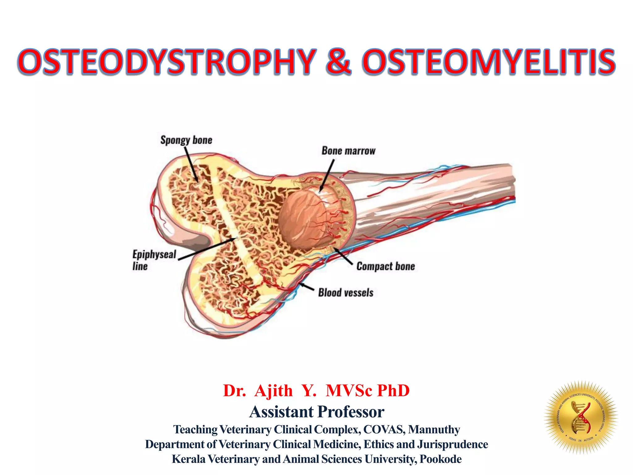
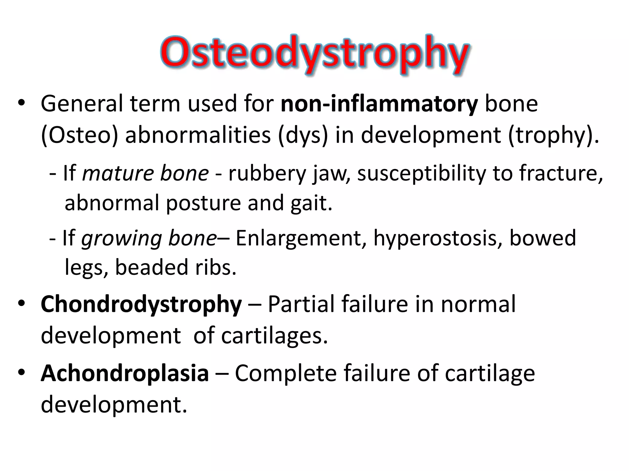
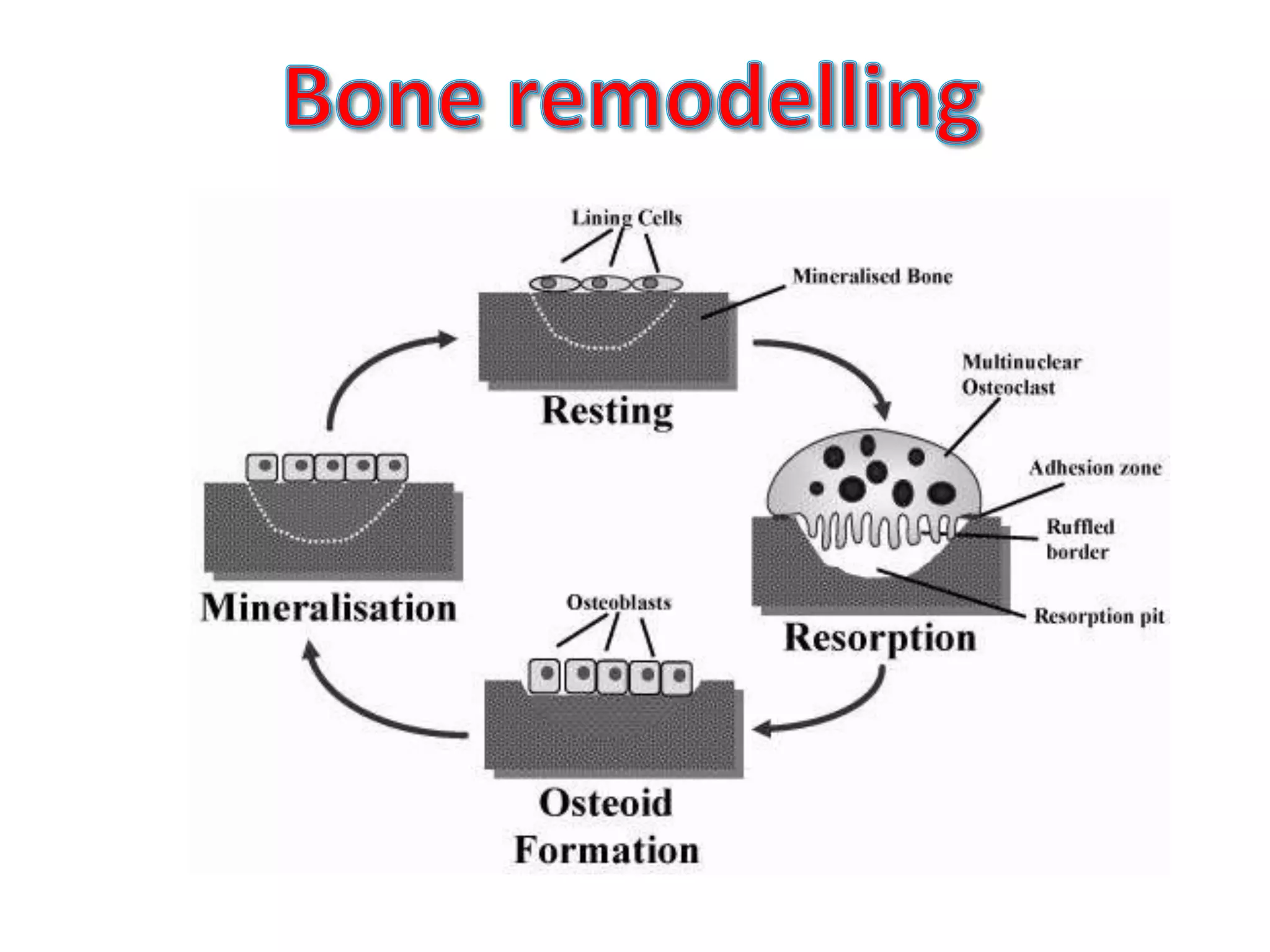
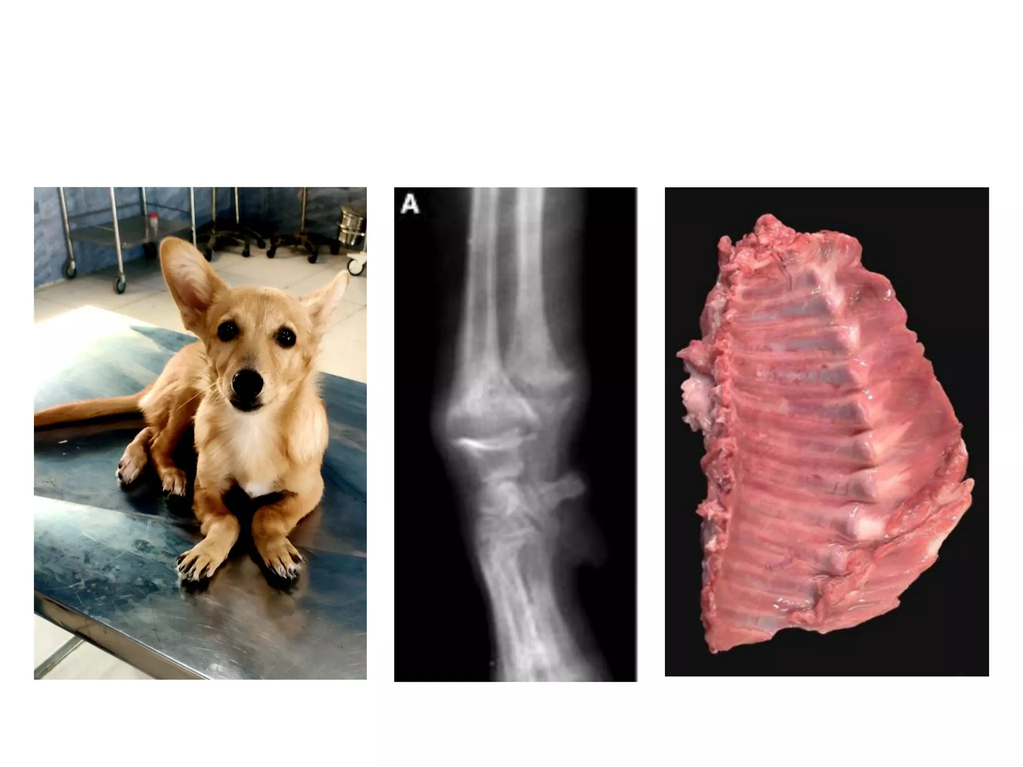
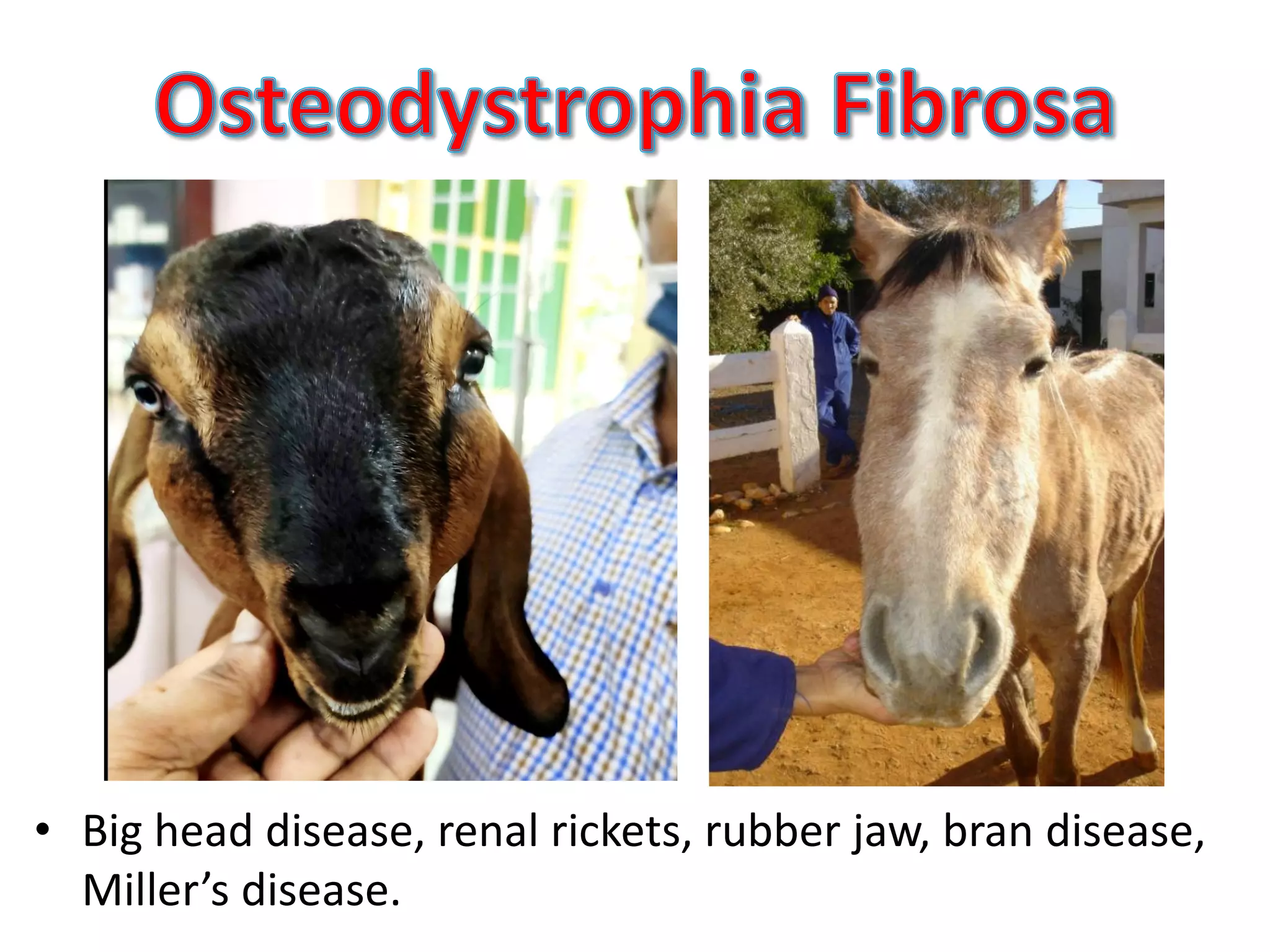
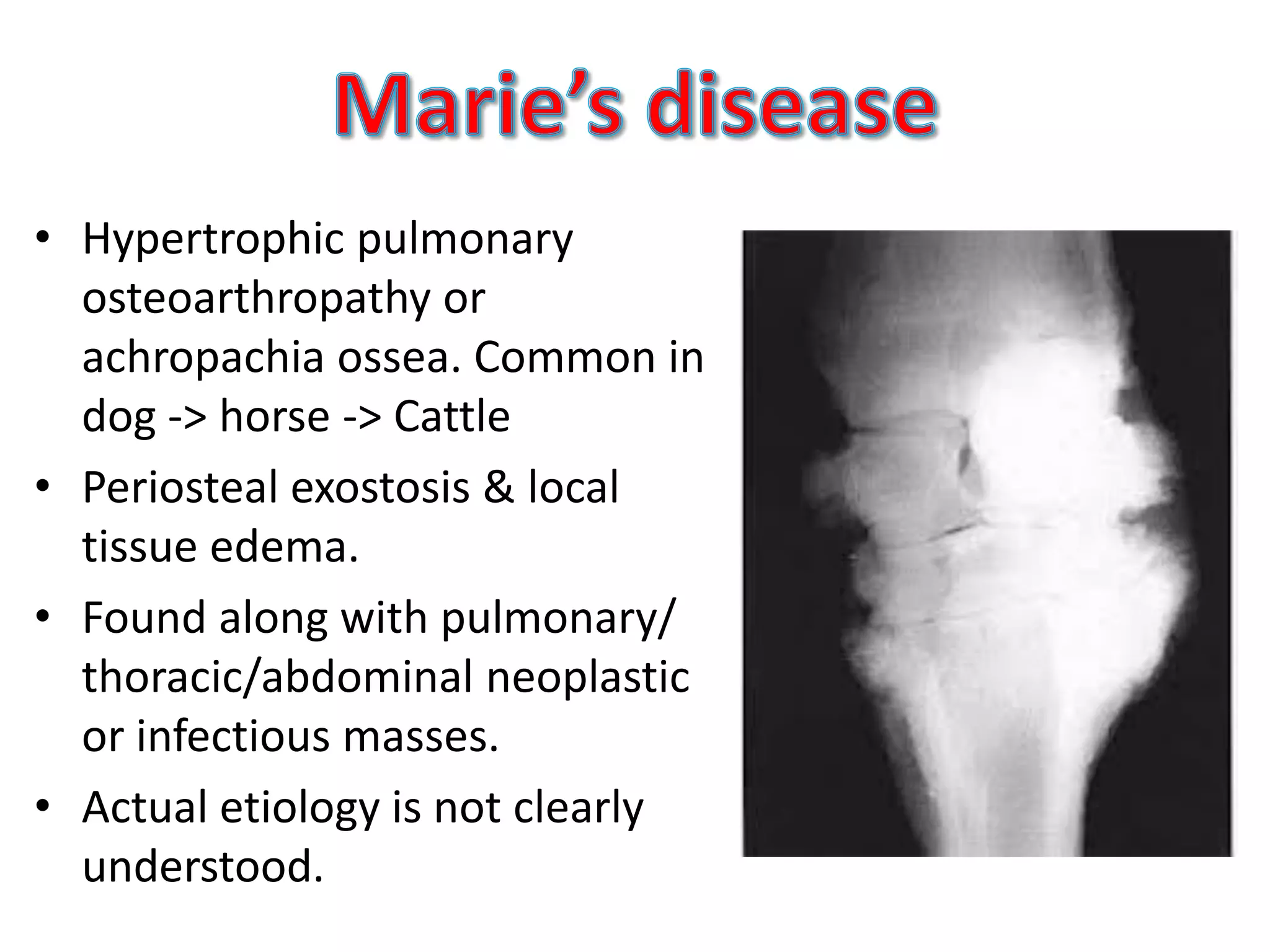
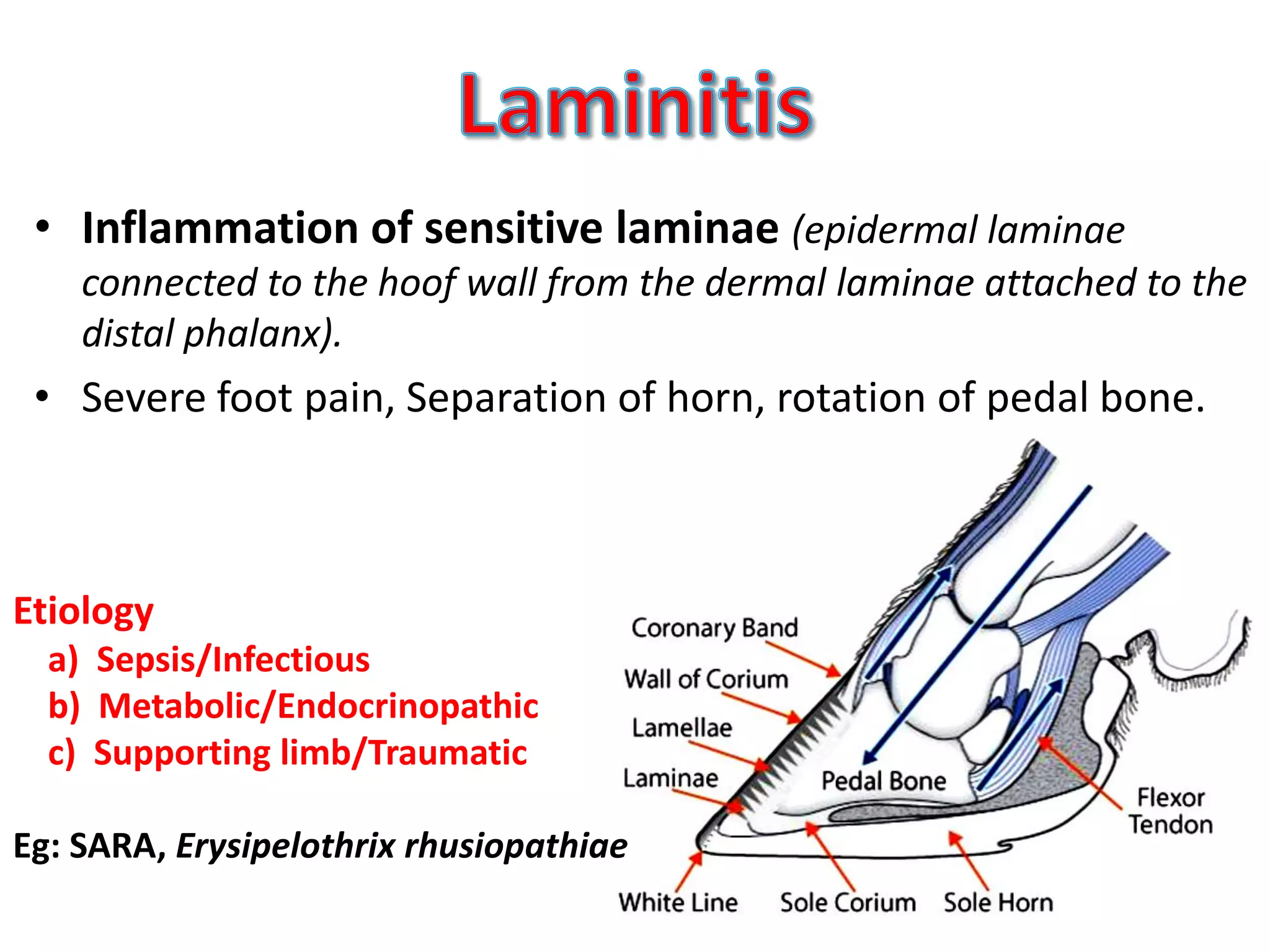
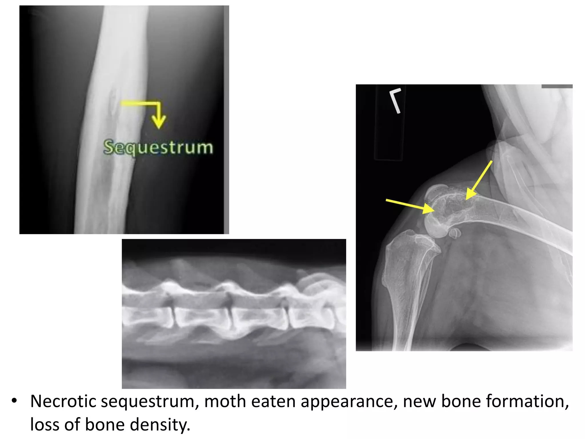
Dr. Ajith Y. MVSc PhD
Assistant Professor
TeachingVeterinary ClinicalComplex, COVAS, Mannuthy
Departmentof VeterinaryClinicalMedicine, Ethics and Jurisprudence
KeralaVeterinaryandAnimal Sciences University, Pookode
• General term used for non-inflammatory bone
(Osteo) abnormalities (dys) in development (trophy).
- If mature bone - rubbery jaw, susceptibility to fracture,
abnormal posture and gait.
- If growing bone– Enlargement, hyperostosis, bowed
legs, beaded ribs.
• Chondrodystrophy – Partial failure in normal
development of cartilages.
• Achondroplasia – Complete failure of cartilage
development.
• Nutritional
– Ca, P, Vit D deficiency/imbalances: ODF, Rickets
– Copper deficiency: Osteoporosis, epiphysitis
– Protein deficiency: Osteoporosis
– Chronic parasitism
– Hypo (low osteoblasts) or hyper (high osteoclasts) vitaminosis A
– Lead and fluride poisoning (deposits in bone/teeth)
• Physical/environmental causes
– Trauma
– Anything affects biomechanics like slatted concrete floors
• Tumors
– Osteosarcomas
• Failure of mineralization of developing bones in Young
animals.
• Overgrowth of fibrous tissue, epiphysitis, hemmorrhages.
• Stunted growth, bowed legs, string of beads (rachetic
rosary) in costochondral jns, enlargement of joints and
bone ends.
• Distorted mandible & maxillae, irregular teeth eruptions,
irregular dental sockets.
• Stiff gait, Pica and unthriftiness
• Osteomalacia: Demineralization/resorption results in
softening of mature bones in adult animals leading to
Spontaneous fracture.
• Osteoporosis: Failure in formation of organic matrix
(osteoid) ie, reduced osteoblatic and increased osteoclastic
activity. Copper deficiency, under nutrition, lead poisoning,
hormonal imbalances etc are common causes.
• Osteodystrophy due to chronic fluorosis: Periosteal
hyperostosis and exostosis on shaft of long bones.
• Big head disease, renal rickets, rubber jaw, bran disease,
Miller’s disease.
• Condition similar to rickets & osteomalacia, but ODF
mostly due to hyperparathyroidism.
(a) Primary hyperparathyroidism: Functional lesion on
Parathyroid gland
(b) Secondary renal hyperparathyroidism: In older animals
(Common in dogs) (http://tru.uni-sz.bg/bjvm/BJVM%20September%202009%20p.212-218.pdf)
(c) Secondary nutritional hyperparathyroidism: In younger
animals. Increased P or decreased Ca diet. Common in goats
and horses.
(Exceptional puppy case ==> https://doi.org/10.1007/s00580-020-03193-x)
• Radiography: Thinning of bone/low density , Epiphyseal
plate widening (Some times moth eaten appearance in
epiphysis also ==>Differentiate with osteomyelitis)
• Ca & P levels: Ratio to be considered (Compensatory changes)
• Nutritional History: Grain diet, Boneless meat
• Treatment history: Ca chelators (OTC, PO4)
• Alkaline Phosphatase Elevated, Not specific
• Clinical signs of renal failure
• Vitamin D injection: 10,000 IU per Kg single injection or
lower dose in alternate days.
• Ca & P supplementation: 10-20 times requirement of Ca.
Based on Ca: P ratio.
• Adequate bedding to prevent inflammation and pain.
• Phosphate binders: Al salts (Kaolin), Ca salts (CaCO3),
Sevelamer………………… Content of IPAKITINE®
• Sunlight & Exercise ==> Improve Vit D activity
• Ecto and Endo parasite control
• Corticosteroids and Anabolic steroids??? (Anecdotal use)
• Hypertrophic pulmonary
osteoarthropathy or
achropachia ossea. Common in
dog -> horse -> Cattle
• Periosteal exostosis & local
tissue edema.
• Found along with pulmonary/
thoracic/abdominal neoplastic
or infectious masses.
• Actual etiology is not clearly
understood.
• Inflammation of sensitive laminae (epidermal laminae
connected to the hoof wall from the dermal laminae attached to the
distal phalanx).
• Severe foot pain, Separation of horn, rotation of pedal bone.
Etiology
a) Sepsis/Infectious
b) Metabolic/Endocrinopathic
c) Supporting limb/Traumatic
Eg: SARA, Erysipelothrix rhusiopathiae
• Inflammation of bone (Infectious or neoplasm or trauma).
• Routes of infection entry
- Hematogenous: Specific => Actinomycosis, Brucellosis,
Non-specific => Omphalitis, TB
- Extension of adjacent infection foci.
- Direct inoculation by trauma/surgery
• Clinical findings: Lameness, inflammatory swelling, pain
on palpation, draining of pus/abscess, secondary muscle
atrophy.
• Necrotic sequestrum, moth eaten appearance, new bone formation,
loss of bone density.
Radiography- Bone tumors
• Moth eaten pattern ??? Osteolytic lesion
• Sunburst appearance??? Osteosarcoma

Osteodystrophy and osteomyelitis in domestic animals

  • 1.
    Dr. Ajith Y.MVSc PhD Assistant Professor TeachingVeterinary ClinicalComplex, COVAS, Mannuthy Departmentof VeterinaryClinicalMedicine, Ethics and Jurisprudence KeralaVeterinaryandAnimal Sciences University, Pookode
  • 2.
    • General termused for non-inflammatory bone (Osteo) abnormalities (dys) in development (trophy). - If mature bone - rubbery jaw, susceptibility to fracture, abnormal posture and gait. - If growing bone– Enlargement, hyperostosis, bowed legs, beaded ribs. • Chondrodystrophy – Partial failure in normal development of cartilages. • Achondroplasia – Complete failure of cartilage development.
  • 4.
    • Nutritional – Ca,P, Vit D deficiency/imbalances: ODF, Rickets – Copper deficiency: Osteoporosis, epiphysitis – Protein deficiency: Osteoporosis – Chronic parasitism – Hypo (low osteoblasts) or hyper (high osteoclasts) vitaminosis A – Lead and fluride poisoning (deposits in bone/teeth) • Physical/environmental causes – Trauma – Anything affects biomechanics like slatted concrete floors • Tumors – Osteosarcomas
  • 5.
    • Failure ofmineralization of developing bones in Young animals. • Overgrowth of fibrous tissue, epiphysitis, hemmorrhages. • Stunted growth, bowed legs, string of beads (rachetic rosary) in costochondral jns, enlargement of joints and bone ends. • Distorted mandible & maxillae, irregular teeth eruptions, irregular dental sockets. • Stiff gait, Pica and unthriftiness
  • 7.
    • Osteomalacia: Demineralization/resorptionresults in softening of mature bones in adult animals leading to Spontaneous fracture. • Osteoporosis: Failure in formation of organic matrix (osteoid) ie, reduced osteoblatic and increased osteoclastic activity. Copper deficiency, under nutrition, lead poisoning, hormonal imbalances etc are common causes. • Osteodystrophy due to chronic fluorosis: Periosteal hyperostosis and exostosis on shaft of long bones.
  • 8.
    • Big headdisease, renal rickets, rubber jaw, bran disease, Miller’s disease.
  • 9.
    • Condition similarto rickets & osteomalacia, but ODF mostly due to hyperparathyroidism. (a) Primary hyperparathyroidism: Functional lesion on Parathyroid gland (b) Secondary renal hyperparathyroidism: In older animals (Common in dogs) (http://tru.uni-sz.bg/bjvm/BJVM%20September%202009%20p.212-218.pdf) (c) Secondary nutritional hyperparathyroidism: In younger animals. Increased P or decreased Ca diet. Common in goats and horses. (Exceptional puppy case ==> https://doi.org/10.1007/s00580-020-03193-x)
  • 10.
    • Radiography: Thinningof bone/low density , Epiphyseal plate widening (Some times moth eaten appearance in epiphysis also ==>Differentiate with osteomyelitis) • Ca & P levels: Ratio to be considered (Compensatory changes) • Nutritional History: Grain diet, Boneless meat • Treatment history: Ca chelators (OTC, PO4) • Alkaline Phosphatase Elevated, Not specific • Clinical signs of renal failure
  • 11.
    • Vitamin Dinjection: 10,000 IU per Kg single injection or lower dose in alternate days. • Ca & P supplementation: 10-20 times requirement of Ca. Based on Ca: P ratio. • Adequate bedding to prevent inflammation and pain. • Phosphate binders: Al salts (Kaolin), Ca salts (CaCO3), Sevelamer………………… Content of IPAKITINE® • Sunlight & Exercise ==> Improve Vit D activity • Ecto and Endo parasite control • Corticosteroids and Anabolic steroids??? (Anecdotal use)
  • 12.
    • Hypertrophic pulmonary osteoarthropathyor achropachia ossea. Common in dog -> horse -> Cattle • Periosteal exostosis & local tissue edema. • Found along with pulmonary/ thoracic/abdominal neoplastic or infectious masses. • Actual etiology is not clearly understood.
  • 13.
    • Inflammation ofsensitive laminae (epidermal laminae connected to the hoof wall from the dermal laminae attached to the distal phalanx). • Severe foot pain, Separation of horn, rotation of pedal bone. Etiology a) Sepsis/Infectious b) Metabolic/Endocrinopathic c) Supporting limb/Traumatic Eg: SARA, Erysipelothrix rhusiopathiae
  • 14.
    • Inflammation ofbone (Infectious or neoplasm or trauma). • Routes of infection entry - Hematogenous: Specific => Actinomycosis, Brucellosis, Non-specific => Omphalitis, TB - Extension of adjacent infection foci. - Direct inoculation by trauma/surgery • Clinical findings: Lameness, inflammatory swelling, pain on palpation, draining of pus/abscess, secondary muscle atrophy.
  • 15.
    • Necrotic sequestrum,moth eaten appearance, new bone formation, loss of bone density.
  • 16.
    Radiography- Bone tumors •Moth eaten pattern ??? Osteolytic lesion • Sunburst appearance??? Osteosarcoma