5. Dentin
It is the main bulk of tooth.
Composition:
Inorganic: 60-65% hydroxyapatite crystals.
Organic: 35-40% collagen and non-collagenous proteins
6. Dentinogenesis
Ondotoblast differentiation
Formation of mantle dentin
Vascular supply
Control of mineralization
Pattern of mineralization.
Root dentin formation
Secondary and tertiary dentin formation
7. Odontoblast formation
The cells responsible for the formation of dentin are ondontoblasts.
Ectomesenchyme of dental papilla under the influence of inner enamel epithelium.
Signaling molecules and growth factors (Bone morhogenic proteins BMP, Insulin
growth factor Igf, Transforming growth factors tgf).
8.
9. Cells of dental papilla:
Undifferentiated ectomesenchyme cells
small, centrally placed nucleus and a few oraganelles.
10. Acellular zone:
Initially dental papilla is separated from the inner enamel epithelium by a zone
which does not have cells but few collagen fibers.
11.
12. Inner enamel epithelial cells reverse their polarity, the ectomesenchymal cells
adjacent to acellular zone become enlarged in size.
Number of cell organelles also increased.
The acellular zone diminished and is filled by the odontoblasts.
Odontoblast: Columnar shaped cells, polarized.
13.
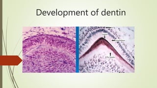
14. Formation of mantle dentin
The first step towards the formation of dentin is the secretion of organic matrix.
Vonkorff,s fibers:
initially collagen III fibers are laid down.
large fibers diameter (0.1-0.2um)
Silver stained
Along fibronectin
Fan shaped spread just below the epithelium.
15.
16. Collagen I is the main secretory product of ondontoblast.
Ondotoblastic process (tomes’s fiber):
These cells secrete cytopaspic extensions.
They have few organelles but have abundant cytoskeleton components.
17.
18. The ondotoblast secretes number of matrix vesicles.
These matrix vesicles have collagen I.
They fuses with each other and give rise to a large globule.
Then minerals are added in these vesicles.
thus there is always a layer of un-mineralized dentin just ahead of mineralized
dentin.
19. Pre dentin
Located adjacent to pulp tissue.
2-6um depending on the activity of odontoblasts.
First form dentin and non-mineralized.
20.
21.
22. Vascular supply
During the mantle dentin formation the capillaries are underneath of odontoblasts.
Dentinogenesis become rapid during the formation of circumpulpal dentin, these
capillaries migrate among the ondontoblasts.
For rapid diffusion fenestrations appear among endothelial cells.
After the completion of primary dentin formation, they retreat to their previous
position.
23.
24. Control of mineralization
Initiated by the small crystallites with in matrix vesicles budded by the odontoblasts.
Increased concentration of phosphate combine with calcium to form hydroxyl apatite
crystals.
Crystals grow and matrix vesicles rupture.
Fuse with adjacent vesicles and form mineralization matrix.
Initially deposited on the surface of collagen fibrils and ground substance and later with
in fibrillis.
25. The minerals are added by three mechanisms
Leaky junctions between odontoblasts for calcium and phosphate
L shaped calcium channel
The presence of alkaline phosphatase and calcium adenotriphosphatase
26.
27. Pattern of minerlization
Globular pattern:
the first formed predentin begins its mineralization in a globular pattern, where a
small centers of calcification spread concentrically until they fuse with each other to
form homogenously calcified dentin.
28.
29. Linear mineralization:
Mineralization of pre dentin appears primary by crystal deposition in the form of
fine plates of hydroxyapatite on the surface of collagen fibrillis and the ground
substance.
Circumpulpal dentin have both type of mineralization.
30.
31. Formation of root dentin
Begins when the formation of enamel and dentin reach at the future
cementoenamel junction.
The epithelial cell of hertwig’s root sheath initiates the cells of dental papilla to
form odontoblasts for the formation of root dentin.
The collagen fibrils of root dentin parallel to the cementodentinal junction
Less mineralized and less number of tubules.
32.
33.
34. Secondary and tertiary dentinogenesis
Secondary dentin:
After root formation
Slower pace
Tertiary dentin:
At specific sites due to injury
Source:
Ondotoblast at the pulp periphery
Undiffereniated cells in the pulp