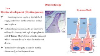
1. Dentine development (Dentinogenesis)
Dentinogenesis starts at the late bell
stage, and occurs in the crown as well as
root regions.
Differentiated odontoblasts are secretory
cells with characteristic apical cytoplasmic
called Tomes fibers (odontoblastic process)
which connect the cells with the surface of
dentin.
Tomes fibers elongate as dentin matrix
formation (predentin) continues.
Dr.Ana’am Mahdi
Oral Histology
Lec.4
2. Dentinogenesis occurs prenatally as well as
postnatally, and can be seen during the whole
life when secondary and tertiary dentin is
formed.
Dentin formation continues throughout the
life of the tooth, and its formation results in
a gradual but progressive reduction in the
size of the pulp cavity.
3. Odontoblasts
Odontoblasts differentiate from the
peripheral cells of the dental papilla by
signaling molecules and growth factors
of the inner enamel epithelium (IEE) .
Odontoblasts first appear at sites of
tooth development at 17–18 weeks in
utero and remain present until death
unless killed by bacterial or chemical
attack, or indirectly through heat or
trauma (e.g. during dental procedures).
4. Histologically:
odontoblasts are large columnar cells in the
crown to flatter cells near the apex of root,
whose cell bodies are arranged along the
interface between dentin and pulp.
The cell is rich in endoplasmic reticulum and
Golgi complex, especially during primary
dentin formation, which allows it to have a
high secretory capacity.
As more dentine matrix is deposited, the
odontoblast cells retreat in the direction of
the pulp leaving an elongated process known
as the odontoblastic process or Tomes fibers.
5. During secretion , its nucleus is aligned away
from the newly formed dentin (toward the pulp
side), with its Golgi complex and endoplasmic
reticulum towards the dentin.
Shape of the odontoblasts also reflect the
functional activity of the cell.
secretory phase, cells show increase in length
about 40μm and 7μm in width with increase
endoplasmic reticulum, Golgi apparatus and
secretory vesicles.
Quiescent phase: resting phase cells are
flattened with little cytoplasm condensed
chromatin and decrease number of
endoplasmic reticulum.
6. Numerous junctions such as gap
junctions, tight junction and
desmosomes are found between
odontoblasts indicating exchange of
ions and small molecules and also
promote cell to cell adhesion
7.
8.
9. Dentinogenesis occur in two stages:
1-Secretion of dentin matrix (predentin):
The first unmineralized organic matrix
secreted by odontoblasts.
A narrow layer of predentin is always
present on the surface of the pulp adjacent
to odontoblasts.
Odontoblasts form approximately 4 μm of
predentin daily during tooth development.
The odontoblasts form the main components
of the dentin matrix, the collagen fibers and
non-collagenous protein which forms the
ground substance.
10. The main non-collagenous proteins in the predentin are:
1. Bone morphogenic proteis (BMP 2,4 ,7)
2. Dentin phosphoprotein /phosphoryn (DPP)
3. Osteocalcin, Osteonectin and Osteopontin
4. Dentin sialoproteins (DSP)
The first indication of forming predentin is the development
of the vonKorff ’s fibers which are bundles of type III
collagen fibers secreted by odontoblasts and they are
perpendicular to the basement membrane.
This fibers is is present in Mantle dentin (first type mineralized
primary dentin).
Then smaller collagen fibril (type I) form a network in the
dentin adjacent to the mantle dentin, which is called
Circumpulpal dentin(second type mineralized primary dentin).
11.
12.
13. 2-Mineralization of the predentin
• It occurs parallel to predentin formation.
• It begins at the tip of the crown and then it
proceeds in a rhythmic pattern to gradually complete cervically.
• The first layer of predentin begins its mineralization in a
globular pattern, where small centers of calcification(crystals)
which come from matrix vesicle(electrone microscopic vesicle
attached to cell membrane of odontoblast which contain first
Hydroxyapatite crystals and alkaline phosphatase enzyme).
• When the crystals grow the matrix vesicles rupture and their
content spread in predentin until they fuse together and form
globules.
14. • Mineralization of predentin or maturation of dentin takes place in two
phases: primary and secondary.
primary mineralization phase: the calcium hydroxyapatite crystals form as
globules, or calcospherules, in the collagen fibers of the predentin, which allows
for both the expansion and fusion
Secondary mineralization phase: new areas of mineralization occur as
globules form in the partially mineralized predentin. These new areas of
crystal formation are more or less regularly layered on the initial crystals,
allowing them to expand.
15. In areas where both primary and secondary mineralization
have occurred with complete crystalline fusion, these appear
as lighter rounded areas on a stained section of dentin and
are considered globular dentin.
In contrast, the darker arclike areas in a stained section of
dentin are considered interglobular dentin.
In interglobular area, only primary mineralization has
occurred within the predentin, and the globules of dentin
do not fuse completely.
Thus, interglobular dentin is slightly less mineralized than
globular dentin.
Interglobular dentin is especially evident in coronal dentin,
near the DEJ.
16.
17. Pattern of mineralization in dentin:
• Histologically, two patterns of dentin mineralization globular and
linear mineralization that depend on the rate of dentin
formation.
• Globular (or calcospheric) calcification involves the deposition
of crystals in several discrete areas of predentin.
• Mantle dentin mineralization occur in a globular pattern (matrix
vesicles)
18. Polarization of odontoblasts, including cell-to-cell junctions that trigger activation of genes for proteins secreted into dentin.
Legend: Green = cell-cell junctions help establish cell polarity by triggering intra-cellular signals. Yellow= secretion of collagen-
filled vesicles to form pre-dentin (light blue layer) occurs at the apical surface of the cell body. Brown = secretion of enzyme-
filled vesicles from the odontoblastic process triggers mineralization of pre-dentin into dentin (darker blue layer). Pink = pulp.
19. • The mineralization goes then in linear or
occasionally globular pattern in the
remnant or bulk thickness of dentin
which is called circumpulpal dentin.
• The mineralization begins by crystal
deposition in form of fine plates of
hydroxyapatite crystals on the surface of
the collagen fibrils. The long axes of the
crystals are paralleling to the collagen
fibrils.
A - interglobular
dentin
B - globular dentin
C - dentinal tubules
20. Pattern of Dentin formation
Dentin of the crown( coronal dentin)
begins at the bell stage of tooth development in the
papillary tissue adjacent to the inner enamel
epithelium, the site where cuspal development begins.
From that point, dentin formation spreads down the
cusp slope as far as the cervical loop of the enamel
organ, and the dentin thickens until all the coronal
dentin is formed.
In multicusped teeth, dentin formation begins
independently at the sites of each future cusp tip and
again spreads down the flanks of the cusp slopes.
21. Root dentin:-
• Forms at a slightly later stage of
development and
requires the proliferation of epithelial
cells
(Hertwig’s epithelial root sheath) from the
cervical loop of the enamel organ to initiate
the differentiation of root odontoblasts.
• Root dentin is different from dentin of
the crown because of:
different orientation of collagen fibers, as
well as less mineralization.
22. • The onset of root formation precedes the onset of
tooth eruption, and by the time the tooth reaches
its functional position, about two thirds of the root
dentin will have been formed. Completion of root
dentin formation does not occur in the deciduous
tooth until about 18 months after it erupts and in
the permanent tooth until 2 to 3 years after it
erupts. During this period the tooth is said to have
an open apex.
23. Dentinogenesis imperfecta(DGI):
Hereditary condition associated with abnormal
dentin mineralization and varying degrees of
changes in tooth morphology.
DGI type I is associated with osteogenesis
imperfecta(brittle bone disease), whereas the
clinically similar DGI type II is not associated
with a syndrome and is caused by mutations in
the gene encoding dentin sialophosphoprotein
(DSPP).
The DGI tooth phenotype is highly variable and
is characterized by blue-gray to yellow-brown
coloration .