
Recommended
More Related Content
What's hot
What's hot (20)
Similar to Periodontal Ligament.ppt
Similar to Periodontal Ligament.ppt (20)
More from madhusudhan reddy
More from madhusudhan reddy (20)
Recently uploaded
Recently uploaded (20)
Periodontal Ligament.ppt
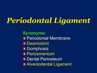
- 1. Periodontal Ligament Synonyms: Periodontal Membrane Desmodont Gomphosis Pericementum Dental Periosteum Alveolodental Ligament
- 2. Periodontium The word Periodontium refers to the attachment apparatus of teeth and consists : – Cementum – Alveolar bone lining tooth socket – Periodontal ligament – Part of Gingiva facing the tooth
- 3. Periodontal Ligament The space occupied by PDL is called Periodontal Space Coronally :- Continuous with Gingiva Apically :- Continuous with Pulp Is the connective tissue which surrounds the roots of the teeth and Attaches tooth root to the bony alveolus
- 4. The width of PDL ranges from 0.15 – 0.38mm Thinnest around middle 1/3rd of root and has “Hour-Glass” appearance Reduced in nonfunctional and unerupted teeth. Increased in teeth subjected to heavy functional stresses. Thicker in deciduous teeth than permanent teeth. The principal fibers run a wavy course from Cementum to alveolar bone
- 5. Periodontal Ligament consists of: – Cells – Extracellular substance: Fibers Ground substance Structure and Composition
- 7. Cells in Periodontal Ligament A – Alveolar Bone B - Cementum
- 8. The cells of Periodontal Ligament: 1. Synthetic cells: Osteoblasts Fibroblasts Cementoblasts 2. Resorptive cells: Osteoclasts Fibroblasts Cementoclasts 3. Progenitor cells 4. Epithelial Cell rests of Malassez 5. Defense cells: Mast cells Macrophages Eosinophils
- 9. Synthetic cells Unique characteristics of Synthesizing cells: Vesicular nucleus containing prominent nuclei Abundant cytoplasm containing numerous RER and Increased Golgi bodies Large numbers of Mitochondria Cells with above description present on Alveolar surface of PDL Osteoblasts Such cells lying in body of soft connective tissue of PDL Fibroblasts Cells found on cemental surface Cementoblasts
- 10. Osteoblasts Bone forming cells lining the alveolar bone of tooth socket closely resembling cementoblasts Cuboid in shape with prominent spherical and large nucleus placed at basal end of cell Large amounts of RER gives the cytoplasm of active osteoblasts a slightly basophilic hue Active osteoblast produces an enormous quantity of collagen type-I Alkaline phosphatase is present and is mostly found on the apical cell membrane
- 12. Fibroblasts A – Alveolar Bone B – Fibroblasts C – Cementum D – Dentin
- 13. Fibroblasts Predominant cells in PDL Capable of Both Secretion and Degradation of Collagen Fibroblasts near cementum are derived from investing layer of dental papilla and fibroblasts near Alveolar bone are derived from Perivascular Mesenchyme of Dental Follicle and show abundant Alkaline phosphatase activity Fibroblasts are large cells with extensive cytoplasm and characteristics of synthesizing cells are seen prominently
- 14. Fibroblasts are of various shapes: – Fusiform – Tripolar – Stellate Fibroblasts of PDL have Cilia and the Cilia is associated with Control of Cell cycle or Inhibition of Centriolar Activity These cells produce growth factors and cytokines such as IGF-1, BMP’s, PDGF, IL-1, etc TGF-β stimulates synthesis of collagen and Inhibits the synthesis of collagenase
- 16. Functions of Fibroblasts To produce structural connective tissue proteins: Collagen Elastin Proteoglycans Glycoproteins Glycosaminoglycans Collagenolytic enzymes: Matrix Metalloproteinases (MMP’s) Fibroblasts are responsible for formation and remodelling of PDL They have a signaling system to maintain width of PDL by preventing encroachment of bone or cementum into the PDL space
- 17. Differences between PDL Fibroblasts Ectomesenchymal origin Expression of Alkaline phosphatase and cyclic AMP is MORE Motile and Contractile and hence can generate a force for tooth eruption Gingival Fibroblasts Mesodermal origin Expression of Alkaline phosphatase and cyclic AMP is LESS Non-contractile and no role to play in tooth eruption
- 18. Cementoblasts
- 19. Cementoblasts Cuboidal cells with large vesicular nucleus Active in cementum formation found adjacent to the surface of cementum and show all the prominent features of synthesizing cells Cells depositing cellular cementum show abundant basophilic cytoplasm and cytoplasmic processes and nuclei are folded and irregularly shaped Cells depositing - acellular cementum – DO NOT SHOW prominent cytoplasmic processes
- 20. Cementoblsts
- 21. Resorptive Cells Osteoclasts & Cementoclasts: Found in areas of resorption. Multinucleated Originate from undifferentiated mesenchymal cells in periodontal ligament.
- 23. Cytoplasm of the cells produce a substance which dissolves the organic components of bone and a chelating agent capable of bringing calcium salts into solution. Where ever their cytoplasm comes into contact with bone - hollows or grooves called 'Howship's Lacunae' are formed. When bone resorption ceases - they disappear.
- 24. Progenitor cells These cells have the capacity to undergo mitotic division. They give rise to all of the specialized synthetic cells. Epithelial cell rests of Malassez: Remanents of Hertwig’s Epithelial Root Sheath. Epithelial double strands and islands limited by basement membrane of reticulin.
- 26. Mast cells These are small round or oval cells. 12 - 15μm. Characterized by numerous cytoplasmic granules. These granules contain heparin and histamine. Histamine plays and important role in inflammatory reactions and also in antigen antibody reaction.
- 28. Macrophages These can be visualized by the presence of phagocytosed material in their cytoplasm. They are derived from blood monocytes.
- 30. Extracellular Substance Comprises of: –Fibers: Collagen Oxytalan. –Ground substance: Proteoglycans Glycoproteins.
- 32. Fibers Made up of collagen and Oxytalan. Elastic fibers are restricted to the walls of blood vessels. The majority of fibers are collagen. Mostly made up of type I collagen and some amount of type III collagen. The collagen fibers are gathered in bundles having a clear orientation relative to the periodontal space - “Principal fibers”.
- 34. Principal Fibers Arranged in five groups: 1. Alveolar crest group. 2. Horizontal group. 3. Oblique group. 4. Apical group. 5. Inter-radicular group.
- 35. Principal Fibers 1. Alveolar crest 2. Horizontal 3. Oblique 4. Apical 5. Interradicular
- 36. Principal Fibers Alveolar Crest group: They radiate from the crest of the alveolar process and attach themselves to the cervical part of the Cementum. These fibers resist tilting, intrusive, extrusive and rotational forces. Most often confused with Dentoperiosteal fibers
- 38. Horizontal group: They run at right angles to long axis of tooth from Cementum to Bone. Roughly parallel to the occlusal plane of the arch They are found immediately apical to the alveolar crest fiber group. They are limited mostly to the coronal one-fourth of the periodontal ligament space. These fibers resist horizontal and tipping forces.
- 39. Oblique group: Most numerous and occupy nearly 2/3rd of the ligament. They are attached to cementum apically from their attachment to the bone thereby resulting in their oblique orientation within the periodontal space These fibers constitute the main attachment. These fibers resist vertical and intrusive forces.
- 40. Apical group: They radiate from the cementum at the root tip to become anchored into the fundus of the bony socket. The apical fibers resist the forces of luxation, may prevent tooth tipping and protect delicate blood and lymph vessels and nerves traversing the PDL space at the root apex. These fibers are not seen on incompletely formed roots.
- 41. Interradicular Group: From the crest of interradicular septum, bundles extend to furcation of multirooted teeth. These fibers resist tooth tipping, torquing and luxation. These fibers are lost, if age- related gingival recession proceeds to the extent, that the furcation area is exposed. Total loss of these fibers occurs in chronic inflammatory periodontal disease.
- 42. Oblique set
- 43. Inter-radicular
- 44. Elastic Fibers There are three types of elastic fibers, which are histochemically and ultrastructurally different. They are, the mature elastic fibers, (elastin) fibers, the elaunin fibers, and the oxytalan fibers. Elaunin and oxytalan fibers have been described as immature elastic fibers. Mature elastic fibers consist of a microfibrillar component surrounding an amorphous core of elastin protein.
- 45. Elaunin Fibers Elaunin fibers are seen as bundles of microfibrils embedded in a relatively small amount of amorphous elastin. These fibers may be found within the fibers of the gingival ligament.
- 46. Oxytalan Fibers These are immature elastic fibers. Found in the periodontal ligament. Largely restricted to the walls of blood vessels. The orientation of these fibers is quite different. They tend to run axially - one end being embedded into cementum or possibly in bone and the other - into the wall of a blood vessel. In the vicinity of apex they form a complex network.
- 47. Reticular Fibers These are fine immature collagen fibers with argyrophilic staining properties and are related to basement membrane of blood vessels and epithelial cells which lie within the periodontal ligament. These fibers are composed of type III collagen.
- 50. Ground Substance Glycosaminoglycans: – Hyaluronic acid – Dermatan sulfate – Chondroitin sulfate. Carbohydrates, proteins, lipids and Glycoproteins. Other Structures Present in connectinve tissue: – Blood vessels – Lymphatics – Nerves and Cementicles.
- 51. Functions 1. Formative 2. Supportive 3. Sensory 4. Nutritive 5. Occlusion 6. Reparative 7. Protective
- 53. Sharpey’s Fibers A – Alveolar Bone B - Cementum
- 54. Sharpey's Fibers: The collagen fibers are embedded into the cementum on one side of the periodontal space and into alveolar bone on the other. Intermediate Plexus: When examined under light microscope the fibers appear to be joined in the mid region of the periodontal space giving rise to a zone of distinct appearance, so called "Intermediate Plexus" – believed to be site for rapid remodeling of fibers.
- 57. Blood Supply: From three sources - 1. From the apical vessels supplying the dental pulp. 2. Branches from intra-alveolar vessels. 3. Branches form gingival vessels. Lymphatics: Net work of lymphatic vessels following the path of blood vessels.
- 58. Nerve Supply
- 59. Nerves are usually associated with blood vessels Pass through foramina in the alveolar bone to enter the periodontal ligament. Myelinated or non myelinated. Either large or small in diameter: - larger diameter - concerned with touch. - smaller diameter - concerned with pain. Cementicles: Calcified bodies. Origin is probably from degenerated epithelial cells – forming a nidus for calcification.
- 60. Nerve Bundles A – Alveolar bone B - Dentin