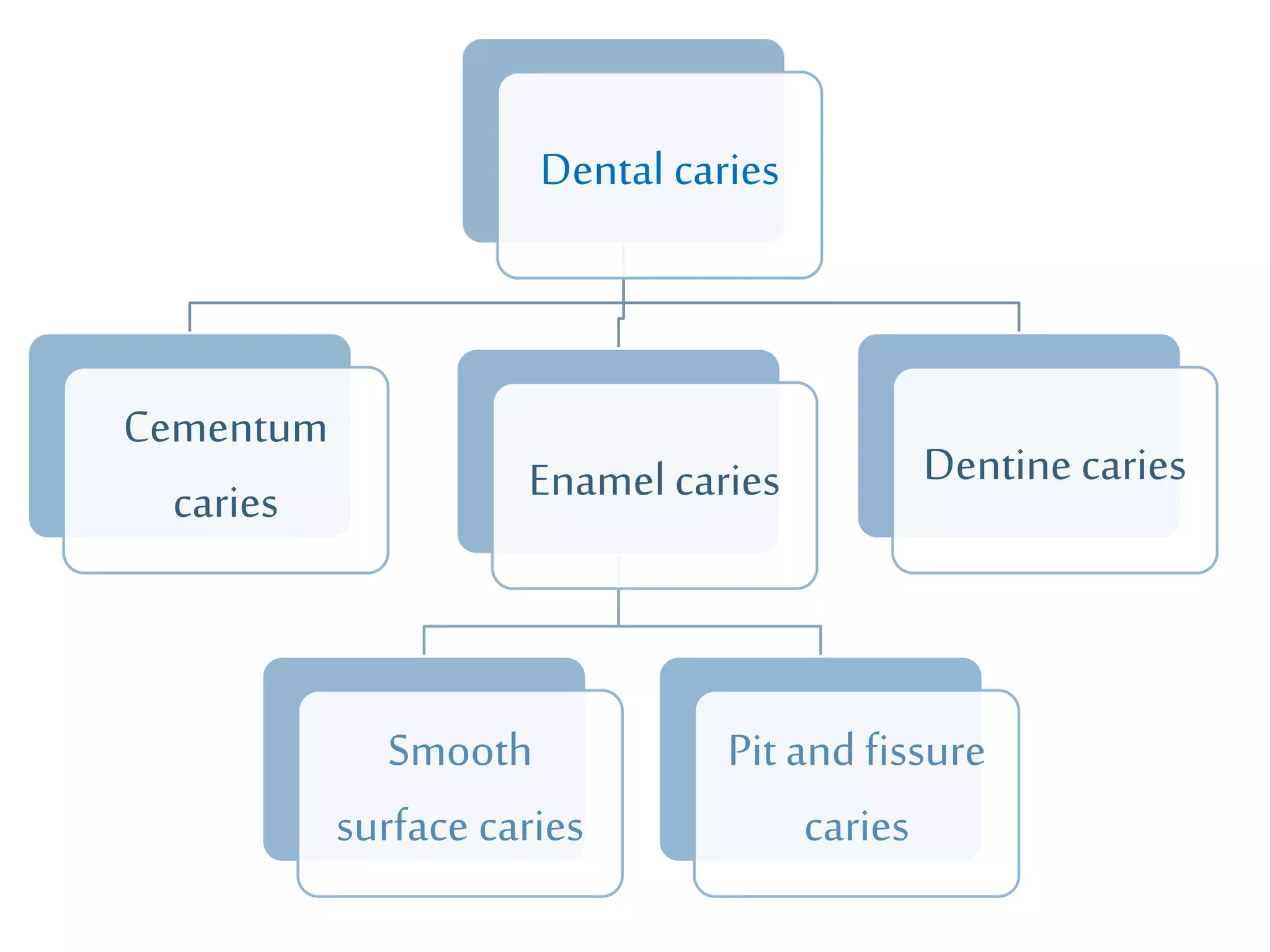
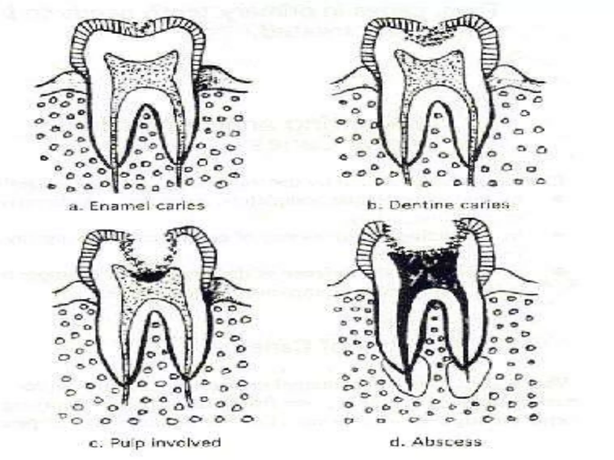
DENTAL CARIES
Preparedby- ShreejaNair
 Definition: Dental caries is an infectious microbiologic disease of the
teeth that results in localized dissolution and destruction of the
calcifiedtissues.
 Carious lesions occur only under a mass of bacteria capable of
producing a sufficiently acidicenvironment to demineralize tooth
structure.
 Organisms that cause caries are cariogenic.
 Thedegreeto which a tooth is likely to becomecarious is described as its
cariogenicity potential.
Organisms responsible
1. Streptococcus mutans (MS)
2. Lactobacilli
 MSare associated withtheonset of caries.
 Lactobacilliare associated withactive progression of cavitated
lesions.
Typeof caries Microorganism
Pit and Fissure S. Mutans
S. Sanguis
Lactobacillus species
Actinomycesspecies
Smooth Surface S. Mutans
S. Salivarius
Root surfaces A. Viscosus
A. Naeslundii
S. Mutans
S. Sanguis
Deep dentinal caries Lactobacillus species
A. Naeslundii
Other filamentous rods
THEORIES OFDENTALCARIES
1. ChemicoparasiticTheory
(Miller W.D 1890 )
 This states that oral bacteria actsonsugars to release acidsthat
demineralize the inorganicportion of enamel, resulting inthe
development of dental caries.
2. Proteolytictheory
(Bodekar C.F.1948)
 This theory states theorganiccomponent of the enamel is first
brokendownby proteolyticenzymes, openingup pathwaysfor
bacteria to attackthe enamel by other processes such as by acidor
chelation.
3. Chelationtheory
(ShatzAet al1957)
 This states that the enamel is demineralized by chelatingagents at
neutral pH.Protein breakdownproducts and lacticacidare some
chelatingagents present innature.
 This is the most accepted theory.
 The factors responsible for this delicate balanceare:
 pHofplaque.
 Calciumandphosphateionconcentrationattheinterfacebetween
enamelandplaque.
 Fluorideionconcentration.
Etiologic agentsof caries
Pathogenic bacterialplaque
 Bacterialplaque:The soft, translucent, andtenaciously adherent
material accumulatingonthe surface of teeth is commonly called
bacterial plaque.
 The accumulationof plaqueonteeth is a highly organizedand
ordered sequence of events.
 Caries causes damageby demineralized andthe dissolution of tooth
structure, resulting from:
1. AhighlylocalizeddecreaseinthepHattheplaque-toothinterface.
2. Toothdemineralization.
Dentalplaqueon toothsurface.
Factors affecting
I. PRIMARY FACTORS
1. Tooth
2. Dentalplaque
3. Diet
4. Time
II. MODIFYING FACTORS
1. Saliva
2. Systemichealth
3. Sex
4. Heredity
5. Race
6. Geographic environment
7. Occupation
CLINICALCHARACTERISTICSOF LESION
 The characteristics vary with the natureof surface onwhichthe
lesions develops.
 There are 3 different clinicalsites:
1. Pitsandfissures.
2. Smoothenamelsurfaces.
3. Rootsurface.
Dental caries
Cementum
caries Enamel caries
Smooth
surface caries
Pit and fissure
caries
Dentine caries
1. Pits and fissures.
 Bacteria rapidly colonizethepits andfissures andform a “bacterial
plug.”
 They:
1. endblindly.
2. opennear thedentin.
3. penetrateentirelythroughenamel.
 Incross section : invertedV with narrow entrance and
progressively widerarea of involvementcloserto theDEJ.
LS showing caries on occlusal surface.
2. Smooth enamelsurfaces.
 The smoothenamel surfaces of teeth are less favorable site for
plaque attachment.
 Lesions have abroad area of originand aconicalor pointed
extension towardDEJ.
 Cross section : V shaped witha widearea of origin and the apex
of the vdirectedtowards DEJ.
LS showing initiation and progression of caries on interproximal surfaces.
3. Rootsurfaces.
 Root surfaces are rougher thanenamel and readily allowsplaque
formation.
 Cementum coveringthe root surface is extremely thin and provides
little resistance to caries attack.
 Cross section: U shapedwith less welldefined margins .
Histopathology ofcaries
Enamelcaries
 Histology ofenamel:
 Accordingtochelationtheorythemovementofionsthroughcarious
enamelcanresultinaciddissolutionoftheunderlyingdentinbefore
actualcavitationoftheenamelsurface.
 Cariespreferentiallyattacksthecoresoftherodsandthemore
permeablestriaeofRetzius,whichpromoteslateral spreadingand
underminingoftheadjacentenamel.
 Clinical& histologicalcharacteristicsof enamelcaries:
 Anearlyenamellesionseenunderpolarizedlightreveals4distinctzones
ofmineralization.
1. Translucentzone
2. Darkzone
3. Bodyof lesion
4. Surface zone
 This is the deepest zone
whichrepresents the
advancingfront of the
enamel caries.
 This zone is translucent due
to demineralizationwhich
creates a structureless
appearanceof the enamel.
 This lies deeper to the bodyof
the lesion andit represents
some remineralization.
1. Translucent zone Darkzone
 This is the largest portion of
enamel caries.
 It is poorlymineralized.
 The striae of Retzius are well
marked.
 The caries spreads alongthe
striae of Retzius and
interprismatic areas and then
attacksthe prism cores.
 Bacteria are present inthis
zone.
 This outermost zone is
relatively unaffected by caries
attack.
 It is well mineralized by
replacement ionsfrom
plaque and saliva.
Bodyoflesion Surfacezone
Dentinalcaries
 Histology ofdentin:
 Caries of the dentinbegins with the spread of
process along the DEJ.
 There is a rapidinvolvement of great numbers
of dentinaltubules whichacts as a tract leading
to dental pulpalong with the microorganisms.
 Clinical& histologicalcharacteristicsof dentin caries:
 Progressionofcariesindentinisdifferentfromprogressioninthe
overlyingenamelbecauseofthestructuraldifferenceofdentin.
 Episodesofpainmay befeltwhichisduetostimulationofunderlying
pulp.
 Cariesadvancementindentinproceedsthrough3changes.
1. Weakorganicacidsdemineralizethe dentin.
2. Collagendegeneratesand dissolves.
3. Lossof structuralintegrityfollowedby invasionof bacteria.
 5differentzonesare observed:
1. Normaldentin
2. Subtransparentdentin
3. Transparentdentin
4. Turbiddentin
5. Infecteddentin
 The deepest zone of carious
dentinis normal withnormal
collagen, odontoblastic
processes andintertubular
dentin.
 The intertubular dentin is
demineralized , odontoblastic
processes are damagedand
fine crystals are seen inthe
lumenof dentinal tubules.
 No bacteria is foundin this
zone.
Normaldentin Subtransparentdentin
 Superficial to the
subtransparent layer.
 Softer thannormal dentin
and exhibits mineral loss in
theintertubular dentin.
 No bacteria is seen and the
collagencross-linking is
intact.
 Capableof remineralization.
 Next superficiallayer.
 Dentinal tubules arewidenedand
distorted due tobacterial
penetration.
 There is considerable
demineralizationand collagenis
irreversiblydenatured.
 This zone is incapable of
remineralizationand must be
removed.
Transparent dentin Turbid dentin
 Outermost zone.
 It has decomposed dentin
with destruction of dentinal
tubules andcollagen.
 A highconcentration of
bacteria are seen.
 This zone has to be removed
to prevent thespread of the
infection.
Infecteddentin
Histopathology of Dental Caries
Enamel Caries Dentin Caries
Radiograph showing cariesin dentin
Classificationof caries
BASED ON LOCATION
1. Pit andfissure caries
2. Smooth surface caries
3. Root surface caries
BASEDON RATE OFPROGRESSION
1. Acute / rampant caries
 Rapidlyinvadingcariesinvolvingseveral teeth.
 Appearssoftor lightcolored.
 Ifunattendedcausesearlypulpinvolvement.
2. Chroniccaries
 Slowlyprogressing,longstandingcaries.
 Hard consistencyanddark colored.
3. Arrested caries
 Sometimesochroniccariouslesioncanbecomearresteddueto
changeinlocalenvironment.
BASEDON EXTENT OF CARIES
1. Incipient caries
 Firstevidenceofcariesactivityinenamel.
 DemineralizedenamelnotextendedtoDEJ.
 Enamelsurfaceishard andstillintact.
 Can beremineralized.
2. Cavitatedcaries
 Carieshasspreadbeyondenamelintodentin.
 Enamelisbrokendownandremineralizationisnotpossible.
BASEDON NEW OR RECURRENT
1. Initial/primary caries
 Firstattackofcariesontooth.
2. Recurrent /secondary caries
 Cariesseenaroundthemarginsofrestoration.
 Occursduetomicroleakageandother favorableconditions.
BASEDON PATHWAYOF CARIESSPREAD
1. Forward caries
 Whenever the caries cone in enamel is larger or the same size as
that in dentin its referred to asforward caries.
2. Backwardcaries
 Whenever the spread of caries along the DEJ exceeds the caries
cone in enamel, the caries extends into enamel from the junction.
 Spread of caries isin backward direction.
FORWARDCARIES BACKWARDCARIES
BASED ON NO.OF TOOTH SURFACES INVOLVED
1. Simple caries
 Involving1surface.
2. Compoundcaries
 Involving2surfaces.
3. Complexcaries
 Involving3 ormoresurfaces.
BASEDONWHETHERCARIESIS COMPLETELY
REMOVEDOR NOT
 Residual caries
 Cariesthatremainsinthepreparedcavityevenafter therestorationis
completed.
 Thismay beleftbehindeitherbyaccident,neglector intension.
BASEDON AGE OFPERSON
1. Nursing bottle caries
 Duringearlyinfancy,bottlefedbabiesdeveloprapidlyspreading
caries.
 Usuallymaxillaryincisors.
2. Adolescent caries
 Acutecariesseeninadultsduetodietaryhabits.
3. Senile caries
 Cariesoccurringinelderlyinvolvingrootsurfaces.
 Duetogingivalrecessionandpoororalhygiene.
BASEDON TOOTH SURFACESINVOLVED
1. O occlusal surface.
2. M mesial surface.
3. D distal surface.
4. F facialsurface.
5. B buccal surface.
6. L lingualsurface.
WHO SYSTEM
1. D1clinicallydetectable enamel lesions with intact
surfaces.
2. D2clinicallydetectable cavities limited to enamel.
3. D3clinicallydetectable cavities in dentin.
4. D4lesions extending into the pulp.
GRAHAMMOUNT’S CLASSIFICATION
Cavitysite Size 1 Size 2 Size 3 Size 4
Minimal Moderate Enlarged Extensive
Site 1
Pit and fissure
1.1 1.2 1.3 1.4
Site 2
Approximal surface/ contact
surface
2.1 2.2 2.3 2.4
Site 3
Cervical region
3.1 3.2 3.3 3.4
BASED ONRESTORATION
(G.V.BLACK CLASSIFICATION)
1. Class I caries
2. ClassII caries
3. ClassIIIcaries
4. Class IVcaries
5. ClassV caries
6. ClassVI caries
CLASSI CAVITYPERPARATION
 Restorations on occlusal surfaces of premolars and molars
 Occlusal 2/3rd of facial and lingual surfaces of molars
 Lingual surfaces of maxillaryincisors.
CLASSII CAVITYPERPARATION
 Restorations on proximal surfaces of posterior teeth.
CLASSIII CAVITYPERPARATION
 Restorations on proximal surfaces of anterior teeth that do not involve incisal angle.
CLASSIV CAVITYPERPARATION
 Restorations on proximal surfaces of anterior teeth that involve incisal edge.
CLASSV CAVITYPERPARATION
 Restorations on gingival 1/3rd of facial or lingualsurfaces of all teeth.
CLASSVI CAVITYPERPARATION
 Restorations on incisal edge of anterior teeth or occlusal cusp heights of posterior teeth.
Dental caries ppt

Dental caries ppt

  • 1.
  • 2.
     Definition: Dentalcaries is an infectious microbiologic disease of the teeth that results in localized dissolution and destruction of the calcifiedtissues.  Carious lesions occur only under a mass of bacteria capable of producing a sufficiently acidicenvironment to demineralize tooth structure.
  • 3.
     Organisms thatcause caries are cariogenic.  Thedegreeto which a tooth is likely to becomecarious is described as its cariogenicity potential.
  • 4.
    Organisms responsible 1. Streptococcusmutans (MS) 2. Lactobacilli  MSare associated withtheonset of caries.  Lactobacilliare associated withactive progression of cavitated lesions.
  • 5.
    Typeof caries Microorganism Pitand Fissure S. Mutans S. Sanguis Lactobacillus species Actinomycesspecies Smooth Surface S. Mutans S. Salivarius Root surfaces A. Viscosus A. Naeslundii S. Mutans S. Sanguis Deep dentinal caries Lactobacillus species A. Naeslundii Other filamentous rods
  • 6.
  • 7.
    1. ChemicoparasiticTheory (Miller W.D1890 )  This states that oral bacteria actsonsugars to release acidsthat demineralize the inorganicportion of enamel, resulting inthe development of dental caries.
  • 8.
    2. Proteolytictheory (Bodekar C.F.1948) This theory states theorganiccomponent of the enamel is first brokendownby proteolyticenzymes, openingup pathwaysfor bacteria to attackthe enamel by other processes such as by acidor chelation.
  • 9.
    3. Chelationtheory (ShatzAet al1957) This states that the enamel is demineralized by chelatingagents at neutral pH.Protein breakdownproducts and lacticacidare some chelatingagents present innature.  This is the most accepted theory.  The factors responsible for this delicate balanceare:  pHofplaque.  Calciumandphosphateionconcentrationattheinterfacebetween enamelandplaque.  Fluorideionconcentration.
  • 10.
  • 11.
    Pathogenic bacterialplaque  Bacterialplaque:Thesoft, translucent, andtenaciously adherent material accumulatingonthe surface of teeth is commonly called bacterial plaque.  The accumulationof plaqueonteeth is a highly organizedand ordered sequence of events.  Caries causes damageby demineralized andthe dissolution of tooth structure, resulting from: 1. AhighlylocalizeddecreaseinthepHattheplaque-toothinterface. 2. Toothdemineralization.
  • 12.
  • 13.
    Factors affecting I. PRIMARYFACTORS 1. Tooth 2. Dentalplaque 3. Diet 4. Time II. MODIFYING FACTORS 1. Saliva 2. Systemichealth 3. Sex 4. Heredity 5. Race 6. Geographic environment 7. Occupation
  • 15.
  • 16.
     The characteristicsvary with the natureof surface onwhichthe lesions develops.  There are 3 different clinicalsites: 1. Pitsandfissures. 2. Smoothenamelsurfaces. 3. Rootsurface.
  • 17.
    Dental caries Cementum caries Enamelcaries Smooth surface caries Pit and fissure caries Dentine caries
  • 18.
    1. Pits andfissures.  Bacteria rapidly colonizethepits andfissures andform a “bacterial plug.”  They: 1. endblindly. 2. opennear thedentin. 3. penetrateentirelythroughenamel.  Incross section : invertedV with narrow entrance and progressively widerarea of involvementcloserto theDEJ.
  • 19.
    LS showing carieson occlusal surface.
  • 20.
    2. Smooth enamelsurfaces. The smoothenamel surfaces of teeth are less favorable site for plaque attachment.  Lesions have abroad area of originand aconicalor pointed extension towardDEJ.  Cross section : V shaped witha widearea of origin and the apex of the vdirectedtowards DEJ.
  • 21.
    LS showing initiationand progression of caries on interproximal surfaces.
  • 22.
    3. Rootsurfaces.  Rootsurfaces are rougher thanenamel and readily allowsplaque formation.  Cementum coveringthe root surface is extremely thin and provides little resistance to caries attack.  Cross section: U shapedwith less welldefined margins .
  • 23.
  • 24.
    Enamelcaries  Histology ofenamel: Accordingtochelationtheorythemovementofionsthroughcarious enamelcanresultinaciddissolutionoftheunderlyingdentinbefore actualcavitationoftheenamelsurface.  Cariespreferentiallyattacksthecoresoftherodsandthemore permeablestriaeofRetzius,whichpromoteslateral spreadingand underminingoftheadjacentenamel.
  • 25.
     Clinical& histologicalcharacteristicsofenamelcaries:  Anearlyenamellesionseenunderpolarizedlightreveals4distinctzones ofmineralization. 1. Translucentzone 2. Darkzone 3. Bodyof lesion 4. Surface zone
  • 26.
     This isthe deepest zone whichrepresents the advancingfront of the enamel caries.  This zone is translucent due to demineralizationwhich creates a structureless appearanceof the enamel.  This lies deeper to the bodyof the lesion andit represents some remineralization. 1. Translucent zone Darkzone
  • 27.
     This isthe largest portion of enamel caries.  It is poorlymineralized.  The striae of Retzius are well marked.  The caries spreads alongthe striae of Retzius and interprismatic areas and then attacksthe prism cores.  Bacteria are present inthis zone.  This outermost zone is relatively unaffected by caries attack.  It is well mineralized by replacement ionsfrom plaque and saliva. Bodyoflesion Surfacezone
  • 31.
    Dentinalcaries  Histology ofdentin: Caries of the dentinbegins with the spread of process along the DEJ.  There is a rapidinvolvement of great numbers of dentinaltubules whichacts as a tract leading to dental pulpalong with the microorganisms.
  • 32.
     Clinical& histologicalcharacteristicsofdentin caries:  Progressionofcariesindentinisdifferentfromprogressioninthe overlyingenamelbecauseofthestructuraldifferenceofdentin.  Episodesofpainmay befeltwhichisduetostimulationofunderlying pulp.  Cariesadvancementindentinproceedsthrough3changes. 1. Weakorganicacidsdemineralizethe dentin. 2. Collagendegeneratesand dissolves. 3. Lossof structuralintegrityfollowedby invasionof bacteria.
  • 33.
     5differentzonesare observed: 1.Normaldentin 2. Subtransparentdentin 3. Transparentdentin 4. Turbiddentin 5. Infecteddentin
  • 34.
     The deepestzone of carious dentinis normal withnormal collagen, odontoblastic processes andintertubular dentin.  The intertubular dentin is demineralized , odontoblastic processes are damagedand fine crystals are seen inthe lumenof dentinal tubules.  No bacteria is foundin this zone. Normaldentin Subtransparentdentin
  • 35.
     Superficial tothe subtransparent layer.  Softer thannormal dentin and exhibits mineral loss in theintertubular dentin.  No bacteria is seen and the collagencross-linking is intact.  Capableof remineralization.  Next superficiallayer.  Dentinal tubules arewidenedand distorted due tobacterial penetration.  There is considerable demineralizationand collagenis irreversiblydenatured.  This zone is incapable of remineralizationand must be removed. Transparent dentin Turbid dentin
  • 36.
     Outermost zone. It has decomposed dentin with destruction of dentinal tubules andcollagen.  A highconcentration of bacteria are seen.  This zone has to be removed to prevent thespread of the infection. Infecteddentin
  • 38.
    Histopathology of DentalCaries Enamel Caries Dentin Caries
  • 39.
  • 40.
  • 41.
    BASED ON LOCATION 1.Pit andfissure caries 2. Smooth surface caries 3. Root surface caries
  • 42.
    BASEDON RATE OFPROGRESSION 1.Acute / rampant caries  Rapidlyinvadingcariesinvolvingseveral teeth.  Appearssoftor lightcolored.  Ifunattendedcausesearlypulpinvolvement. 2. Chroniccaries  Slowlyprogressing,longstandingcaries.  Hard consistencyanddark colored. 3. Arrested caries  Sometimesochroniccariouslesioncanbecomearresteddueto changeinlocalenvironment.
  • 43.
    BASEDON EXTENT OFCARIES 1. Incipient caries  Firstevidenceofcariesactivityinenamel.  DemineralizedenamelnotextendedtoDEJ.  Enamelsurfaceishard andstillintact.  Can beremineralized. 2. Cavitatedcaries  Carieshasspreadbeyondenamelintodentin.  Enamelisbrokendownandremineralizationisnotpossible.
  • 44.
    BASEDON NEW ORRECURRENT 1. Initial/primary caries  Firstattackofcariesontooth. 2. Recurrent /secondary caries  Cariesseenaroundthemarginsofrestoration.  Occursduetomicroleakageandother favorableconditions.
  • 45.
    BASEDON PATHWAYOF CARIESSPREAD 1.Forward caries  Whenever the caries cone in enamel is larger or the same size as that in dentin its referred to asforward caries. 2. Backwardcaries  Whenever the spread of caries along the DEJ exceeds the caries cone in enamel, the caries extends into enamel from the junction.  Spread of caries isin backward direction.
  • 46.
  • 47.
    BASED ON NO.OFTOOTH SURFACES INVOLVED 1. Simple caries  Involving1surface. 2. Compoundcaries  Involving2surfaces. 3. Complexcaries  Involving3 ormoresurfaces.
  • 48.
    BASEDONWHETHERCARIESIS COMPLETELY REMOVEDOR NOT Residual caries  Cariesthatremainsinthepreparedcavityevenafter therestorationis completed.  Thismay beleftbehindeitherbyaccident,neglector intension.
  • 49.
    BASEDON AGE OFPERSON 1.Nursing bottle caries  Duringearlyinfancy,bottlefedbabiesdeveloprapidlyspreading caries.  Usuallymaxillaryincisors. 2. Adolescent caries  Acutecariesseeninadultsduetodietaryhabits. 3. Senile caries  Cariesoccurringinelderlyinvolvingrootsurfaces.  Duetogingivalrecessionandpoororalhygiene.
  • 50.
    BASEDON TOOTH SURFACESINVOLVED 1.O occlusal surface. 2. M mesial surface. 3. D distal surface. 4. F facialsurface. 5. B buccal surface. 6. L lingualsurface.
  • 51.
    WHO SYSTEM 1. D1clinicallydetectableenamel lesions with intact surfaces. 2. D2clinicallydetectable cavities limited to enamel. 3. D3clinicallydetectable cavities in dentin. 4. D4lesions extending into the pulp.
  • 52.
    GRAHAMMOUNT’S CLASSIFICATION Cavitysite Size1 Size 2 Size 3 Size 4 Minimal Moderate Enlarged Extensive Site 1 Pit and fissure 1.1 1.2 1.3 1.4 Site 2 Approximal surface/ contact surface 2.1 2.2 2.3 2.4 Site 3 Cervical region 3.1 3.2 3.3 3.4
  • 53.
    BASED ONRESTORATION (G.V.BLACK CLASSIFICATION) 1.Class I caries 2. ClassII caries 3. ClassIIIcaries 4. Class IVcaries 5. ClassV caries 6. ClassVI caries
  • 55.
    CLASSI CAVITYPERPARATION  Restorationson occlusal surfaces of premolars and molars  Occlusal 2/3rd of facial and lingual surfaces of molars  Lingual surfaces of maxillaryincisors.
  • 56.
    CLASSII CAVITYPERPARATION  Restorationson proximal surfaces of posterior teeth.
  • 57.
    CLASSIII CAVITYPERPARATION  Restorationson proximal surfaces of anterior teeth that do not involve incisal angle.
  • 58.
    CLASSIV CAVITYPERPARATION  Restorationson proximal surfaces of anterior teeth that involve incisal edge.
  • 59.
    CLASSV CAVITYPERPARATION  Restorationson gingival 1/3rd of facial or lingualsurfaces of all teeth.
  • 60.
    CLASSVI CAVITYPERPARATION  Restorationson incisal edge of anterior teeth or occlusal cusp heights of posterior teeth.