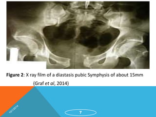
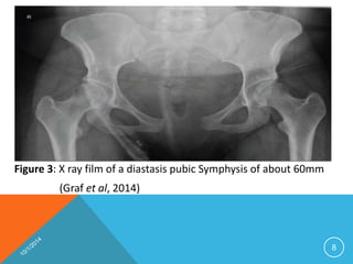
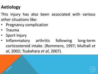
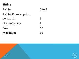
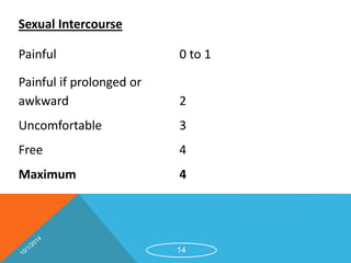
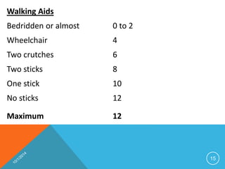
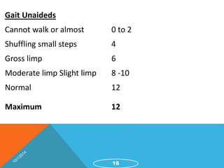
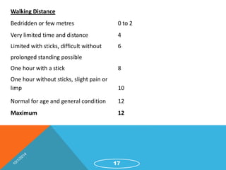
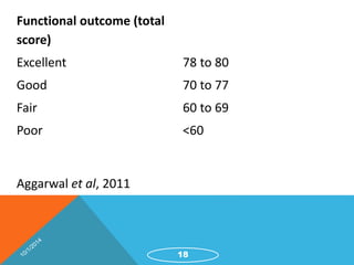
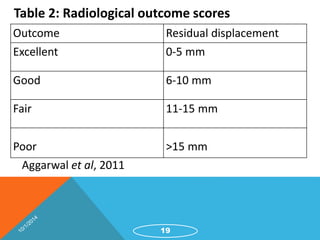
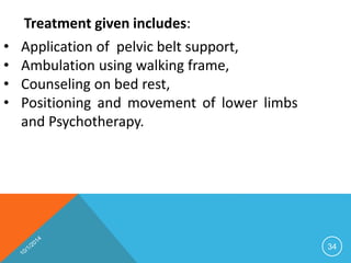
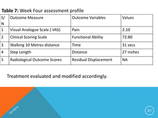
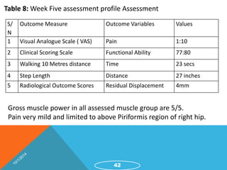
MANAGEMENT OF DIASTASIS PUBIC SYMPHYSIS: CASE 
REPORT 
By 
Bakare, Akeem 
1
Outline 
2 
Introduction 
Epidemiology 
Aetiology 
Management 
Prognosis 
Case Study 
Conclusion 
Reference
Introduction 
• Rupture of pubic symphysis is uncommon 
• Reported incidence: 1 in 300 deliveries (Snow and 
Neubert, 1997) 
• Mild diastasis: less than 10 mm is considered 
physiological in pregnancy 
• Greater separation results in tenderness and difficulty 
with ambulation (Joosoph and Kwek, 2007). 
3
Introduction: 
Diagnosis can be confirmed rapidly by: 
ď‚§ Pelvic X-ray. 
ď‚§ Additionally, MRI serves to exclude soft tissue 
injury (Graf et al, 2014). 
4
Figure 1: Normal anatomical structure of a pelvic bone with intact pubic symphysis 
5
Definition: 
Diastasis symphysis pubis is the separation 
of normally joined pubic bones, as in the 
dislocation of the bones, without a 
fracture. According to Kelly et al (2002). 
6
Figure 2: X ray film of a diastasis pubic Symphysis of about 15mm 
(Graf et al, 2014) 
7
Figure 3: X ray film of a diastasis pubic Symphysis of about 60mm 
(Graf et al, 2014) 
8
Epidemiology 
• The incidence of pubic diastasis is 1 out of 800 
patients in post partum stage (Scriven et al, 
1995). 
• In the work of Wu et al (2004), a diastasis of the 
symphysis pubis is a cause of pelvic girdle pain 
(PGP). Overall, about 45% of all pregnant women 
and 25% of all women postpartum suffers from 
PGP. 
9
Aetiology 
This injury has also been associated with various 
other situations like: 
• Pregnancy complication 
• Trauma 
• Sport Injury 
• Inflammatory arthritis following long-term 
corticosteroid intake. (Rommens, 1997; Mulhall et 
al, 2002; Tsukahara et al, 2007). 
10
Severity Grading and Outcome Measure 
Patient can be assessed and graded pre and 
post management using the Clinical Scoring 
scale designed by Majeed (1986). The scale is 
described below: 
11
Table 1: Clinical scoring Scale 
Patient ability score 
Pain 
Intense, continuous at rest 0 to 5 
Intense with activity 10 
Tolerable, but limits activity 15 
With moderate activity, abolished by rest 20 
Mild, intermittent, normal activity 25 
Slight, occasional or no pain 30 
Maximum 30 
12
Sitting 
Painful 0 to 4 
Painful if prolonged or 
awkward 6 
Uncomfortable 8 
Free 10 
Maximum 10 
13
Sexual Intercourse 
Painful 0 to 1 
Painful if prolonged or 
awkward 2 
Uncomfortable 3 
Free 4 
Maximum 4 
14
Walking Aids 
Bedridden or almost 0 to 2 
Wheelchair 4 
Two crutches 6 
Two sticks 8 
One stick 10 
No sticks 12 
Maximum 12 
15
Gait Unaideds 
Cannot walk or almost 0 to 2 
Shuffling small steps 4 
Gross limp 6 
Moderate limp Slight limp 8 -10 
Normal 12 
Maximum 12 
16
Walking Distance 
Bedridden or few metres 0 to 2 
Very limited time and distance 4 
Limited with sticks, difficult without 6 
prolonged standing possible 
One hour with a stick 8 
One hour without sticks, slight pain or 
limp 10 
Normal for age and general condition 12 
Maximum 12 
17
Functional outcome (total 
score) 
Excellent 78 to 80 
Good 70 to 77 
Fair 60 to 69 
Poor <60 
Aggarwal et al, 2011 
18
Table 2: Radiological outcome scores 
Outcome Residual displacement 
Excellent 0-5 mm 
Good 6-10 mm 
Fair 11-15 mm 
Poor >15 mm 
19 
Aggarwal et al, 2011
Management 
Management includes: 
ď‚§ Conservative management 
ď‚§ Use of medications 
ď‚§ Surgery 
20
Management: 
Typically, a conservative treatment is performed 
comprising: 
• Pelvic girdle, 
• Analgesia, 
• Bed rest in lateral decubitus i.e. lying on his or 
her side, and 
• Physical therapy ( Dunbar and Ries, 2002; Jain 
and Sternber, 2005; Nouta et al, 2011). 
21
Rehabilitation 
1. Bed rest 
2. Deep breathing exercises 
3. Isometric quadriceps contraction exercises 
4. Ankle pump exercises 
5. Cryotherapy 
6. Soft tissue manipulation to the low back and hip 
regions 
7. Transcutaneous electrical nerve stimulation to the 
low back and hip regions. (Okafor and Shokunbi, 
2009). 
22
Prognosis 
Prognosis depends on severity of injury and it may 
resolve in weeks. The condition can take from 11 
weeks, 6 months or even up to 2 years postpartum 
to subside. If detected on time and proper 
management channelled, prognosis is good 
according to Larsen et al, (2001). 
23
A Case Report 
Mrs Y was referred on account of severe pain, 
inability to stand unaided and inability to neither sit 
nor walk due to pain around the pelvic and gluteal 
region. The history indicated that she underwent a 
caesarean section after a prolonged labour at the 
traditional birth attendance clinic. 
24
A Case Report: 
The surgery was done two months before 
presentation at the hospital, however, several 
interventions had been sought to help in the post 
partum symptom of functional loss, which include 
medications and help from the traditional bone 
setters but to no avail. 
25
A Case Report: 
At presentation, she was helped into the cubicle 
carried by two individuals with excruciating pain. 
She underwent five weeks intensive physiotherapy. 
After the fifth week, the pain had significantly 
reduced (VAS: 1/10) and had significant functional 
ability with Majeed Scoring Scale increasing to 77/ 
80. 
26
Presenting Complaints: 
ď‚· Severe pain on the lower limbs especially the RLL 
for 2 months 
ď‚· Inability to sit and rest on the right side of the 
buttocks for 2 months 
ď‚· Inability to stand and walk on the right lower limb 
ď‚· Extreme difficulty in lying supine, prefers to lie in 
side position especially on the left 
27
Assessment revealed: 
ď‚· Antalgic gait with very short steps, nil foot drop 
observed 
ď‚· Visual analogue scale (VAS): 10/10 
ď‚· Gluteal tenderness greatest on the right 
ď‚· Tenderness on the pubic symphysis 
ď‚· Marked hypotonicity of the right thigh muscles and 
gluteal muscles. 
28
Assessment revealed: 
ď‚· Marked atrophy of the thigh muscles and gluteal 
muscles 
 Range of motion: PROM – Hip flexion/extension limited 
with pain 
oHip abduction/adduction limited because of 
pain 
oAROM – Not possible due to pain in all 
ranges 
ď‚· Strength: not assessed because of pain. 
29
Tests: 
ď‚§ Walking 10 metres distance: 11 minutes 
ď‚§ Hip Compression test: + 
ď‚§ Hip Distraction Test: patient unable to lie supine because 
of pain, laid on the left side of the body 
ď‚§ Hip log roll: not assessed because of her position 
 Gaeslens’ test: not assessed 
 Thomas and Patrick’s test: not assessed 
 Flamingo’s test: not done. 
30
Radiological Investigation 
ď‚§ X-ray: Pelvic x ray revealed widening of 
the pubic symphysis to 15mm: (normal > 
7mm) 
ď‚§ Hip joint spaces are preserved. 
31
Summary of assessment at first visit 
Table 3:Week One assessment profile 
S/ 
N 
Outcome Measure Outcome Variables Values 
1 Visual Analogue Scale ( VAS) Pain 10:10 
2 Clinical Scoring Scale Functional Ability 28:80 
3 Walking 10 Metres distance Time 11 minutes 
4 Step Length Distance 6 inches 
5 Radiological Outcome Scores Residual Displacement 15mm 
32
Treatment given includes: 
• Cryotherapy, 
• TENS, 
• Muscle setting for quadriceps, hamstrings and 
gluteal muscles, ankle pump exercises, 
• Soft tissue manipulation using voltaren emulgel, 
33
34 
Treatment given includes: 
• Application of pelvic belt support, 
• Ambulation using walking frame, 
• Counseling on bed rest, 
• Positioning and movement of lower limbs 
and Psychotherapy.
Treatment given includes: 
Treatment was progressed according to patient 
tolerance and level of improvement. Patient 
improved progressively as shown in the assessment 
profile column in tables 4, 5, 6,7and 8. During the 
week two of treatment, the gross muscle power of 
the lower limbs group of muscles were assessed and 
resistance exercises was commenced for all the 
weak muscles. 
35
Treatment given includes: 
At the end of the third week, the walking frame was 
discontinued and she ambulated unaided with 
lesser degree of difficulty; also the pelvic support 
was discontinued. At the end of the fourth week, 
patient was referred for a check x ray which 
revealed reduction in the diastasis gap to 4mm. 
36
Treatment given includes: 
The patient became more stable and highly 
independent at the end of the fifth week of 
management, and her appointment was 
spaced out to once in a month and contact 
was kept via the mobile phone. 
37
Table 4: Week Two assessment profile 
S/ 
N 
Outcome Measure Outcome Variables Values 
1 Visual Analogue Scale ( VAS) Pain 6:10 
2 Clinical Scoring Scale Functional Ability 59:80 
3 Walking 10 Metres distance Time 6min,58 secs 
4 Step Length Distance 9 inches 
5 Radiological Outcome Scores Residual Displacement NA 
Further assessment of muscle power was carried out because patient 
could move limbs more actively with lesser pain. 
38
Table 5: Gross Muscle Power chart for the lower limbs 
Group of Muscle Tested Lower Limbs 
Right Left 
Hip Adductors 3:5 3:5 
Hip Abductors 1:5 1:5 
Hip Flexors 3:5 3:5 
Hip Extensors 3:5 3:5 
Knee Flexors 3:5 3:5 
Knee Extensors 3:5 3:5 
Ankle Dorsiflexors 5:5 5:5 
Ankle Plantarflexors 5:5 5:5 
Management: 
Strengthening exercise program was included. 
39
Table 6: Week Three assessment profile Assessment: 
S/N Outcome Measure Outcome Variables Values 
1 Visual Analogue Scale ( VAS) Pain 4:10 
2 Clinical Scoring Scale Functional Ability 68:80 
3 Walking 10 Metres distance Time 38 secs 
4 Step Length Distance 27 inches 
5 Radiological Outcome Scores Residual Displacement NA 
All the assessed gross muscle power increased to 5/5, except knee 
flexors, hip abductors, flexors and extensors. 
Pain localized only to the anterior pelvic and above the Piriformis 
region of the right hip. 
40
Table 7: Week Four assessment profile 
S/ 
N 
Outcome Measure Outcome Variables Values 
1 Visual Analogue Scale ( VAS) Pain 2:10 
2 Clinical Scoring Scale Functional Ability 72:80 
3 Walking 10 Metres distance Time 31 secs 
4 Step Length Distance 27 inches 
5 Radiological Outcome Scores Residual Displacement NA 
41 
Treatment evaluated and modified accordingly.
Table 8: Week Five assessment profile Assessment 
S/ 
N 
Outcome Measure Outcome Variables Values 
1 Visual Analogue Scale ( VAS) Pain 1:10 
2 Clinical Scoring Scale Functional Ability 77:80 
3 Walking 10 Metres distance Time 23 secs 
4 Step Length Distance 27 inches 
5 Radiological Outcome Scores Residual Displacement 4mm 
Gross muscle power in all assessed muscle group are 5/5. 
Pain very mild and limited to above Piriformis region of right hip. 
42
Conclusion: 
Pubic symphysis rupture is an uncommon but often 
underestimated injury after vaginal delivery that can 
lead to significant chronic disability. Therefore, in case 
of peripartum suprapubic pain, it is important to 
consider a pubic symphyseal diastasis that requires 
interdisciplinary treatment. 
43
44 
Conclusion: 
It is pertinent that clinicians should consider it 
when assessing patients in the ante-natal or post-natal 
period who complain of pain along the 
suprapubic, sacroiliac or thigh regions. Though the 
symptoms and clinical presentation are gross and 
may be incapacitating, conservative medical 
rehabilitation approaches are very effective.
References 
Aggarwal S, Bali K Krishnan V, Kumar V, Meena D, Sen RK (2011). Management outcomes in pubic 
diastasis: our experience with 19 patients. Journal of Orthopeadic and Surgical Research: Vol. 6. pp 21 
Alessio P, Roberto B, Remo B, Dante S, Aldo G (2005). Post partum diastasis of the pubic 
symphysis: a case report. ACTA Bio Medical; 76; 49-52 
Becker I, Woodley SJ, Stringer MD (2010). The adult human pubic symphysis: a systematic 
review. Journal of Anatomy. 217(5):475-487 
Dhar S, Anderton JM. (1992). Rupture of the symphysis pubis during labour. Journal of 
Clinical Orthopeadics; 283: 252-257 
Diagnosis of Pelvic Girdle Pain. Available @ www.acpwh.org.uk. Accessed on 6/2/2013 
Dunbar RP. (2002). Puerperal diastasis of the public symphysis. A case report. Journal of 
Reproductive Medicine; 47: 581-3 
45
References 
Dunbar RP, Ries AM (2002). “Puerperal diastasis of the pubic symphysis: a case report.” 
Journal of Reproductive Medicine for the Obstetrician and Gynecologist, vol. 47; no. 7, pp. 
581–583 
Exercise for Symphysis Pubis Dysfunction @www. mutusystem.com. Accessed on 20/6/2014 
Gamble JG, Simmons SC. (1986). The Symphysis Pubis: Anatomic and Pathologic 
Considerations . Clinical Orthopaedics and Related Research Feb; No. 203; 261-272 
Gräf C, Sellei RM, Schrading S, Bauerschlag DO (2014). Treatment of Parturition-Induced 
Rupture of Pubic Symphysis after Spontaneous Vaginal Delivery. Case Reports in Obstetrics 
and Gynecology Volume 2014, Article ID 485916, 3 
Jain N, Sternberg LB (2005). “Symphyseal separation.” Obstetrics and Gynecology, vol. 105, 
no. 5, pp. 1229–1232 
46
References 
Joosoph J, Kwek, K (2007). “Symphysis pubis diastasis afternormal vaginal birth: a case 
report.” Annals of the Academy of Medicine Singapore, vol. 36; no. 1, pp. 83–85 
Journal of Orthopaedic Surgery and Research (2011). Available @ www.josr-online.com. 
Accessed on 20/06/2014 
Kelly O, Anne P, Gerald M (2002). Pubic symphysis separation. In: Foetal and Maternal 
Medicine Review (13th edition) pp 141-155. London, Butterworth-Heinemann 
Kharrazi FD, Rodgers WB, Kennedy JG, Lhowe DW (1997). “Parturition-induced pelvic 
dislocation: a report of four cases.” Journal of Orthopaedic Trauma, vol. 11, no. 4, pp. 277– 
282 
Larsen EC, Wilken-Jensen C, Hansen A, Jensen DV, Johansen S, Minck H, Wormslev M, 
Davidsen M, Hansen TM (1999). Symptom-Giving Pelvic Girdle Relaxation in Pregnancy: 
Prevalence and Risk Factors. Acta Obstetrics Gynecology Scandinavian 78(2):105-110 
47
References 
Lebel DE, Levy A, Holcberg G, Sheiner E (2010). Symphysiolysis as an independent risk factor 
for cesarean delivery. Journal of Maternal-Foetal and Neonatal Medicine 23 (5): 417–420 
Majeed SA: (1989). Grading the outcome of pelvic fractures. Journal of Bone Joint 
Surgery 71(2):304-6 
Mulhall KJ, Khan Y, Ahmed A, O'Farrell D, Burke TE, Moloney M (2002). Diastasis of the pubic 
symphysis peculiar to horse riders: modern aspects of pelvic pommel injuries. British Journal 
of Sports Medicine 36(1):74-5 
Musumeci R, Villa E (1994). Symphysis pubis separation during vaginal delivery with epidural 
anaesthesia. Journal of Regional Anaesthesia 19: 289-91 
48
References 
Niederhauser, A, Magann EF, Mullin PM, Morrison JC (2008). “Resolution of infant shoulder 
dystocia with maternal spontaneous symphyseal separation: a case report.” Journal of 
Reproductive Medicine for the Obstetrician and Gynecologist, vol. 53, no. 1, pp. 62–64 
Nouta KA, Rhee MV, Van Langelaan EJ ( 2011).“Symphysis rupture during partus.” Nederlands 
Tijdschrift voor Geneeskunde, vol. 155; p. A2802 
Okafor UAC, Shokunbi TF (2009). Physiotherapy Management of Sub-acute Postpartum 
Diastasis of Pubic Symphysis: A case report. Journal of the Nigeria Society of Physiotherapy 17: 
37-40 
Omololu AB, Alonge TO, Salawu SA (2001). Spontaneus pubic symphysial diastasis following 
vaginal delivery. Africa Journal of Medical Science 30: 133-5 
49
Panditrao SA, Eknathrao BP, Popat GU, Ramkrishna MA (2005). Pubic Symphysial Diastasis 
During Normal Vaginal Delivery. Journal of Obstetrics India 55 No.4 July/August pgs:365-366 
Rodrigo CG, Renato PC (2004). Nutrition pathways to the symphysis pubis. Journal Anatomy 
204(3): 209–215 
Rommens PM (1997). Internal fixation in postpartum symphysis pubis rupture: report of three 
cases. Journal of Orthopaedic Trauma 11(4):273-6 
Samet T, Cem L, Memduh D, Suleyman EA, Recep H, Ilkhur C, Sinan B (2006). Pubic symphysis 
diastasis: Imaging and clinical features. European Journal of Radiology Extra 59(3): 127-129 
Scicluna JK, Alderson JD, Webster VJ, Whiting P (2004). Epidural analgesia for acute symphysis 
pubis dysfunction in the 2nd trimester. International Journal of Obstetrics Anaesthesia 13(1): 
50-52 
50
Scriven MW, Jones DA, McKnight L. (1995). The importance of pubic pain following childbirth: 
a clinical ultrasonographic study of diastasis of the pubic symphysis. Journal of Social Medicine 
22: 48-52 
Snow RE, Neubert, AG ( 1997). “Peripartum pubic symphysis separation: a case series and 
review of the literature.” Obstetrical and Gynecological Survey, vol. 52; no. 7, pp. 438–443 
Symphysis Pubis Dysfunction. Available @ www.acpwh.org.uk. Accessed on 18/06/2014 
Tsukahara S, Momohara S, Ikari K, Murakoshi K, Mochizuki T, Kawamura K, Kobayashi S, 
Nishimoto K, Okamoto H, Tomatsu T (2007): Disturbances of the symphysis pubis in 
rheumatoid arthritis: report of two cases. Mod Rheumatology 17(4):344-7 
Wu WH, Meijer OG, Uegaki JM, Mens JH, van Dieën PI, Wuisman JM, Östgaard HC (2004). 
Pregnancy-related pelvic girdle pain (PPP), I: Terminology, clinical presentation, and 
prevalence. European Spine Journal 13, No. 7 / Nov 
51

Diatesis Pubic Symphysis - Case Presentation

  • 1.
    MANAGEMENT OF DIASTASISPUBIC SYMPHYSIS: CASE REPORT By Bakare, Akeem 1
  • 2.
    Outline 2 Introduction Epidemiology Aetiology Management Prognosis Case Study Conclusion Reference
  • 3.
    Introduction • Ruptureof pubic symphysis is uncommon • Reported incidence: 1 in 300 deliveries (Snow and Neubert, 1997) • Mild diastasis: less than 10 mm is considered physiological in pregnancy • Greater separation results in tenderness and difficulty with ambulation (Joosoph and Kwek, 2007). 3
  • 4.
    Introduction: Diagnosis canbe confirmed rapidly by: ď‚§ Pelvic X-ray. ď‚§ Additionally, MRI serves to exclude soft tissue injury (Graf et al, 2014). 4
  • 5.
    Figure 1: Normalanatomical structure of a pelvic bone with intact pubic symphysis 5
  • 6.
    Definition: Diastasis symphysispubis is the separation of normally joined pubic bones, as in the dislocation of the bones, without a fracture. According to Kelly et al (2002). 6
  • 7.
    Figure 2: Xray film of a diastasis pubic Symphysis of about 15mm (Graf et al, 2014) 7
  • 8.
    Figure 3: Xray film of a diastasis pubic Symphysis of about 60mm (Graf et al, 2014) 8
  • 9.
    Epidemiology • Theincidence of pubic diastasis is 1 out of 800 patients in post partum stage (Scriven et al, 1995). • In the work of Wu et al (2004), a diastasis of the symphysis pubis is a cause of pelvic girdle pain (PGP). Overall, about 45% of all pregnant women and 25% of all women postpartum suffers from PGP. 9
  • 10.
    Aetiology This injuryhas also been associated with various other situations like: • Pregnancy complication • Trauma • Sport Injury • Inflammatory arthritis following long-term corticosteroid intake. (Rommens, 1997; Mulhall et al, 2002; Tsukahara et al, 2007). 10
  • 11.
    Severity Grading andOutcome Measure Patient can be assessed and graded pre and post management using the Clinical Scoring scale designed by Majeed (1986). The scale is described below: 11
  • 12.
    Table 1: Clinicalscoring Scale Patient ability score Pain Intense, continuous at rest 0 to 5 Intense with activity 10 Tolerable, but limits activity 15 With moderate activity, abolished by rest 20 Mild, intermittent, normal activity 25 Slight, occasional or no pain 30 Maximum 30 12
  • 13.
    Sitting Painful 0to 4 Painful if prolonged or awkward 6 Uncomfortable 8 Free 10 Maximum 10 13
  • 14.
    Sexual Intercourse Painful0 to 1 Painful if prolonged or awkward 2 Uncomfortable 3 Free 4 Maximum 4 14
  • 15.
    Walking Aids Bedriddenor almost 0 to 2 Wheelchair 4 Two crutches 6 Two sticks 8 One stick 10 No sticks 12 Maximum 12 15
  • 16.
    Gait Unaideds Cannotwalk or almost 0 to 2 Shuffling small steps 4 Gross limp 6 Moderate limp Slight limp 8 -10 Normal 12 Maximum 12 16
  • 17.
    Walking Distance Bedriddenor few metres 0 to 2 Very limited time and distance 4 Limited with sticks, difficult without 6 prolonged standing possible One hour with a stick 8 One hour without sticks, slight pain or limp 10 Normal for age and general condition 12 Maximum 12 17
  • 18.
    Functional outcome (total score) Excellent 78 to 80 Good 70 to 77 Fair 60 to 69 Poor <60 Aggarwal et al, 2011 18
  • 19.
    Table 2: Radiologicaloutcome scores Outcome Residual displacement Excellent 0-5 mm Good 6-10 mm Fair 11-15 mm Poor >15 mm 19 Aggarwal et al, 2011
  • 20.
    Management Management includes: ď‚§ Conservative management ď‚§ Use of medications ď‚§ Surgery 20
  • 21.
    Management: Typically, aconservative treatment is performed comprising: • Pelvic girdle, • Analgesia, • Bed rest in lateral decubitus i.e. lying on his or her side, and • Physical therapy ( Dunbar and Ries, 2002; Jain and Sternber, 2005; Nouta et al, 2011). 21
  • 22.
    Rehabilitation 1. Bedrest 2. Deep breathing exercises 3. Isometric quadriceps contraction exercises 4. Ankle pump exercises 5. Cryotherapy 6. Soft tissue manipulation to the low back and hip regions 7. Transcutaneous electrical nerve stimulation to the low back and hip regions. (Okafor and Shokunbi, 2009). 22
  • 23.
    Prognosis Prognosis dependson severity of injury and it may resolve in weeks. The condition can take from 11 weeks, 6 months or even up to 2 years postpartum to subside. If detected on time and proper management channelled, prognosis is good according to Larsen et al, (2001). 23
  • 24.
    A Case Report Mrs Y was referred on account of severe pain, inability to stand unaided and inability to neither sit nor walk due to pain around the pelvic and gluteal region. The history indicated that she underwent a caesarean section after a prolonged labour at the traditional birth attendance clinic. 24
  • 25.
    A Case Report: The surgery was done two months before presentation at the hospital, however, several interventions had been sought to help in the post partum symptom of functional loss, which include medications and help from the traditional bone setters but to no avail. 25
  • 26.
    A Case Report: At presentation, she was helped into the cubicle carried by two individuals with excruciating pain. She underwent five weeks intensive physiotherapy. After the fifth week, the pain had significantly reduced (VAS: 1/10) and had significant functional ability with Majeed Scoring Scale increasing to 77/ 80. 26
  • 27.
    Presenting Complaints: ď‚·Severe pain on the lower limbs especially the RLL for 2 months ď‚· Inability to sit and rest on the right side of the buttocks for 2 months ď‚· Inability to stand and walk on the right lower limb ď‚· Extreme difficulty in lying supine, prefers to lie in side position especially on the left 27
  • 28.
    Assessment revealed: ď‚·Antalgic gait with very short steps, nil foot drop observed ď‚· Visual analogue scale (VAS): 10/10 ď‚· Gluteal tenderness greatest on the right ď‚· Tenderness on the pubic symphysis ď‚· Marked hypotonicity of the right thigh muscles and gluteal muscles. 28
  • 29.
    Assessment revealed: Marked atrophy of the thigh muscles and gluteal muscles  Range of motion: PROM – Hip flexion/extension limited with pain oHip abduction/adduction limited because of pain oAROM – Not possible due to pain in all ranges  Strength: not assessed because of pain. 29
  • 30.
    Tests:  Walking10 metres distance: 11 minutes  Hip Compression test: +  Hip Distraction Test: patient unable to lie supine because of pain, laid on the left side of the body  Hip log roll: not assessed because of her position  Gaeslens’ test: not assessed  Thomas and Patrick’s test: not assessed  Flamingo’s test: not done. 30
  • 31.
    Radiological Investigation ď‚§X-ray: Pelvic x ray revealed widening of the pubic symphysis to 15mm: (normal > 7mm) ď‚§ Hip joint spaces are preserved. 31
  • 32.
    Summary of assessmentat first visit Table 3:Week One assessment profile S/ N Outcome Measure Outcome Variables Values 1 Visual Analogue Scale ( VAS) Pain 10:10 2 Clinical Scoring Scale Functional Ability 28:80 3 Walking 10 Metres distance Time 11 minutes 4 Step Length Distance 6 inches 5 Radiological Outcome Scores Residual Displacement 15mm 32
  • 33.
    Treatment given includes: • Cryotherapy, • TENS, • Muscle setting for quadriceps, hamstrings and gluteal muscles, ankle pump exercises, • Soft tissue manipulation using voltaren emulgel, 33
  • 34.
    34 Treatment givenincludes: • Application of pelvic belt support, • Ambulation using walking frame, • Counseling on bed rest, • Positioning and movement of lower limbs and Psychotherapy.
  • 35.
    Treatment given includes: Treatment was progressed according to patient tolerance and level of improvement. Patient improved progressively as shown in the assessment profile column in tables 4, 5, 6,7and 8. During the week two of treatment, the gross muscle power of the lower limbs group of muscles were assessed and resistance exercises was commenced for all the weak muscles. 35
  • 36.
    Treatment given includes: At the end of the third week, the walking frame was discontinued and she ambulated unaided with lesser degree of difficulty; also the pelvic support was discontinued. At the end of the fourth week, patient was referred for a check x ray which revealed reduction in the diastasis gap to 4mm. 36
  • 37.
    Treatment given includes: The patient became more stable and highly independent at the end of the fifth week of management, and her appointment was spaced out to once in a month and contact was kept via the mobile phone. 37
  • 38.
    Table 4: WeekTwo assessment profile S/ N Outcome Measure Outcome Variables Values 1 Visual Analogue Scale ( VAS) Pain 6:10 2 Clinical Scoring Scale Functional Ability 59:80 3 Walking 10 Metres distance Time 6min,58 secs 4 Step Length Distance 9 inches 5 Radiological Outcome Scores Residual Displacement NA Further assessment of muscle power was carried out because patient could move limbs more actively with lesser pain. 38
  • 39.
    Table 5: GrossMuscle Power chart for the lower limbs Group of Muscle Tested Lower Limbs Right Left Hip Adductors 3:5 3:5 Hip Abductors 1:5 1:5 Hip Flexors 3:5 3:5 Hip Extensors 3:5 3:5 Knee Flexors 3:5 3:5 Knee Extensors 3:5 3:5 Ankle Dorsiflexors 5:5 5:5 Ankle Plantarflexors 5:5 5:5 Management: Strengthening exercise program was included. 39
  • 40.
    Table 6: WeekThree assessment profile Assessment: S/N Outcome Measure Outcome Variables Values 1 Visual Analogue Scale ( VAS) Pain 4:10 2 Clinical Scoring Scale Functional Ability 68:80 3 Walking 10 Metres distance Time 38 secs 4 Step Length Distance 27 inches 5 Radiological Outcome Scores Residual Displacement NA All the assessed gross muscle power increased to 5/5, except knee flexors, hip abductors, flexors and extensors. Pain localized only to the anterior pelvic and above the Piriformis region of the right hip. 40
  • 41.
    Table 7: WeekFour assessment profile S/ N Outcome Measure Outcome Variables Values 1 Visual Analogue Scale ( VAS) Pain 2:10 2 Clinical Scoring Scale Functional Ability 72:80 3 Walking 10 Metres distance Time 31 secs 4 Step Length Distance 27 inches 5 Radiological Outcome Scores Residual Displacement NA 41 Treatment evaluated and modified accordingly.
  • 42.
    Table 8: WeekFive assessment profile Assessment S/ N Outcome Measure Outcome Variables Values 1 Visual Analogue Scale ( VAS) Pain 1:10 2 Clinical Scoring Scale Functional Ability 77:80 3 Walking 10 Metres distance Time 23 secs 4 Step Length Distance 27 inches 5 Radiological Outcome Scores Residual Displacement 4mm Gross muscle power in all assessed muscle group are 5/5. Pain very mild and limited to above Piriformis region of right hip. 42
  • 43.
    Conclusion: Pubic symphysisrupture is an uncommon but often underestimated injury after vaginal delivery that can lead to significant chronic disability. Therefore, in case of peripartum suprapubic pain, it is important to consider a pubic symphyseal diastasis that requires interdisciplinary treatment. 43
  • 44.
    44 Conclusion: Itis pertinent that clinicians should consider it when assessing patients in the ante-natal or post-natal period who complain of pain along the suprapubic, sacroiliac or thigh regions. Though the symptoms and clinical presentation are gross and may be incapacitating, conservative medical rehabilitation approaches are very effective.
  • 45.
    References Aggarwal S,Bali K Krishnan V, Kumar V, Meena D, Sen RK (2011). Management outcomes in pubic diastasis: our experience with 19 patients. Journal of Orthopeadic and Surgical Research: Vol. 6. pp 21 Alessio P, Roberto B, Remo B, Dante S, Aldo G (2005). Post partum diastasis of the pubic symphysis: a case report. ACTA Bio Medical; 76; 49-52 Becker I, Woodley SJ, Stringer MD (2010). The adult human pubic symphysis: a systematic review. Journal of Anatomy. 217(5):475-487 Dhar S, Anderton JM. (1992). Rupture of the symphysis pubis during labour. Journal of Clinical Orthopeadics; 283: 252-257 Diagnosis of Pelvic Girdle Pain. Available @ www.acpwh.org.uk. Accessed on 6/2/2013 Dunbar RP. (2002). Puerperal diastasis of the public symphysis. A case report. Journal of Reproductive Medicine; 47: 581-3 45
  • 46.
    References Dunbar RP,Ries AM (2002). “Puerperal diastasis of the pubic symphysis: a case report.” Journal of Reproductive Medicine for the Obstetrician and Gynecologist, vol. 47; no. 7, pp. 581–583 Exercise for Symphysis Pubis Dysfunction @www. mutusystem.com. Accessed on 20/6/2014 Gamble JG, Simmons SC. (1986). The Symphysis Pubis: Anatomic and Pathologic Considerations . Clinical Orthopaedics and Related Research Feb; No. 203; 261-272 Gräf C, Sellei RM, Schrading S, Bauerschlag DO (2014). Treatment of Parturition-Induced Rupture of Pubic Symphysis after Spontaneous Vaginal Delivery. Case Reports in Obstetrics and Gynecology Volume 2014, Article ID 485916, 3 Jain N, Sternberg LB (2005). “Symphyseal separation.” Obstetrics and Gynecology, vol. 105, no. 5, pp. 1229–1232 46
  • 47.
    References Joosoph J,Kwek, K (2007). “Symphysis pubis diastasis afternormal vaginal birth: a case report.” Annals of the Academy of Medicine Singapore, vol. 36; no. 1, pp. 83–85 Journal of Orthopaedic Surgery and Research (2011). Available @ www.josr-online.com. Accessed on 20/06/2014 Kelly O, Anne P, Gerald M (2002). Pubic symphysis separation. In: Foetal and Maternal Medicine Review (13th edition) pp 141-155. London, Butterworth-Heinemann Kharrazi FD, Rodgers WB, Kennedy JG, Lhowe DW (1997). “Parturition-induced pelvic dislocation: a report of four cases.” Journal of Orthopaedic Trauma, vol. 11, no. 4, pp. 277– 282 Larsen EC, Wilken-Jensen C, Hansen A, Jensen DV, Johansen S, Minck H, Wormslev M, Davidsen M, Hansen TM (1999). Symptom-Giving Pelvic Girdle Relaxation in Pregnancy: Prevalence and Risk Factors. Acta Obstetrics Gynecology Scandinavian 78(2):105-110 47
  • 48.
    References Lebel DE,Levy A, Holcberg G, Sheiner E (2010). Symphysiolysis as an independent risk factor for cesarean delivery. Journal of Maternal-Foetal and Neonatal Medicine 23 (5): 417–420 Majeed SA: (1989). Grading the outcome of pelvic fractures. Journal of Bone Joint Surgery 71(2):304-6 Mulhall KJ, Khan Y, Ahmed A, O'Farrell D, Burke TE, Moloney M (2002). Diastasis of the pubic symphysis peculiar to horse riders: modern aspects of pelvic pommel injuries. British Journal of Sports Medicine 36(1):74-5 Musumeci R, Villa E (1994). Symphysis pubis separation during vaginal delivery with epidural anaesthesia. Journal of Regional Anaesthesia 19: 289-91 48
  • 49.
    References Niederhauser, A,Magann EF, Mullin PM, Morrison JC (2008). “Resolution of infant shoulder dystocia with maternal spontaneous symphyseal separation: a case report.” Journal of Reproductive Medicine for the Obstetrician and Gynecologist, vol. 53, no. 1, pp. 62–64 Nouta KA, Rhee MV, Van Langelaan EJ ( 2011).“Symphysis rupture during partus.” Nederlands Tijdschrift voor Geneeskunde, vol. 155; p. A2802 Okafor UAC, Shokunbi TF (2009). Physiotherapy Management of Sub-acute Postpartum Diastasis of Pubic Symphysis: A case report. Journal of the Nigeria Society of Physiotherapy 17: 37-40 Omololu AB, Alonge TO, Salawu SA (2001). Spontaneus pubic symphysial diastasis following vaginal delivery. Africa Journal of Medical Science 30: 133-5 49
  • 50.
    Panditrao SA, EknathraoBP, Popat GU, Ramkrishna MA (2005). Pubic Symphysial Diastasis During Normal Vaginal Delivery. Journal of Obstetrics India 55 No.4 July/August pgs:365-366 Rodrigo CG, Renato PC (2004). Nutrition pathways to the symphysis pubis. Journal Anatomy 204(3): 209–215 Rommens PM (1997). Internal fixation in postpartum symphysis pubis rupture: report of three cases. Journal of Orthopaedic Trauma 11(4):273-6 Samet T, Cem L, Memduh D, Suleyman EA, Recep H, Ilkhur C, Sinan B (2006). Pubic symphysis diastasis: Imaging and clinical features. European Journal of Radiology Extra 59(3): 127-129 Scicluna JK, Alderson JD, Webster VJ, Whiting P (2004). Epidural analgesia for acute symphysis pubis dysfunction in the 2nd trimester. International Journal of Obstetrics Anaesthesia 13(1): 50-52 50
  • 51.
    Scriven MW, JonesDA, McKnight L. (1995). The importance of pubic pain following childbirth: a clinical ultrasonographic study of diastasis of the pubic symphysis. Journal of Social Medicine 22: 48-52 Snow RE, Neubert, AG ( 1997). “Peripartum pubic symphysis separation: a case series and review of the literature.” Obstetrical and Gynecological Survey, vol. 52; no. 7, pp. 438–443 Symphysis Pubis Dysfunction. Available @ www.acpwh.org.uk. Accessed on 18/06/2014 Tsukahara S, Momohara S, Ikari K, Murakoshi K, Mochizuki T, Kawamura K, Kobayashi S, Nishimoto K, Okamoto H, Tomatsu T (2007): Disturbances of the symphysis pubis in rheumatoid arthritis: report of two cases. Mod Rheumatology 17(4):344-7 Wu WH, Meijer OG, Uegaki JM, Mens JH, van Dieën PI, Wuisman JM, Östgaard HC (2004). Pregnancy-related pelvic girdle pain (PPP), I: Terminology, clinical presentation, and prevalence. European Spine Journal 13, No. 7 / Nov 51