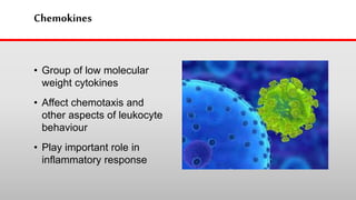
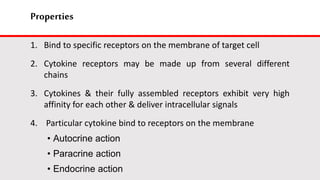
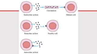
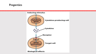
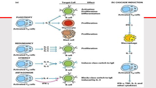
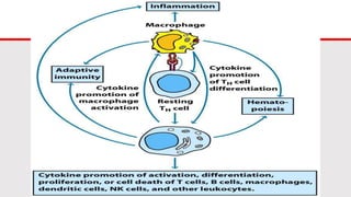
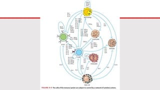
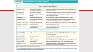
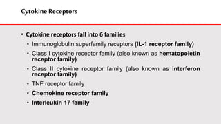
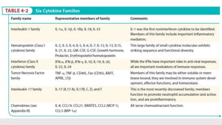
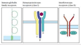
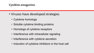
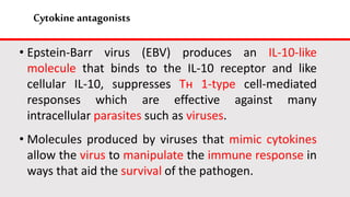
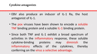
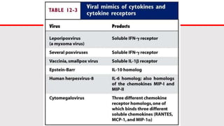
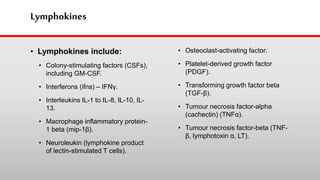
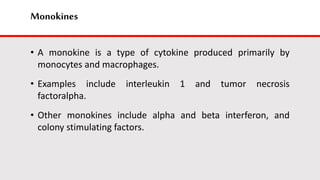
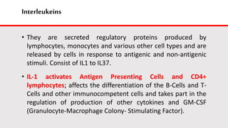
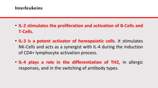
SlideShare a Scribd company logo
CYTOKINES
By Zainab O. Hamid Ph.D. Student, 2017 Ref. Kuby, Immunology, 2013
Objective
• General Properties of Cytokines and Chemokines
• Families of Cytokines and Associated Receptor Molecules
• Cytokine Antagonists
• Cytokine-Related Diseases
• Cytokine-Based Therapies
Introduction
• The development of an effective immune response
involves lymphoid cells, inflammatory cells, and
hematopoietic cells.
• The complex interactions among these cells are mediated
by a group of proteins collectively designated as
cytokines to indicate their role in cell-to-cell
communication.
Introduction
• Cytokines are low molecular weight regulatory proteins
or glycoproteins secreted by white blood cells and various
other cells in the body in response to a number of stimuli.
• These proteins assist in regulating the development of
immune effector cells
• And some cytokines possess direct effector functions of
their own.
Introduction
Many referred as
interleukins
Secreted by
leukocytes and act
on other leukocytes
IL-1 through IL 29
have been described
Chemokines
• Group of low molecular
weight cytokines
• Affect chemotaxis and
other aspects of leukocyte
behaviour
• Play important role in
inflammatory response
Properties
1. Bind to specific receptors on the membrane of target cell
2. Cytokine receptors may be made up from several different
chains
3. Cytokines & their fully assembled receptors exhibit very high
affinity for each other & deliver intracellular signals
4. Particular cytokine bind to receptors on the membrane
• Autocrine action
• Paracrine action
• Endocrine action
Properties
Properties
5. Cytokines regulate the intensity & duration of immune
response
6. Binding of a given cytokine to responsive target cells
generally stimulates increased expression of cytokine
receptors and secretion of other cytokines
7. Exhibit attributes of pleiotropy, redundancy, synergy,
antagonism and cascade induction.
8. Share many properties with hormones
Cytokines families
• Falls in the following families
• Hematopoietin family
• Interferon family
• Interleukin family
• Tumor necrosis factor family
• All have molecular mass less than 30kDa
• All have similarities and few rarely act alone
Cytokines belong to four families
• The amino acid sequences of these family members differ
considerably
• All have high degree of α helical structure and little or no β sheet
structure
• Molecules have similar polypeptide fold, with for α helical regions
(A-D)
• In which the 1st and 2nd helices & the 3rd and 4th helices run
roughly parallel to one another & are connected by loops
Interleukin 4
Cytokines havenumerous biological functions
• Although a variety of cells can secrete cytokines, the
principal producers are Tн cells, dendritic cells, and
macrophages
• Cytokines released from these cell types activate an entire
network of interacting cells
Cytokines havenumerous biological functions
• Among numerous physiological responses that require
cytokine involvement are
• Development of cellular and humoral immune
responses
• Induction of inflammatory response
• Regulation of hematopoiesis
• Control of cellular proliferation
• Differentiation
• Healing of wounds
Cytokines havenumerous biological functions
• What keeps cytokines from activating cells in a non
specific fashion during the immune response?
• Specificity is maintained by careful regulation of the
expression of cytokine receptors on cells
• Cytokine receptors are expressed on a cell only after that
cell has interacted with antigen, limiting cytokine response
to antigen activated lymphocytes
Cytokines havenumerous biological functions
• Specificity maintained if cytokine secretion occurs only when
the cytokine-producing cell interacts directly with target cell,
thus ensuring that effective concentrations of the cytokine
occur in the vicinity of the intended target.
• In case of Tн cell, a major producer of cytokines, cellular
interactions occurs when the T-cell receptor recognizes an
antigen-MHC complex on an appropriate antigen-presenting
cell, such as a macrophage, dendritic cell, or B lymphocyte.
Cytokines havenumerous biological functions
• The concentration of cytokines secreted at the junction of
these interacting cells reaches high enough local
concentration to affect the target APC, but not more distant
cells.
• Half-life of cytokines in the blood stream or other
extracellular fluids into which they are secreted is usually very
short, ensuring that they act for only a limited period and
thus over a short distance.
Cytokine Receptors
• Cytokine receptors fall into 6 families
• Immunoglobulin superfamily receptors (IL-1 receptor family)
• Class I cytokine receptor family (also known as hematopoietin
receptor family)
• Class II cytokine receptor family (also known as interferon
receptor family)
• TNF receptor family
• Chemokine receptor family
• Interleukin 17 family
Cytokine Receptors
• Immunoglobulin superfamily
• Cytokines of the interleukin 1 (IL-1) family are typically
secreted very early in the immune response by dendritic
cells and monocytes or macrophages. IL-1 secretion is
stimulated by recognition of viral, parasitic, or bacterial
antigens by innate immune receptors. IL-1 family members
are generally pro-inflammatory, meaning that they induce
an increase in the capillary permeability at the site of
cytokine secretion, along with an amplification of the level
of leukocyte migration into the infected tissues.
Cytokine Receptors
• Hematopoietin (Class I) cytokine family
• Members are small, soluble cytokines that
communicate between and among cells of the
immune system. Their name is somewhat misleading
in that not all members of this family are
implicated in hematopoietic (blood-cell forming)
functions . However, some of the earliest members
of this family to be characterized indeed have
hematopoietic functions, and the cytokine family was
then defined on the basis of structural similarities
among all the participants. Because the
hematopoietin family contains some of the
earliest cytokines to be structurally characterized,
it is sometimes also referred to as the Class I
cytokine family.
Cytokine Receptors
• Class II cytokine receptor family
• Named “interferon” because of its ability to
“interfere” with the growth of the live virus. Their
more straightforward in vitro assay system
enabled them to rapidly characterize the
biological effects of the molecule involved, and
they wrote a series of papers describing the
biological effects of interferon(s).
• There are two major types of interferons, Types 1
and 2, and that
• Type 1 interferons can be subdivided into two
subgroups.
Cytokine Receptors
• Type I interferons are composed of Interferons α, a family of
about 20 related proteins, and interferon-β, which are secreted by
activated macrophages and dendritic cells, as well as by virus-
infected cells.
• Interferons α and β are also secreted by virally infected cells
after recognition of viral components by pattern recognition
receptors (PRRs) located either at the cell surface, or inside the
cell.
Cytokine Receptors
• Type II interferon, otherwise known as interferon-γ, is produced
by activated T and NK cells. Interferon-γ is a powerful modulator
of the adaptive immune response,
• directing T cell help toward the TH1 type
• inducing the activation of macrophages
• subsequent destruction of any intracellular pathogens
• and the differentiation of cytotoxic T cells.
• All three interferons increase the expression of MHC complex
proteins on the surface of cells, thus enhancing their antigen-
presentation capabilities.
Cytokine Receptors
• Interferon-γ originally discovered because of its ability to
induce cells to block or inhibit the replication of a wide
variety of viruses
• Antiviral activity is a property it shares with IFN-α and IFN-β
• IFN-γ plays a central role in many immunoregulatory proteins
including
• Regulation of mononuclear phagocytes
• B cell switching to certain IgG classes
• Support or inhibition of the development of Tн cell subsets
TNF receptors
• The Tumor Necrosis Family (TNF) family of
cytokines regulates the development,
effector function, and homeostasis of cells
participating in the skeletal, neuronal, and
immune systems, among others.
• Cytokines of the TNF Family Can Be Soluble
or Membrane Bound.
• There are two eponymous (having the same
name as) members of the TNF family: TNF-
α and TNF-β.
Cytokine Receptors
• TNF-α (Lymphotoxin-α) is a pro-inflammatory cytokine,
produced primarily by activated macrophages, but also by
other cell types including lymphocytes, fibroblasts, and
keratinocytes (skin cells), in response to infection,
inflammation, and environmental stressors.
• Binding of TNF-α to neutrophils, endothelial cells, and
osteoclasts can lead to increased expression of MHC
glycoproteins and of adhesion molecules.
Cytokine Receptors
• TNF-β (Lymphotoxin-β), a membrane-bound cytokine, is important
in lymphocyte differentiation.
• CD40L is a cytokine expressed on the surface of T cells that is
required to signal for B-cell differentiation.
• Fas ligand (FasL), or CD95L, induces apoptosis on binding to its
receptor, Fas, or CD95.
Cytokine Receptors
• Chemokine receptors
• Chemokines Direct the Migration of Leukocytes through the Body.
• Chemokines are a structurally related family of small cytokines.
• that bind to cell-surface receptors and induce the movement of leukocytes up a
concentration gradient and toward the chemokine source.
• This soluble factor-directed cell movement is known as chemotaxis, and molecules
that can elicit such movement are referred to as chemo-attractants.
Cytokine Receptors
• Some chemokines display innate affinity for the
carbohydrates named glycosaminoglycans, located on the
surfaces of endothelial cells, a property that enables them
to bind to the inner surfaces of blood vessels. Directing
leukocyte movement.
Cytokine Receptors
• Chemokine receptors
• Threads through the membrane and
transduces the ligand signal via interactions
with a polymeric GTP/GDP-binding “G protein.”
This class of G-Protein– Coupled Receptors
(GPCRs) is used in the recognition of many
types of signals, including those mediated by
chemokines.
Cytokine Receptors
• The number of cysteines as well as the positions of the disulfide bonds
determine the subclass into six different structural categories of these
cytokines as shown.
• The GPCRs are classified according to the type of chemokine
they bind. For example, the CC receptors (CCRs) recognize CC
chemokines, the CXCRs recognize CXCL chemokines, and so
on.
• Interestingly, the intrinsic specificity of the receptors is
balanced by the capacity of many receptors to bind more
than one chemokine from a particular family and of several
chemokines to bind to more than one receptor.
• For example, the receptor CXCR2 recognizes seven different
chemokines, and CCL5 can bind to both CCR3 and CCR5.
Cytokine Receptors
• The IL-17 Family
• Is a Recently Discovered, Pro-inflammatory Cytokine Cluster
• The most recently described family of cytokines, the IL-17 family,
includes interleukins 17A, 17B, 17C, 17D, and 17F. Signaling
through most members of this family culminates in the generation
of inflammation.
• IL-17 receptors are found on neutrophils, keratinocytes, and other
nonlymphoid cells.
Cytokine receptors initiate signaling
• Although some cytokine receptors lie outside the class I and
class II families, majority are included within these two
families.
• Class I and class II cytokine receptors lack signaling motifs.
• Unifying model emerged from studies of the molecular events
triggered by binding of IFN-γ to its receptor, a member of the
Class II family
Cytokine receptors initiate signaling
• Although some cytokine receptors lie outside the class I and
class II families, majority are included within these two
families.
• Class I and class II cytokine receptors lack signaling motifs
• Unifying model emerged from studies of the molecular events
triggered by binding of IFN-γ to its receptor, a member of the
Class II family
Cytokine receptors initiate signaling
• The cytokine receptor is composed of separate subunits
• Different inactive protein kinases are associated with different
subunits of the receptor.
• Cytokine binding induces the association of the two separate
cytokine receptors subunits and activation of the receptor
associated JAKs (Janus Kinase).
Cytokine receptors initiate signaling
• Activated JAKs create docking sites for the STAT (Signal
Transducer and Activator of Transcription) transcription
factors by phosphorylation of specific tyrosine residues on
cytokine receptor subunits.
• After undergoing JAK-mediated phosphorylation, STAT
transcription factors translocate from receptor docking sites
at the membrane to the nucleus, where they initiate the
transcription of specific genes.
Cytokine receptors initiate signaling
• In addition to IFN-γ, a number of other class I and class II ligands
have been shown to cause dimerization of their receptors.
• An important element of cytokine specificity derives from the
specificity of the matching between cytokine and their receptors.
• Another aspect of cytokine specificity is that each particular
cytokine induces transcription of a specific subset of genes in a
given cell type; the resulting gene products then mediate the
various effects typical of that cytokine.
Cytokine receptors initiate signaling
• Specificity is traceable to three factors
• Particular cytokine receptors start particular JAK-STAT
pathways
• Transcriptional activity of activated STATs is specific because a
particular STAT homodimer/heterodimer will only recognize
certain sequence motifs & thus can interact only with the
promoters of certain genes.
• Only those target genes whose expression is permitted by a
particular cell type can be activated within that variety of cell
Cytokine receptors initiate signaling
• i.e., in any given cell type only subset of the potential target
genes of a particular STAT may be permitted expression.
• For eg., IL-4 induces one set of genes in T cells, another in B
cells and third in eosinophils.
• IL-1 does not signal via the JAK-STAT pathway but utilizes a
kinase designated IL-1 receptor-associated kinase, or IRAK.
• IRAK proteins also utilized by TLRs for signal transduction.
Cytokine antagonists
• Number of proteins can inhibit cytokine activity
• Can bind to receptor, fail to activate the cell, OR
• Can bind directly to cytokine, inhibiting it attachment
• enzymatic cleavage of receptors and release of these can
bind cytokines in the blood.
Marker of chronic T cell activation (transplant rejection, AIDS)
Cytokine antagonists
• Viruses have developed strategies
• Cytokine homologs
• Soluble cytokine binding proteins
• Homologs of cytokine receptors
• Interference with intracellular signaling
• Interference with cytokine secretion
• Induction of cytokine inhibitors in the host cell
Cytokine antagonists
• Epstein-Barr virus (EBV) produces an IL-10-like
molecule that binds to the IL-10 receptor and like
cellular IL-10, suppresses Tн 1-type cell-mediated
responses which are effective against many
intracellular parasites such as viruses.
• Molecules produced by viruses that mimic cytokines
allow the virus to manipulate the immune response in
ways that aid the survival of the pathogen.
Cytokine antagonists
• EBV also produce an inducer of IL-1 Ra, the host
antagonist of IL-1.
• The pox viruses have been shown to encode a soluble
TNF binding protein and a soluble IL-1 binding protein.
• Since both TNF and IL-1 exhibit a broad spectrum of
activities in the inflammatory response, these soluble
cytokine-binding proteins may diminish the
inflammatory effects of the cytokines, thereby
conferring on the virus a selective advantage.
Cytokine secretion by Tн1 andTн2 subsets
• CD4⁺ TH cells exert most of helper functions through secreted
cytokines, which either act on the cells that produce them in an
autocrine fashion or modulate the responses of other cells through
paracrine pathways.
• Although CD8⁺ CTLs also secrete cytokines, their array of cytokines
generally is more restricted than that of CD4⁺ Tн cells.
• Two CD4⁺ Tн cell subpopulations designated Tн1 and Tн2 can be
distinguished in vitro by the cytokines they secrete.
• Both subsets secrete IL-3 and GM-CSF but differ in the other cytokines
they produce.
Cytokine secretion by Tн1 andTн2 subsets
• TH1 and Tн2 cells are characterized by the following functional differences:
• TH1 subset is responsible for many cell-mediated functions, such as delayed
type hypersensitivity & activation of TC cells, & for the production of
opsonization-promoting IgG antibodies, that is, Ab that bind to the high
affinity Fc receptors of phagocytes and interact with the complement system.
• Associated with promotion of excessive inflammation & tissue injury.
• TH2 subset stimulates eosinophil activation, and differentiation, provides
help to B cells, promotes production of large amounts of IgM, IgE, and
noncomplement activating IgG isotypes
• Supports allergic reactions
Cytokine secretion by Tн1 andTн2 subsets
• Differences in the cytokine secreted by Tн1 and Tн2 cells
determine the different biological functions of these subsets.
• A defining cytokine of the Tн1 subset, INF-γ, activates
macrophages, stimulating these cells to increase microbicidal
activity, up-regulate the level of class II MHC, secrete cytokines
such as IL-12, which induces Tн cells to differentiate into the Tн1
subset.
• IFN-γ secretion by Tн1 cells also induces antibody class switching
to IgG classes that support phagocytosis and fixation of
complement.
Cytokine secretion by Tн1 andTн2 subsets
• TNF-β and IFN-γ are cytokines that mediate inflammation, and it is
their secretion that accounts for the association of Tн1 cells with
inflammatory phenomena such as delayed hypersensitivity.
• Tн1 cells produce IL-2 and IFN-γ cytokines that promote the
differentiation of fully cytotoxic Tс cells from CD8+ precursors.
• This pattern of cytokine production makes the Tн1 subset
particularly suited to respond to viral infections and intracellular
pathogens.
• Finally, IFN-γ inhibits the expansion of the Tн2 population.
Development of Tн1 and Tн2 subsets determination
• The cytokine environment in which antigen-primed Tн cells
differentiate determines the subset that develops.
• IL-4 is essential for the development of a Tн2 response
• IFN-γ, IL-12, and IL-18 all are important in the physiology of
the development of Tн1 cells.
Development of Tн1and Tн2subsets determination
• Tн1 development is also critically dependent on IFN-γ, which
induces a number of changes, including
• the up-regulation of IL-12 production by macrophages and
dendritic cells.
• and the activation of the IL-12 receptor on activated T cells,
which it accomplishes by up-regulating expression of the
chain of the IL-12 receptor.
Development of Tн1 and Tн2 subsets determination
• IL-18, promotes proliferation and IFN-γ production by both
developing and fully differentiated Tн1 cells and by NK cells.
• So a regulatory network of cytokines positively controls the
generation of Tн1 cells.
Development of Tн1 and Tн2 subsets determination
• The generation of Tн2 cells depends critically on IL-4.
• Exposing naive helper cells to IL-4 at the beginning of an
immune response causes them to differentiate into Tн2 cells.
• This influence of IL-4 is predominant in directing Tн cells to
the Tн2 route.
Cytokine profiles are cross regulated
• • The critical cytokines produced by Tн1 and Tн2 subsets have
two characteristic effects on subset development.
1. Promote the growth of the subset that produces them
2. Inhibit the development and activity of the opposite subset, an
effect known as cross-regulation
• IFN- γ (secreted by the Tн1 subset) preferentially inhibits
proliferation of the Tн2 subset, and IL-4 and IL-10 (secreted by the
Tн2 subset) down-regulate secretion of IL-12, one of the critical
cytokines for Tн1 differentiation, by both macrophages and
dendritic cells.
NaïveT helper cell divelopement
Cytokine profiles are cross regulated
• Cross regulation- when antibody production is high, cell mediated
immunity is low, and vice versa.
• Two transcription factors, T-Bet and GATA-3, are key elements in
determining subset commitment and cross-regulation.
• The expression of T-Bet drives cells to differentiate into Tн1 cells
and suppresses their differentiation along the Tн2 pathway.
• Expression of GATA-3 does the opposite, promoting the
development of naive T cells into Tн2 cells while suppressing their
differentiation into Tн cells.
TH1/TH2balancedetermines disease outcomes
• The progression of some diseases may depend on the balance between
the Tн1 and Tн2 subsets.
• In humans, a well-studied example of this phenomenon is leprosy,
which is caused by Mycobacterium leprae, an intracellular pathogen
that can survive within the phagosomes of macrophages.
• In tuberculoid leprosy, a cell-mediated immune response forms
granulomas, resulting in the destruction of most of the mycobacteria,
so that only a few organisms remain in the tissues.
• Although skin and peripheral nerves are damaged, tuberculoid leprosy
progresses slowly and patients usually survive.
TH1/TH2balancedetermines disease outcomes
• In lepromatous leprosy, the cell-mediated response is
depressed and, instead, humoral antibodies are formed,
sometimes resulting in hyper-gamma-globulin-emia.
• The mycobacteria are widely disseminated in macrophages,
often reaching numbers as high as 1010 per gram of tissue.
• Lepromatous leprosy progresses into disseminated infection
of the bone and cartilage with extensive nerve damage.
Cytokine Related Diseases
• Defects in the complex regulatory networks governing the
expression of cytokines and cytokine receptors have been
implicated in a number of diseases.
• Genetic defects in cytokines, their receptors, or the molecules
involved in signal transduction following receptor-cytokine
interaction lead to immunodeficiencies such as severe combined
immunodeficiency (SCID).
• Other defects in the cytokine network can cause inability to
defend against specific families of pathogens.
Cytokine Related Diseases
• For eg., people with a defective receptor for INF-γ are
susceptible to mycobacterial infections that rarely occur in
the normal population.
• In addition to the diseases rooted in the genetic defects in
cytokine activity, a number of disease conditions result from
over-expression or under-expression of the cytokine or
cytokine receptors.
• Therapies aimed at preventing the potential harm caused by
cytokine activity.
1.Septic shock
• Bacterial infections remain a major cause of septic
shock, which may develop a few hours after
infection by certain Gram negative bacteria
including E.coli, Klebsiella pneumoniae,
Pseudomonas aeruginosa, Enterobacter aerogenes,
and Neisseria meningitidis .
• Symptom- drop of blood pressure, fever, diarrhea
and wide spread clotting of blood in various organs.
1.Septic shock
• Bacterial septic shock apparently develops because
bacterial cell wall endotoxins bind TLRs on dendritic
cellls and macrophages, causing them to
overproduce IL-1 and TNF-α to levels that cause
septic shock.
• A common feature of sepsis is an overwhelming
production of proinflammatory cytokines such as
TNF-α and IL-1β.
1.Septic shock
• The cytokine imbalance often causes very
abnormal body temperature and respiratory rate
and high white blood cell counts, followed by
capillary leakage, tissue injury, and lethal organ
failure.
• The increases in TNF-α and IL-1 occur rapidly in
early sepsis, so neutralizing these cytokines is most
beneficial early in the process.
1.Septic shock
• Cytokines critical in the later stages may include IL-
6, MIF, and IL-8.
2. Bacterial toxic shock is caused by super antigens
• A variety of M.O. produce toxins that act as
superantigens.
• Superantigens bind simultaneously to class II MHC
molecule and to the variable domain Vβ domain of the T-
cell receptor, activating a particular Vβ domain.
• Because of their unique binding ability, superantigens can
activate large numbers of T cells irrespective of their
antigenic specificity.
2. Bacterial toxic shock is caused by super antigens
• Bacterial superantigens have been implicated as the
causative agent of several diseases, such as bacterial toxic
shock and food poisoning.
• Included among these bacterial superantigens are several
enterotoxins, exfoliating toxins, and toxic shock syndrome
toxin (TSST) from S aureus and Mycoplasma arthritidis
supernatant (MAS).
2. Bacterial toxic shock is caused by super antigens
• TSST, shown to induce extremely high levels of TNF-α
& IL-1
3. Lymphoid and myeloid cancers
• Abnormalities in the production of cytokines or their
receptors have been associated with some types of cancer.
• For eg, abnormally high levels of IL-6 are secreted by cardiac
myxoma cells, myeloma and plastocystoma cells, and cervical
and bladder cancer cells.
• In myeloma & plastocytoma cells, IL-6 appears to operate in
an autocrine manner to stimulate cell proliferation.
• When Mabs to IL-6 are added to invitro cultures of myeloma
cells, their growth is inhibited.
4.Chaga’s disease
• Causative agent- Trypanosoma cruzi, characterised by severe
immune suppression
• Evidence that soluble factor produced by T. cruzi leads to
reduction in T cell IL-2 (CD25) receptor
Cytokine-basedTherapies
• Problems with cytokine therapies:
• Effective dose levels
• Short half-life
• Potent biological response modifiers
• Can cause unpredictable side effects
Cytokine-based Therapies
Cytokines in hematopoiesis
• Many cytokines have been shown to play essential roles in
hematopoiesis.
• During hematopoiesis, cytokines act as developmental signals
that direct commitment of progenitor cells into and through
particular lineages.
• Suitable concentrations of a group of cytokines including IL-3,
GM-CSF, IL-1 and IL-6 will cause it to enter differentiation
pathways that lead to the generation of monocytes,
neutrophils and other leukocytes of the myeloid group
Cytokines in hematopoiesis
• The participation f leukocytes in immune response often
results in their death and removal.
• Hematopoetic cytokines that stimulate production of
neutrophils (G-CSF), myleoid cells (GM-CSF), platelets (IL-
11), and RBCs (erythropoietin) have been used in clinical
applications, most often as supportive therapy for
patients with immunodeficiency resulting from a genetic
defect or from cancer chemotherapy.
Classification
• Major cytokines include:
• Lymphokines
• Interleukins (IL)
• Monokines
• Interferons (IFN)
• Colony stimulating factors (CSF)
• Tumor Necrosis Factors-Alpha and Beta (TNF)
Lymphokines
• Lymphokines include:
• Colony-stimulating factors (CSFs),
including GM-CSF.
• Interferons (ifns) – IFNγ.
• Interleukins IL-1 to IL-8, IL-10, IL-
13.
• Macrophage inflammatory protein-
1 beta (mip-1β).
• Neuroleukin (lymphokine product
of lectin-stimulated T cells).
• Osteoclast-activating factor.
• Platelet-derived growth factor
(PDGF).
• Transforming growth factor beta
(TGF-β).
• Tumour necrosis factor-alpha
(cachectin) (TNFα).
• Tumour necrosis factor-beta (TNF-
β, lymphotoxin α, LT).
Monokines
• A monokine is a type of cytokine produced primarily by
monocytes and macrophages.
• Examples include interleukin 1 and tumor necrosis
factoralpha.
• Other monokines include alpha and beta interferon, and
colony stimulating factors.
Interleukeins
• They are secreted regulatory proteins produced by
lymphocytes, monocytes and various other cell types and are
released by cells in response to antigenic and non-antigenic
stimuli. Consist of IL1 to IL37.
• IL-1 activates Antigen Presenting Cells and CD4+
lymphocytes; affects the differentiation of the B-Cells and T-
Cells and other immunocompetent cells and takes part in the
regulation of production of other cytokines and GM-CSF
(Granulocyte-Macrophage Colony- Stimulating Factor).
Interleukeins
• IL-2 stimulates the proliferation and activation of B-Cells and
T-Cells.
• IL-3 is a potent activator of hemopoietic cells. It stimulates
NK-Cells and acts as a synergist with IL-4 during the induction
of CD4+ lymphocyte activation process.
• IL-4 plays a role in the differentiation of TH2, in allergic
responses, and in the switching of antibody types.
Interleukeins
• IL-5 stimulates the production and maturation of eosinophils
during inflammation.
• IL-7 is known as the growth factor of the immature B-Cells
and T Cells. It induces apoptosis of tumor cells and causes
differentiation of cells from a subgroup of acute myeloblastic
leukemia.
• IL-8 acts as a chemotactic factor that attracts neutrophils,
basophils and T-Cells to sites of inflammation.
Interleukeins
• IL-9 stimulates the excretion of IL-2, IL-4, IL-6, IL-11, and
takes part in a stimulation of cytotoxicity of T-killers and
NKCells, inducing apoptosis.
• IL-10 acts to repress secretion of pro-inflammatory
cytokines.
• IL-11 is a pro-inflammative factor, which regulates the
functions of B-Cells and T-Cells. It also takes part in the
induction of various killer cells activities and acts as an
autocrine factor for the proliferation of megacaryocytes.
Interleukeins
• IL-12 is a critical linker between the innate immunity and
adaptive immunity, capable of TH1 (T Helper Type-1)
differentiation and IFN Gamma release by T-Cells and NK
cells.
• IL-13 is very sensitive to monocytes and B-Cells. IL-13 does
not act on T-Cells but inhibits the proliferation of leukemic
pro-B-Cells.
Interleukeins
• IL-14 is a BCGF (B-Cell Growth Factor) and the hyper
production of this interleukin enables the progression of NHL-
B (B-cell Type Non Hodgkin's lymphoma).
• IL-15 is analogous to IL-2 and increases the anti-tumor
activities of T-killers and NK-Cells, and the production of
cytokines CD4+ lymphocytes.
Interleukeins
• IL-17 is principally produced by CD4+ T-Cells, which induces
granulopoiesis via GMCSF. It takes part in the regulation of
many cytokines and can reinforce the antibody dependant
tumor cell destruction.
• IL-18 acts as a synergist with IL-12, especially in the
induction of IFN-Gamma production and inhibition of
angiogenesis.
Interleukeins
• IL-19 is produced mainly by monocytes and is similar to IL-10
in its function. It is stimulated by GM-CSF and regulates the
functions of macrophages, and also suppresses the activities
of TH1 and TH2.
• IL-21 executes an important role in the regulation of
hematopoiesis and immune response. It promotes a high
production of T-Cells, fast growth and maturation of NKCells
and B-Cells population.
Interleukeins
• IL-22 is produced by activated T-Cells in acute inflammation.
It is similar to IL-10 in function, but does not prohibit the
production of pro-inflammatory cytokines through
monocytes.
Interferons
• Based on the type of receptor through which they signal,
human interferons have been classified into three major
types.
1. Interferon type I: All type I IFNs bind to a specific cell surface receptor complex
known as the IFN α receptor (IFNAR) that consists of IFNAR1 and IFNAR2 chains.
The type I interferons present in humans are IFN-α, IFN-β and IFN-ω.
2. Interferon type II: Binds to IFNGR that consists of IFNGR1 and IFNGR2 chains.
In humans this is IFN-γ.
3. Interferon type III: Signal through a receptor complex consisting of IL10R2 (also
called CRF2-4) and IFNLR1 (also called CRF2-12).
Chemokines
• Chemokines have been classified into four main subfamilies :
1. CXC Chemokines (contain CXL1 to CXL17)
2. CC Chemokines (contain CCL1 to CCL28)
3. CX3C Chemokines (contain CX3CL1)
4. XC Chemokines (contain XCL1 & XCL2)
• All of these proteins exert their biological effects by interacting
with G protein-linked transmembrane receptors called chemokine
receptors, that are selectively found on the surfaces of their target
cells.
Colony stimulating factors
• Colony-stimulating factors (CSFs) are secreted glycoproteins that
bind to receptor proteins on the surfaces of hemotopoietic stem
cells, thereby activating intracellular signaling pathways that can
cause the cells to proliferate and differentiate into a specific kind
of blood cell.
• The colony-stimulating factors are soluble, in contrast to other,
membrane-bound substances of the hematopoietic
microenvironment.
• They transduce by paracrine, endocrine, or autocrine signaling.
Colony stimulating factors
• Colony-stimulating factors include:
– CSF1 - Macrophage colony-stimulating factor(MCSF)
– CSF2 - Granulocyte macrophage colony stimulating factors(GMCSF).
– CSF3 - Granulocyte colony-stimulating factors(GCSF)
Tumor necrosis factors
• Tumor necrosis factors (or the TNF family) refer to a group of
cytokines that can cause cell death (apoptosis).
• Nineteen cytokines have been identified as part of the TNF
family on the basis of sequence, functional, and structural
similarities. They include:
• Tumor necrosis factor (TNF), formerly known as TNFα or TNF
alpha, is the best-known member of this class. TNF is a
monocyte-derived cytotoxin that has been implicated in
tumor regression, septic shock, and cachexia.
Tumor necrosis factors
• Lymphotoxin-alpha, formerly known as Tumor necrosis
factor-beta (TNF-β), is a cytokine that is inhibited by
interleukin 10.
• Lymphotoxin-alpha (LT-alpha) and lymphotoxin-beta (LT-
beta), two related cytokines produced by lymphocytes
that are cytotoxic for a wide range of tumor cells in vitro
and in vivo.
Tumor necrosis factors
• T cell antigen gp39 (CD40L), a cytokine that seems to be
important in B-cell development and activation.
• CD27L, a cytokine that plays a role in T-cell activation. It
induces the proliferation of co-stimulated T cells and
enhances the generation of cytolytic T cells.
• CD30L, a cytokine that induces proliferation of T cells.
• FASL, a cytokine involved in cell death.
Tumor necrosis factors
• 4-1BBL, an inducible T cell surface molecule that
contributes to T-cell stimulation.
• OX40L, a cytokine that co-stimulates T cell proliferation
and cytokine production.
• TNF-related apoptosis inducing ligand (TRAIL), a cytokine
that induces apoptosis.
Thank you for listening…
Ref: Owen, J. A., Punt, J., Stranford, S. A., & Jones, P. P. (2013). Kuby immunology. 7th. New
York: WH Freeman, 27(692), 109.

More Related Content

What's hot

Interferones
InterferonesInterferones
Antigen antibody interactions
Antigen antibody interactionsAntigen antibody interactions
Antigen antibody interactions
Sufyan Akram
 
Basic immunology
Basic immunologyBasic immunology
Basic immunology
Hema Duddukuri
 
Cytokines in diseases
Cytokines in diseasesCytokines in diseases
Cytokines in diseases
Dr.M.Prasad Naidu
 
Cytokines by Dr. Komal Lohi, LTMMC, Sion
Cytokines by Dr. Komal Lohi, LTMMC, SionCytokines by Dr. Komal Lohi, LTMMC, Sion
Cytokines by Dr. Komal Lohi, LTMMC, Sion
Dr. Komal Lohi
 
Cytokines
CytokinesCytokines
Cytokines
RESHMASOMAN3
 
Organization and expression of immunoglobulin genes
Organization and expression of immunoglobulin genesOrganization and expression of immunoglobulin genes
Organization and expression of immunoglobulin genes
Dr. Dinesh C. Sharma
 
Cytokines
CytokinesCytokines
Cytokines
jyoti arora
 
Immune response to infection by Virus
Immune response to infection by VirusImmune response to infection by Virus
Immune response to infection by Virus
Genetica in Biotechnica
 
Interleukin.pptx
Interleukin.pptxInterleukin.pptx
Interleukin.pptx
FisiologiBiomedikFKU
 
Interferons & interleukines
Interferons & interleukinesInterferons & interleukines
Interferons & interleukines
binu anand
 
02.13.09(b): T-Cell Development
02.13.09(b): T-Cell Development02.13.09(b): T-Cell Development
02.13.09(b): T-Cell Development
Open.Michigan
 
Cytokines
Cytokines Cytokines
Cytokines
Juliet Abisha
 
Hybridoma Technology & its application in fisheries
Hybridoma Technology & its application in fisheriesHybridoma Technology & its application in fisheries
Hybridoma Technology & its application in fisheries
Uday Das
 
Cytokines sk
Cytokines skCytokines sk
Cytokines sk
Sumit Pal
 
Cytokines
CytokinesCytokines
Cytokine signaling pathway
Cytokine signaling pathwayCytokine signaling pathway
Cytokine signaling pathway
Juliet Abisha
 

What's hot (20)

Interferones
InterferonesInterferones
Interferones
 
Antigen antibody interactions
Antigen antibody interactionsAntigen antibody interactions
Antigen antibody interactions
 
Basic immunology
Basic immunologyBasic immunology
Basic immunology
 
Cytokines
CytokinesCytokines
Cytokines
 
Cytokines in diseases
Cytokines in diseasesCytokines in diseases
Cytokines in diseases
 
Cytokines by Dr. Komal Lohi, LTMMC, Sion
Cytokines by Dr. Komal Lohi, LTMMC, SionCytokines by Dr. Komal Lohi, LTMMC, Sion
Cytokines by Dr. Komal Lohi, LTMMC, Sion
 
Cytokines
CytokinesCytokines
Cytokines
 
Organization and expression of immunoglobulin genes
Organization and expression of immunoglobulin genesOrganization and expression of immunoglobulin genes
Organization and expression of immunoglobulin genes
 
Cytokines
CytokinesCytokines
Cytokines
 
Immune response to infection by Virus
Immune response to infection by VirusImmune response to infection by Virus
Immune response to infection by Virus
 
Interleukin.pptx
Interleukin.pptxInterleukin.pptx
Interleukin.pptx
 
Interferons & interleukines
Interferons & interleukinesInterferons & interleukines
Interferons & interleukines
 
02.13.09(b): T-Cell Development
02.13.09(b): T-Cell Development02.13.09(b): T-Cell Development
02.13.09(b): T-Cell Development
 
Cytokines
CytokinesCytokines
Cytokines
 
Cytokines
Cytokines Cytokines
Cytokines
 
Hybridoma Technology & its application in fisheries
Hybridoma Technology & its application in fisheriesHybridoma Technology & its application in fisheries
Hybridoma Technology & its application in fisheries
 
Cytokines
CytokinesCytokines
Cytokines
 
Cytokines sk
Cytokines skCytokines sk
Cytokines sk
 
Cytokines
CytokinesCytokines
Cytokines
 
Cytokine signaling pathway
Cytokine signaling pathwayCytokine signaling pathway
Cytokine signaling pathway
 

Similar to Cytokines

Cytokines and Chemokines.pdf
Cytokines and Chemokines.pdfCytokines and Chemokines.pdf
Cytokines and Chemokines.pdf
TamaraElyas
 
Role of Cytokines in immuneregulation
 Role of Cytokines in immuneregulation Role of Cytokines in immuneregulation
Role of Cytokines in immuneregulation
Praneet Nalla
 
Cytokines
CytokinesCytokines
Cytokines
Kayeen Vadakkan
 
Cytokines il6 tnf alpha ldh ppt
Cytokines il6 tnf alpha ldh pptCytokines il6 tnf alpha ldh ppt
Cytokines il6 tnf alpha ldh ppt
MukulTambe
 
CYTOKINES (Introduction and Description) by Mohammedfaizan Shaikh
CYTOKINES (Introduction and Description) by Mohammedfaizan ShaikhCYTOKINES (Introduction and Description) by Mohammedfaizan Shaikh
CYTOKINES (Introduction and Description) by Mohammedfaizan Shaikh
FaizanShaikh690659
 
4.3. Cytokines.pdf
4.3. Cytokines.pdf4.3. Cytokines.pdf
4.3. Cytokines.pdf
Miki479712
 
Cytokines
CytokinesCytokines
Cytokines
UroojJarral
 
Cytokines
CytokinesCytokines
Cytokines
RamaJumwal2
 
Cytokines.pptx
Cytokines.pptxCytokines.pptx
Cytokines.pptx
malti19
 
lecture series-7-1.pptx
lecture series-7-1.pptxlecture series-7-1.pptx
lecture series-7-1.pptx
PharmTecM
 
Cytokines and hla complex
Cytokines and hla complexCytokines and hla complex
Cytokines and hla complex
dussa vamshikrishna Dr.Vamshikrishna
 
Role of cytokines in CVS.pptx
Role of cytokines in CVS.pptxRole of cytokines in CVS.pptx
Role of cytokines in CVS.pptx
Seema Bansal
 
cytokines presentation 1.pptx
cytokines presentation 1.pptxcytokines presentation 1.pptx
cytokines presentation 1.pptx
BinteHawah1
 
Cytokine
CytokineCytokine
Cytokine
Maliha Firdous
 
Cytokines
Cytokines Cytokines
Cytokines
Navneet Randhawa
 
1.What’s Cytokines.pdf
1.What’s Cytokines.pdf1.What’s Cytokines.pdf
1.What’s Cytokines.pdf
ziyiChen18
 
Immunology by asif iqbal
Immunology by asif iqbalImmunology by asif iqbal
Immunology by asif iqbal
ASIF IQBAL KHAN
 
Immunomodulators
ImmunomodulatorsImmunomodulators
Immunomodulators
Rashi Vasisth
 

Similar to Cytokines (20)

Cytokines and Chemokines.pdf
Cytokines and Chemokines.pdfCytokines and Chemokines.pdf
Cytokines and Chemokines.pdf
 
Role of Cytokines in immuneregulation
 Role of Cytokines in immuneregulation Role of Cytokines in immuneregulation
Role of Cytokines in immuneregulation
 
Cytokines
CytokinesCytokines
Cytokines
 
Cytokines il6 tnf alpha ldh ppt
Cytokines il6 tnf alpha ldh pptCytokines il6 tnf alpha ldh ppt
Cytokines il6 tnf alpha ldh ppt
 
CYTOKINES (Introduction and Description) by Mohammedfaizan Shaikh
CYTOKINES (Introduction and Description) by Mohammedfaizan ShaikhCYTOKINES (Introduction and Description) by Mohammedfaizan Shaikh
CYTOKINES (Introduction and Description) by Mohammedfaizan Shaikh
 
4.3. Cytokines.pdf
4.3. Cytokines.pdf4.3. Cytokines.pdf
4.3. Cytokines.pdf
 
Cytokines
CytokinesCytokines
Cytokines
 
Cytokines
CytokinesCytokines
Cytokines
 
Cytokines.pptx
Cytokines.pptxCytokines.pptx
Cytokines.pptx
 
lecture series-7-1.pptx
lecture series-7-1.pptxlecture series-7-1.pptx
lecture series-7-1.pptx
 
Cytokines and hla complex
Cytokines and hla complexCytokines and hla complex
Cytokines and hla complex
 
cytokines.pptx
cytokines.pptxcytokines.pptx
cytokines.pptx
 
Lecture cytokines
Lecture cytokinesLecture cytokines
Lecture cytokines
 
Role of cytokines in CVS.pptx
Role of cytokines in CVS.pptxRole of cytokines in CVS.pptx
Role of cytokines in CVS.pptx
 
cytokines presentation 1.pptx
cytokines presentation 1.pptxcytokines presentation 1.pptx
cytokines presentation 1.pptx
 
Cytokine
CytokineCytokine
Cytokine
 
Cytokines
Cytokines Cytokines
Cytokines
 
1.What’s Cytokines.pdf
1.What’s Cytokines.pdf1.What’s Cytokines.pdf
1.What’s Cytokines.pdf
 
Immunology by asif iqbal
Immunology by asif iqbalImmunology by asif iqbal
Immunology by asif iqbal
 
Immunomodulators
ImmunomodulatorsImmunomodulators
Immunomodulators
 

Recently uploaded

Hemodialysis: Chapter 3, Dialysis Water Unit - Dr.Gawad
Hemodialysis: Chapter 3, Dialysis Water Unit - Dr.GawadHemodialysis: Chapter 3, Dialysis Water Unit - Dr.Gawad
Hemodialysis: Chapter 3, Dialysis Water Unit - Dr.Gawad
NephroTube - Dr.Gawad
 
Pulmonary Thromboembolism - etilogy, types, medical- Surgical and nursing man...
Pulmonary Thromboembolism - etilogy, types, medical- Surgical and nursing man...Pulmonary Thromboembolism - etilogy, types, medical- Surgical and nursing man...
Pulmonary Thromboembolism - etilogy, types, medical- Surgical and nursing man...
VarunMahajani
 
Alcohol_Dr. Jeenal Mistry MD Pharmacology.pdf
Alcohol_Dr. Jeenal Mistry MD Pharmacology.pdfAlcohol_Dr. Jeenal Mistry MD Pharmacology.pdf
Alcohol_Dr. Jeenal Mistry MD Pharmacology.pdf
Dr Jeenal Mistry
 
Triangles of Neck and Clinical Correlation by Dr. RIG.pptx
Triangles of Neck and Clinical Correlation by Dr. RIG.pptxTriangles of Neck and Clinical Correlation by Dr. RIG.pptx
Triangles of Neck and Clinical Correlation by Dr. RIG.pptx
Dr. Rabia Inam Gandapore
 
Physiology of Chemical Sensation of smell.pdf
Physiology of Chemical Sensation of smell.pdfPhysiology of Chemical Sensation of smell.pdf
Physiology of Chemical Sensation of smell.pdf
MedicoseAcademics
 
Physiology of Special Chemical Sensation of Taste
Physiology of Special Chemical Sensation of TastePhysiology of Special Chemical Sensation of Taste
Physiology of Special Chemical Sensation of Taste
MedicoseAcademics
 
Prix Galien International 2024 Forum Program
Prix Galien International 2024 Forum ProgramPrix Galien International 2024 Forum Program
Prix Galien International 2024 Forum Program
Levi Shapiro
 
Evaluation of antidepressant activity of clitoris ternatea in animals
Evaluation of antidepressant activity of clitoris ternatea in animalsEvaluation of antidepressant activity of clitoris ternatea in animals
Evaluation of antidepressant activity of clitoris ternatea in animals
Shweta
 
How to Give Better Lectures: Some Tips for Doctors
How to Give Better Lectures: Some Tips for DoctorsHow to Give Better Lectures: Some Tips for Doctors
How to Give Better Lectures: Some Tips for Doctors
LanceCatedral
 
For Better Surat #ℂall #Girl Service ❤85270-49040❤ Surat #ℂall #Girls
For Better Surat #ℂall #Girl Service ❤85270-49040❤ Surat #ℂall #GirlsFor Better Surat #ℂall #Girl Service ❤85270-49040❤ Surat #ℂall #Girls
For Better Surat #ℂall #Girl Service ❤85270-49040❤ Surat #ℂall #Girls
Savita Shen $i11
 
Phone Us ❤85270-49040❤ #ℂall #gIRLS In Surat By Surat @ℂall @Girls Hotel With...
Phone Us ❤85270-49040❤ #ℂall #gIRLS In Surat By Surat @ℂall @Girls Hotel With...Phone Us ❤85270-49040❤ #ℂall #gIRLS In Surat By Surat @ℂall @Girls Hotel With...
Phone Us ❤85270-49040❤ #ℂall #gIRLS In Surat By Surat @ℂall @Girls Hotel With...
Savita Shen $i11
 
KDIGO 2024 guidelines for diabetologists
KDIGO 2024 guidelines for diabetologistsKDIGO 2024 guidelines for diabetologists
KDIGO 2024 guidelines for diabetologists
د.محمود نجيب
 
Ophthalmology Clinical Tests for OSCE exam
Ophthalmology Clinical Tests for OSCE examOphthalmology Clinical Tests for OSCE exam
Ophthalmology Clinical Tests for OSCE exam
KafrELShiekh University
 
Surat @ℂall @Girls ꧁❤8527049040❤꧂@ℂall @Girls Service Vip Top Model Safe
Surat @ℂall @Girls ꧁❤8527049040❤꧂@ℂall @Girls Service Vip Top Model SafeSurat @ℂall @Girls ꧁❤8527049040❤꧂@ℂall @Girls Service Vip Top Model Safe
Surat @ℂall @Girls ꧁❤8527049040❤꧂@ℂall @Girls Service Vip Top Model Safe
Savita Shen $i11
 
Ocular injury ppt Upendra pal optometrist upums saifai etawah
Ocular injury  ppt  Upendra pal  optometrist upums saifai etawahOcular injury  ppt  Upendra pal  optometrist upums saifai etawah
Ocular injury ppt Upendra pal optometrist upums saifai etawah
pal078100
 
The POPPY STUDY (Preconception to post-partum cardiovascular function in prim...
The POPPY STUDY (Preconception to post-partum cardiovascular function in prim...The POPPY STUDY (Preconception to post-partum cardiovascular function in prim...
The POPPY STUDY (Preconception to post-partum cardiovascular function in prim...
Catherine Liao
 
24 Upakrama.pptx class ppt useful in all
24 Upakrama.pptx class ppt useful in all24 Upakrama.pptx class ppt useful in all
24 Upakrama.pptx class ppt useful in all
DrSathishMS1
 
The hemodynamic and autonomic determinants of elevated blood pressure in obes...
The hemodynamic and autonomic determinants of elevated blood pressure in obes...The hemodynamic and autonomic determinants of elevated blood pressure in obes...
The hemodynamic and autonomic determinants of elevated blood pressure in obes...
Catherine Liao
 
Maxilla, Mandible & Hyoid Bone & Clinical Correlations by Dr. RIG.pptx
Maxilla, Mandible & Hyoid Bone & Clinical Correlations by Dr. RIG.pptxMaxilla, Mandible & Hyoid Bone & Clinical Correlations by Dr. RIG.pptx
Maxilla, Mandible & Hyoid Bone & Clinical Correlations by Dr. RIG.pptx
Dr. Rabia Inam Gandapore
 
heat stroke and heat exhaustion in children
heat stroke and heat exhaustion in childrenheat stroke and heat exhaustion in children
heat stroke and heat exhaustion in children
SumeraAhmad5
 

Recently uploaded (20)

Hemodialysis: Chapter 3, Dialysis Water Unit - Dr.Gawad
Hemodialysis: Chapter 3, Dialysis Water Unit - Dr.GawadHemodialysis: Chapter 3, Dialysis Water Unit - Dr.Gawad
Hemodialysis: Chapter 3, Dialysis Water Unit - Dr.Gawad
 
Pulmonary Thromboembolism - etilogy, types, medical- Surgical and nursing man...
Pulmonary Thromboembolism - etilogy, types, medical- Surgical and nursing man...Pulmonary Thromboembolism - etilogy, types, medical- Surgical and nursing man...
Pulmonary Thromboembolism - etilogy, types, medical- Surgical and nursing man...
 
Alcohol_Dr. Jeenal Mistry MD Pharmacology.pdf
Alcohol_Dr. Jeenal Mistry MD Pharmacology.pdfAlcohol_Dr. Jeenal Mistry MD Pharmacology.pdf
Alcohol_Dr. Jeenal Mistry MD Pharmacology.pdf
 
Triangles of Neck and Clinical Correlation by Dr. RIG.pptx
Triangles of Neck and Clinical Correlation by Dr. RIG.pptxTriangles of Neck and Clinical Correlation by Dr. RIG.pptx
Triangles of Neck and Clinical Correlation by Dr. RIG.pptx
 
Physiology of Chemical Sensation of smell.pdf
Physiology of Chemical Sensation of smell.pdfPhysiology of Chemical Sensation of smell.pdf
Physiology of Chemical Sensation of smell.pdf
 
Physiology of Special Chemical Sensation of Taste
Physiology of Special Chemical Sensation of TastePhysiology of Special Chemical Sensation of Taste
Physiology of Special Chemical Sensation of Taste
 
Prix Galien International 2024 Forum Program
Prix Galien International 2024 Forum ProgramPrix Galien International 2024 Forum Program
Prix Galien International 2024 Forum Program
 
Evaluation of antidepressant activity of clitoris ternatea in animals
Evaluation of antidepressant activity of clitoris ternatea in animalsEvaluation of antidepressant activity of clitoris ternatea in animals
Evaluation of antidepressant activity of clitoris ternatea in animals
 
How to Give Better Lectures: Some Tips for Doctors
How to Give Better Lectures: Some Tips for DoctorsHow to Give Better Lectures: Some Tips for Doctors
How to Give Better Lectures: Some Tips for Doctors
 
For Better Surat #ℂall #Girl Service ❤85270-49040❤ Surat #ℂall #Girls
For Better Surat #ℂall #Girl Service ❤85270-49040❤ Surat #ℂall #GirlsFor Better Surat #ℂall #Girl Service ❤85270-49040❤ Surat #ℂall #Girls
For Better Surat #ℂall #Girl Service ❤85270-49040❤ Surat #ℂall #Girls
 
Phone Us ❤85270-49040❤ #ℂall #gIRLS In Surat By Surat @ℂall @Girls Hotel With...
Phone Us ❤85270-49040❤ #ℂall #gIRLS In Surat By Surat @ℂall @Girls Hotel With...Phone Us ❤85270-49040❤ #ℂall #gIRLS In Surat By Surat @ℂall @Girls Hotel With...
Phone Us ❤85270-49040❤ #ℂall #gIRLS In Surat By Surat @ℂall @Girls Hotel With...
 
KDIGO 2024 guidelines for diabetologists
KDIGO 2024 guidelines for diabetologistsKDIGO 2024 guidelines for diabetologists
KDIGO 2024 guidelines for diabetologists
 
Ophthalmology Clinical Tests for OSCE exam
Ophthalmology Clinical Tests for OSCE examOphthalmology Clinical Tests for OSCE exam
Ophthalmology Clinical Tests for OSCE exam
 
Surat @ℂall @Girls ꧁❤8527049040❤꧂@ℂall @Girls Service Vip Top Model Safe
Surat @ℂall @Girls ꧁❤8527049040❤꧂@ℂall @Girls Service Vip Top Model SafeSurat @ℂall @Girls ꧁❤8527049040❤꧂@ℂall @Girls Service Vip Top Model Safe
Surat @ℂall @Girls ꧁❤8527049040❤꧂@ℂall @Girls Service Vip Top Model Safe
 
Ocular injury ppt Upendra pal optometrist upums saifai etawah
Ocular injury  ppt  Upendra pal  optometrist upums saifai etawahOcular injury  ppt  Upendra pal  optometrist upums saifai etawah
Ocular injury ppt Upendra pal optometrist upums saifai etawah
 
The POPPY STUDY (Preconception to post-partum cardiovascular function in prim...
The POPPY STUDY (Preconception to post-partum cardiovascular function in prim...The POPPY STUDY (Preconception to post-partum cardiovascular function in prim...
The POPPY STUDY (Preconception to post-partum cardiovascular function in prim...
 
24 Upakrama.pptx class ppt useful in all
24 Upakrama.pptx class ppt useful in all24 Upakrama.pptx class ppt useful in all
24 Upakrama.pptx class ppt useful in all
 
The hemodynamic and autonomic determinants of elevated blood pressure in obes...
The hemodynamic and autonomic determinants of elevated blood pressure in obes...The hemodynamic and autonomic determinants of elevated blood pressure in obes...
The hemodynamic and autonomic determinants of elevated blood pressure in obes...
 
Maxilla, Mandible & Hyoid Bone & Clinical Correlations by Dr. RIG.pptx
Maxilla, Mandible & Hyoid Bone & Clinical Correlations by Dr. RIG.pptxMaxilla, Mandible & Hyoid Bone & Clinical Correlations by Dr. RIG.pptx
Maxilla, Mandible & Hyoid Bone & Clinical Correlations by Dr. RIG.pptx
 
heat stroke and heat exhaustion in children
heat stroke and heat exhaustion in childrenheat stroke and heat exhaustion in children
heat stroke and heat exhaustion in children
 

Cytokines

  • 1. CYTOKINES By Zainab O. Hamid Ph.D. Student, 2017 Ref. Kuby, Immunology, 2013
  • 2. Objective • General Properties of Cytokines and Chemokines • Families of Cytokines and Associated Receptor Molecules • Cytokine Antagonists • Cytokine-Related Diseases • Cytokine-Based Therapies
  • 3. Introduction • The development of an effective immune response involves lymphoid cells, inflammatory cells, and hematopoietic cells. • The complex interactions among these cells are mediated by a group of proteins collectively designated as cytokines to indicate their role in cell-to-cell communication.
  • 4. Introduction • Cytokines are low molecular weight regulatory proteins or glycoproteins secreted by white blood cells and various other cells in the body in response to a number of stimuli. • These proteins assist in regulating the development of immune effector cells • And some cytokines possess direct effector functions of their own.
  • 5. Introduction Many referred as interleukins Secreted by leukocytes and act on other leukocytes IL-1 through IL 29 have been described
  • 6. Chemokines • Group of low molecular weight cytokines • Affect chemotaxis and other aspects of leukocyte behaviour • Play important role in inflammatory response
  • 7. Properties 1. Bind to specific receptors on the membrane of target cell 2. Cytokine receptors may be made up from several different chains 3. Cytokines & their fully assembled receptors exhibit very high affinity for each other & deliver intracellular signals 4. Particular cytokine bind to receptors on the membrane • Autocrine action • Paracrine action • Endocrine action
  • 8.
  • 10. Properties 5. Cytokines regulate the intensity & duration of immune response 6. Binding of a given cytokine to responsive target cells generally stimulates increased expression of cytokine receptors and secretion of other cytokines 7. Exhibit attributes of pleiotropy, redundancy, synergy, antagonism and cascade induction. 8. Share many properties with hormones
  • 11.
  • 12.
  • 13. Cytokines families • Falls in the following families • Hematopoietin family • Interferon family • Interleukin family • Tumor necrosis factor family • All have molecular mass less than 30kDa • All have similarities and few rarely act alone
  • 14. Cytokines belong to four families • The amino acid sequences of these family members differ considerably • All have high degree of α helical structure and little or no β sheet structure • Molecules have similar polypeptide fold, with for α helical regions (A-D) • In which the 1st and 2nd helices & the 3rd and 4th helices run roughly parallel to one another & are connected by loops
  • 16.
  • 17. Cytokines havenumerous biological functions • Although a variety of cells can secrete cytokines, the principal producers are Tн cells, dendritic cells, and macrophages • Cytokines released from these cell types activate an entire network of interacting cells
  • 18. Cytokines havenumerous biological functions • Among numerous physiological responses that require cytokine involvement are • Development of cellular and humoral immune responses • Induction of inflammatory response • Regulation of hematopoiesis • Control of cellular proliferation • Differentiation • Healing of wounds
  • 19. Cytokines havenumerous biological functions • What keeps cytokines from activating cells in a non specific fashion during the immune response? • Specificity is maintained by careful regulation of the expression of cytokine receptors on cells • Cytokine receptors are expressed on a cell only after that cell has interacted with antigen, limiting cytokine response to antigen activated lymphocytes
  • 20. Cytokines havenumerous biological functions • Specificity maintained if cytokine secretion occurs only when the cytokine-producing cell interacts directly with target cell, thus ensuring that effective concentrations of the cytokine occur in the vicinity of the intended target. • In case of Tн cell, a major producer of cytokines, cellular interactions occurs when the T-cell receptor recognizes an antigen-MHC complex on an appropriate antigen-presenting cell, such as a macrophage, dendritic cell, or B lymphocyte.
  • 21. Cytokines havenumerous biological functions • The concentration of cytokines secreted at the junction of these interacting cells reaches high enough local concentration to affect the target APC, but not more distant cells. • Half-life of cytokines in the blood stream or other extracellular fluids into which they are secreted is usually very short, ensuring that they act for only a limited period and thus over a short distance.
  • 22.
  • 23. Cytokine Receptors • Cytokine receptors fall into 6 families • Immunoglobulin superfamily receptors (IL-1 receptor family) • Class I cytokine receptor family (also known as hematopoietin receptor family) • Class II cytokine receptor family (also known as interferon receptor family) • TNF receptor family • Chemokine receptor family • Interleukin 17 family
  • 24.
  • 25.
  • 26.
  • 27. Cytokine Receptors • Immunoglobulin superfamily • Cytokines of the interleukin 1 (IL-1) family are typically secreted very early in the immune response by dendritic cells and monocytes or macrophages. IL-1 secretion is stimulated by recognition of viral, parasitic, or bacterial antigens by innate immune receptors. IL-1 family members are generally pro-inflammatory, meaning that they induce an increase in the capillary permeability at the site of cytokine secretion, along with an amplification of the level of leukocyte migration into the infected tissues.
  • 28. Cytokine Receptors • Hematopoietin (Class I) cytokine family • Members are small, soluble cytokines that communicate between and among cells of the immune system. Their name is somewhat misleading in that not all members of this family are implicated in hematopoietic (blood-cell forming) functions . However, some of the earliest members of this family to be characterized indeed have hematopoietic functions, and the cytokine family was then defined on the basis of structural similarities among all the participants. Because the hematopoietin family contains some of the earliest cytokines to be structurally characterized, it is sometimes also referred to as the Class I cytokine family.
  • 29. Cytokine Receptors • Class II cytokine receptor family • Named “interferon” because of its ability to “interfere” with the growth of the live virus. Their more straightforward in vitro assay system enabled them to rapidly characterize the biological effects of the molecule involved, and they wrote a series of papers describing the biological effects of interferon(s). • There are two major types of interferons, Types 1 and 2, and that • Type 1 interferons can be subdivided into two subgroups.
  • 30. Cytokine Receptors • Type I interferons are composed of Interferons α, a family of about 20 related proteins, and interferon-β, which are secreted by activated macrophages and dendritic cells, as well as by virus- infected cells. • Interferons α and β are also secreted by virally infected cells after recognition of viral components by pattern recognition receptors (PRRs) located either at the cell surface, or inside the cell.
  • 31. Cytokine Receptors • Type II interferon, otherwise known as interferon-γ, is produced by activated T and NK cells. Interferon-γ is a powerful modulator of the adaptive immune response, • directing T cell help toward the TH1 type • inducing the activation of macrophages • subsequent destruction of any intracellular pathogens • and the differentiation of cytotoxic T cells. • All three interferons increase the expression of MHC complex proteins on the surface of cells, thus enhancing their antigen- presentation capabilities.
  • 32. Cytokine Receptors • Interferon-γ originally discovered because of its ability to induce cells to block or inhibit the replication of a wide variety of viruses • Antiviral activity is a property it shares with IFN-α and IFN-β • IFN-γ plays a central role in many immunoregulatory proteins including • Regulation of mononuclear phagocytes • B cell switching to certain IgG classes • Support or inhibition of the development of Tн cell subsets
  • 33. TNF receptors • The Tumor Necrosis Family (TNF) family of cytokines regulates the development, effector function, and homeostasis of cells participating in the skeletal, neuronal, and immune systems, among others. • Cytokines of the TNF Family Can Be Soluble or Membrane Bound. • There are two eponymous (having the same name as) members of the TNF family: TNF- α and TNF-β.
  • 34. Cytokine Receptors • TNF-α (Lymphotoxin-α) is a pro-inflammatory cytokine, produced primarily by activated macrophages, but also by other cell types including lymphocytes, fibroblasts, and keratinocytes (skin cells), in response to infection, inflammation, and environmental stressors. • Binding of TNF-α to neutrophils, endothelial cells, and osteoclasts can lead to increased expression of MHC glycoproteins and of adhesion molecules.
  • 35. Cytokine Receptors • TNF-β (Lymphotoxin-β), a membrane-bound cytokine, is important in lymphocyte differentiation. • CD40L is a cytokine expressed on the surface of T cells that is required to signal for B-cell differentiation. • Fas ligand (FasL), or CD95L, induces apoptosis on binding to its receptor, Fas, or CD95.
  • 36. Cytokine Receptors • Chemokine receptors • Chemokines Direct the Migration of Leukocytes through the Body. • Chemokines are a structurally related family of small cytokines. • that bind to cell-surface receptors and induce the movement of leukocytes up a concentration gradient and toward the chemokine source. • This soluble factor-directed cell movement is known as chemotaxis, and molecules that can elicit such movement are referred to as chemo-attractants.
  • 37. Cytokine Receptors • Some chemokines display innate affinity for the carbohydrates named glycosaminoglycans, located on the surfaces of endothelial cells, a property that enables them to bind to the inner surfaces of blood vessels. Directing leukocyte movement.
  • 38. Cytokine Receptors • Chemokine receptors • Threads through the membrane and transduces the ligand signal via interactions with a polymeric GTP/GDP-binding “G protein.” This class of G-Protein– Coupled Receptors (GPCRs) is used in the recognition of many types of signals, including those mediated by chemokines.
  • 39. Cytokine Receptors • The number of cysteines as well as the positions of the disulfide bonds determine the subclass into six different structural categories of these cytokines as shown.
  • 40. • The GPCRs are classified according to the type of chemokine they bind. For example, the CC receptors (CCRs) recognize CC chemokines, the CXCRs recognize CXCL chemokines, and so on. • Interestingly, the intrinsic specificity of the receptors is balanced by the capacity of many receptors to bind more than one chemokine from a particular family and of several chemokines to bind to more than one receptor. • For example, the receptor CXCR2 recognizes seven different chemokines, and CCL5 can bind to both CCR3 and CCR5.
  • 41. Cytokine Receptors • The IL-17 Family • Is a Recently Discovered, Pro-inflammatory Cytokine Cluster • The most recently described family of cytokines, the IL-17 family, includes interleukins 17A, 17B, 17C, 17D, and 17F. Signaling through most members of this family culminates in the generation of inflammation. • IL-17 receptors are found on neutrophils, keratinocytes, and other nonlymphoid cells.
  • 42. Cytokine receptors initiate signaling • Although some cytokine receptors lie outside the class I and class II families, majority are included within these two families. • Class I and class II cytokine receptors lack signaling motifs. • Unifying model emerged from studies of the molecular events triggered by binding of IFN-γ to its receptor, a member of the Class II family
  • 43. Cytokine receptors initiate signaling • Although some cytokine receptors lie outside the class I and class II families, majority are included within these two families. • Class I and class II cytokine receptors lack signaling motifs • Unifying model emerged from studies of the molecular events triggered by binding of IFN-γ to its receptor, a member of the Class II family
  • 44. Cytokine receptors initiate signaling • The cytokine receptor is composed of separate subunits • Different inactive protein kinases are associated with different subunits of the receptor. • Cytokine binding induces the association of the two separate cytokine receptors subunits and activation of the receptor associated JAKs (Janus Kinase).
  • 45. Cytokine receptors initiate signaling • Activated JAKs create docking sites for the STAT (Signal Transducer and Activator of Transcription) transcription factors by phosphorylation of specific tyrosine residues on cytokine receptor subunits. • After undergoing JAK-mediated phosphorylation, STAT transcription factors translocate from receptor docking sites at the membrane to the nucleus, where they initiate the transcription of specific genes.
  • 46.
  • 47.
  • 48. Cytokine receptors initiate signaling • In addition to IFN-γ, a number of other class I and class II ligands have been shown to cause dimerization of their receptors. • An important element of cytokine specificity derives from the specificity of the matching between cytokine and their receptors. • Another aspect of cytokine specificity is that each particular cytokine induces transcription of a specific subset of genes in a given cell type; the resulting gene products then mediate the various effects typical of that cytokine.
  • 49. Cytokine receptors initiate signaling • Specificity is traceable to three factors • Particular cytokine receptors start particular JAK-STAT pathways • Transcriptional activity of activated STATs is specific because a particular STAT homodimer/heterodimer will only recognize certain sequence motifs & thus can interact only with the promoters of certain genes. • Only those target genes whose expression is permitted by a particular cell type can be activated within that variety of cell
  • 50. Cytokine receptors initiate signaling • i.e., in any given cell type only subset of the potential target genes of a particular STAT may be permitted expression. • For eg., IL-4 induces one set of genes in T cells, another in B cells and third in eosinophils. • IL-1 does not signal via the JAK-STAT pathway but utilizes a kinase designated IL-1 receptor-associated kinase, or IRAK. • IRAK proteins also utilized by TLRs for signal transduction.
  • 51. Cytokine antagonists • Number of proteins can inhibit cytokine activity • Can bind to receptor, fail to activate the cell, OR • Can bind directly to cytokine, inhibiting it attachment • enzymatic cleavage of receptors and release of these can bind cytokines in the blood. Marker of chronic T cell activation (transplant rejection, AIDS)
  • 52. Cytokine antagonists • Viruses have developed strategies • Cytokine homologs • Soluble cytokine binding proteins • Homologs of cytokine receptors • Interference with intracellular signaling • Interference with cytokine secretion • Induction of cytokine inhibitors in the host cell
  • 53. Cytokine antagonists • Epstein-Barr virus (EBV) produces an IL-10-like molecule that binds to the IL-10 receptor and like cellular IL-10, suppresses Tн 1-type cell-mediated responses which are effective against many intracellular parasites such as viruses. • Molecules produced by viruses that mimic cytokines allow the virus to manipulate the immune response in ways that aid the survival of the pathogen.
  • 54. Cytokine antagonists • EBV also produce an inducer of IL-1 Ra, the host antagonist of IL-1. • The pox viruses have been shown to encode a soluble TNF binding protein and a soluble IL-1 binding protein. • Since both TNF and IL-1 exhibit a broad spectrum of activities in the inflammatory response, these soluble cytokine-binding proteins may diminish the inflammatory effects of the cytokines, thereby conferring on the virus a selective advantage.
  • 55.
  • 56. Cytokine secretion by Tн1 andTн2 subsets • CD4⁺ TH cells exert most of helper functions through secreted cytokines, which either act on the cells that produce them in an autocrine fashion or modulate the responses of other cells through paracrine pathways. • Although CD8⁺ CTLs also secrete cytokines, their array of cytokines generally is more restricted than that of CD4⁺ Tн cells. • Two CD4⁺ Tн cell subpopulations designated Tн1 and Tн2 can be distinguished in vitro by the cytokines they secrete. • Both subsets secrete IL-3 and GM-CSF but differ in the other cytokines they produce.
  • 57. Cytokine secretion by Tн1 andTн2 subsets • TH1 and Tн2 cells are characterized by the following functional differences: • TH1 subset is responsible for many cell-mediated functions, such as delayed type hypersensitivity & activation of TC cells, & for the production of opsonization-promoting IgG antibodies, that is, Ab that bind to the high affinity Fc receptors of phagocytes and interact with the complement system. • Associated with promotion of excessive inflammation & tissue injury. • TH2 subset stimulates eosinophil activation, and differentiation, provides help to B cells, promotes production of large amounts of IgM, IgE, and noncomplement activating IgG isotypes • Supports allergic reactions
  • 58. Cytokine secretion by Tн1 andTн2 subsets • Differences in the cytokine secreted by Tн1 and Tн2 cells determine the different biological functions of these subsets. • A defining cytokine of the Tн1 subset, INF-γ, activates macrophages, stimulating these cells to increase microbicidal activity, up-regulate the level of class II MHC, secrete cytokines such as IL-12, which induces Tн cells to differentiate into the Tн1 subset. • IFN-γ secretion by Tн1 cells also induces antibody class switching to IgG classes that support phagocytosis and fixation of complement.
  • 59. Cytokine secretion by Tн1 andTн2 subsets • TNF-β and IFN-γ are cytokines that mediate inflammation, and it is their secretion that accounts for the association of Tн1 cells with inflammatory phenomena such as delayed hypersensitivity. • Tн1 cells produce IL-2 and IFN-γ cytokines that promote the differentiation of fully cytotoxic Tс cells from CD8+ precursors. • This pattern of cytokine production makes the Tн1 subset particularly suited to respond to viral infections and intracellular pathogens. • Finally, IFN-γ inhibits the expansion of the Tн2 population.
  • 60. Development of Tн1 and Tн2 subsets determination • The cytokine environment in which antigen-primed Tн cells differentiate determines the subset that develops. • IL-4 is essential for the development of a Tн2 response • IFN-γ, IL-12, and IL-18 all are important in the physiology of the development of Tн1 cells.
  • 61. Development of Tн1and Tн2subsets determination • Tн1 development is also critically dependent on IFN-γ, which induces a number of changes, including • the up-regulation of IL-12 production by macrophages and dendritic cells. • and the activation of the IL-12 receptor on activated T cells, which it accomplishes by up-regulating expression of the chain of the IL-12 receptor.
  • 62. Development of Tн1 and Tн2 subsets determination • IL-18, promotes proliferation and IFN-γ production by both developing and fully differentiated Tн1 cells and by NK cells. • So a regulatory network of cytokines positively controls the generation of Tн1 cells.
  • 63.
  • 64. Development of Tн1 and Tн2 subsets determination • The generation of Tн2 cells depends critically on IL-4. • Exposing naive helper cells to IL-4 at the beginning of an immune response causes them to differentiate into Tн2 cells. • This influence of IL-4 is predominant in directing Tн cells to the Tн2 route.
  • 65. Cytokine profiles are cross regulated • • The critical cytokines produced by Tн1 and Tн2 subsets have two characteristic effects on subset development. 1. Promote the growth of the subset that produces them 2. Inhibit the development and activity of the opposite subset, an effect known as cross-regulation • IFN- γ (secreted by the Tн1 subset) preferentially inhibits proliferation of the Tн2 subset, and IL-4 and IL-10 (secreted by the Tн2 subset) down-regulate secretion of IL-12, one of the critical cytokines for Tн1 differentiation, by both macrophages and dendritic cells.
  • 66. NaïveT helper cell divelopement
  • 67. Cytokine profiles are cross regulated • Cross regulation- when antibody production is high, cell mediated immunity is low, and vice versa. • Two transcription factors, T-Bet and GATA-3, are key elements in determining subset commitment and cross-regulation. • The expression of T-Bet drives cells to differentiate into Tн1 cells and suppresses their differentiation along the Tн2 pathway. • Expression of GATA-3 does the opposite, promoting the development of naive T cells into Tн2 cells while suppressing their differentiation into Tн cells.
  • 68.
  • 69. TH1/TH2balancedetermines disease outcomes • The progression of some diseases may depend on the balance between the Tн1 and Tн2 subsets. • In humans, a well-studied example of this phenomenon is leprosy, which is caused by Mycobacterium leprae, an intracellular pathogen that can survive within the phagosomes of macrophages. • In tuberculoid leprosy, a cell-mediated immune response forms granulomas, resulting in the destruction of most of the mycobacteria, so that only a few organisms remain in the tissues. • Although skin and peripheral nerves are damaged, tuberculoid leprosy progresses slowly and patients usually survive.
  • 70. TH1/TH2balancedetermines disease outcomes • In lepromatous leprosy, the cell-mediated response is depressed and, instead, humoral antibodies are formed, sometimes resulting in hyper-gamma-globulin-emia. • The mycobacteria are widely disseminated in macrophages, often reaching numbers as high as 1010 per gram of tissue. • Lepromatous leprosy progresses into disseminated infection of the bone and cartilage with extensive nerve damage.
  • 71. Cytokine Related Diseases • Defects in the complex regulatory networks governing the expression of cytokines and cytokine receptors have been implicated in a number of diseases. • Genetic defects in cytokines, their receptors, or the molecules involved in signal transduction following receptor-cytokine interaction lead to immunodeficiencies such as severe combined immunodeficiency (SCID). • Other defects in the cytokine network can cause inability to defend against specific families of pathogens.
  • 72. Cytokine Related Diseases • For eg., people with a defective receptor for INF-γ are susceptible to mycobacterial infections that rarely occur in the normal population. • In addition to the diseases rooted in the genetic defects in cytokine activity, a number of disease conditions result from over-expression or under-expression of the cytokine or cytokine receptors. • Therapies aimed at preventing the potential harm caused by cytokine activity.
  • 73. 1.Septic shock • Bacterial infections remain a major cause of septic shock, which may develop a few hours after infection by certain Gram negative bacteria including E.coli, Klebsiella pneumoniae, Pseudomonas aeruginosa, Enterobacter aerogenes, and Neisseria meningitidis . • Symptom- drop of blood pressure, fever, diarrhea and wide spread clotting of blood in various organs.
  • 74. 1.Septic shock • Bacterial septic shock apparently develops because bacterial cell wall endotoxins bind TLRs on dendritic cellls and macrophages, causing them to overproduce IL-1 and TNF-α to levels that cause septic shock. • A common feature of sepsis is an overwhelming production of proinflammatory cytokines such as TNF-α and IL-1β.
  • 75. 1.Septic shock • The cytokine imbalance often causes very abnormal body temperature and respiratory rate and high white blood cell counts, followed by capillary leakage, tissue injury, and lethal organ failure. • The increases in TNF-α and IL-1 occur rapidly in early sepsis, so neutralizing these cytokines is most beneficial early in the process.
  • 76. 1.Septic shock • Cytokines critical in the later stages may include IL- 6, MIF, and IL-8.
  • 77. 2. Bacterial toxic shock is caused by super antigens • A variety of M.O. produce toxins that act as superantigens. • Superantigens bind simultaneously to class II MHC molecule and to the variable domain Vβ domain of the T- cell receptor, activating a particular Vβ domain. • Because of their unique binding ability, superantigens can activate large numbers of T cells irrespective of their antigenic specificity.
  • 78. 2. Bacterial toxic shock is caused by super antigens • Bacterial superantigens have been implicated as the causative agent of several diseases, such as bacterial toxic shock and food poisoning. • Included among these bacterial superantigens are several enterotoxins, exfoliating toxins, and toxic shock syndrome toxin (TSST) from S aureus and Mycoplasma arthritidis supernatant (MAS).
  • 79. 2. Bacterial toxic shock is caused by super antigens • TSST, shown to induce extremely high levels of TNF-α & IL-1
  • 80. 3. Lymphoid and myeloid cancers • Abnormalities in the production of cytokines or their receptors have been associated with some types of cancer. • For eg, abnormally high levels of IL-6 are secreted by cardiac myxoma cells, myeloma and plastocystoma cells, and cervical and bladder cancer cells. • In myeloma & plastocytoma cells, IL-6 appears to operate in an autocrine manner to stimulate cell proliferation. • When Mabs to IL-6 are added to invitro cultures of myeloma cells, their growth is inhibited.
  • 81. 4.Chaga’s disease • Causative agent- Trypanosoma cruzi, characterised by severe immune suppression • Evidence that soluble factor produced by T. cruzi leads to reduction in T cell IL-2 (CD25) receptor
  • 82. Cytokine-basedTherapies • Problems with cytokine therapies: • Effective dose levels • Short half-life • Potent biological response modifiers • Can cause unpredictable side effects
  • 84. Cytokines in hematopoiesis • Many cytokines have been shown to play essential roles in hematopoiesis. • During hematopoiesis, cytokines act as developmental signals that direct commitment of progenitor cells into and through particular lineages. • Suitable concentrations of a group of cytokines including IL-3, GM-CSF, IL-1 and IL-6 will cause it to enter differentiation pathways that lead to the generation of monocytes, neutrophils and other leukocytes of the myeloid group
  • 85. Cytokines in hematopoiesis • The participation f leukocytes in immune response often results in their death and removal. • Hematopoetic cytokines that stimulate production of neutrophils (G-CSF), myleoid cells (GM-CSF), platelets (IL- 11), and RBCs (erythropoietin) have been used in clinical applications, most often as supportive therapy for patients with immunodeficiency resulting from a genetic defect or from cancer chemotherapy.
  • 86.
  • 87.
  • 88. Classification • Major cytokines include: • Lymphokines • Interleukins (IL) • Monokines • Interferons (IFN) • Colony stimulating factors (CSF) • Tumor Necrosis Factors-Alpha and Beta (TNF)
  • 89. Lymphokines • Lymphokines include: • Colony-stimulating factors (CSFs), including GM-CSF. • Interferons (ifns) – IFNγ. • Interleukins IL-1 to IL-8, IL-10, IL- 13. • Macrophage inflammatory protein- 1 beta (mip-1β). • Neuroleukin (lymphokine product of lectin-stimulated T cells). • Osteoclast-activating factor. • Platelet-derived growth factor (PDGF). • Transforming growth factor beta (TGF-β). • Tumour necrosis factor-alpha (cachectin) (TNFα). • Tumour necrosis factor-beta (TNF- β, lymphotoxin α, LT).
  • 90. Monokines • A monokine is a type of cytokine produced primarily by monocytes and macrophages. • Examples include interleukin 1 and tumor necrosis factoralpha. • Other monokines include alpha and beta interferon, and colony stimulating factors.
  • 91. Interleukeins • They are secreted regulatory proteins produced by lymphocytes, monocytes and various other cell types and are released by cells in response to antigenic and non-antigenic stimuli. Consist of IL1 to IL37. • IL-1 activates Antigen Presenting Cells and CD4+ lymphocytes; affects the differentiation of the B-Cells and T- Cells and other immunocompetent cells and takes part in the regulation of production of other cytokines and GM-CSF (Granulocyte-Macrophage Colony- Stimulating Factor).
  • 92. Interleukeins • IL-2 stimulates the proliferation and activation of B-Cells and T-Cells. • IL-3 is a potent activator of hemopoietic cells. It stimulates NK-Cells and acts as a synergist with IL-4 during the induction of CD4+ lymphocyte activation process. • IL-4 plays a role in the differentiation of TH2, in allergic responses, and in the switching of antibody types.
  • 93. Interleukeins • IL-5 stimulates the production and maturation of eosinophils during inflammation. • IL-7 is known as the growth factor of the immature B-Cells and T Cells. It induces apoptosis of tumor cells and causes differentiation of cells from a subgroup of acute myeloblastic leukemia. • IL-8 acts as a chemotactic factor that attracts neutrophils, basophils and T-Cells to sites of inflammation.
  • 94. Interleukeins • IL-9 stimulates the excretion of IL-2, IL-4, IL-6, IL-11, and takes part in a stimulation of cytotoxicity of T-killers and NKCells, inducing apoptosis. • IL-10 acts to repress secretion of pro-inflammatory cytokines. • IL-11 is a pro-inflammative factor, which regulates the functions of B-Cells and T-Cells. It also takes part in the induction of various killer cells activities and acts as an autocrine factor for the proliferation of megacaryocytes.
  • 95. Interleukeins • IL-12 is a critical linker between the innate immunity and adaptive immunity, capable of TH1 (T Helper Type-1) differentiation and IFN Gamma release by T-Cells and NK cells. • IL-13 is very sensitive to monocytes and B-Cells. IL-13 does not act on T-Cells but inhibits the proliferation of leukemic pro-B-Cells.
  • 96. Interleukeins • IL-14 is a BCGF (B-Cell Growth Factor) and the hyper production of this interleukin enables the progression of NHL- B (B-cell Type Non Hodgkin's lymphoma). • IL-15 is analogous to IL-2 and increases the anti-tumor activities of T-killers and NK-Cells, and the production of cytokines CD4+ lymphocytes.
  • 97. Interleukeins • IL-17 is principally produced by CD4+ T-Cells, which induces granulopoiesis via GMCSF. It takes part in the regulation of many cytokines and can reinforce the antibody dependant tumor cell destruction. • IL-18 acts as a synergist with IL-12, especially in the induction of IFN-Gamma production and inhibition of angiogenesis.
  • 98. Interleukeins • IL-19 is produced mainly by monocytes and is similar to IL-10 in its function. It is stimulated by GM-CSF and regulates the functions of macrophages, and also suppresses the activities of TH1 and TH2. • IL-21 executes an important role in the regulation of hematopoiesis and immune response. It promotes a high production of T-Cells, fast growth and maturation of NKCells and B-Cells population.
  • 99. Interleukeins • IL-22 is produced by activated T-Cells in acute inflammation. It is similar to IL-10 in function, but does not prohibit the production of pro-inflammatory cytokines through monocytes.
  • 100. Interferons • Based on the type of receptor through which they signal, human interferons have been classified into three major types. 1. Interferon type I: All type I IFNs bind to a specific cell surface receptor complex known as the IFN α receptor (IFNAR) that consists of IFNAR1 and IFNAR2 chains. The type I interferons present in humans are IFN-α, IFN-β and IFN-ω. 2. Interferon type II: Binds to IFNGR that consists of IFNGR1 and IFNGR2 chains. In humans this is IFN-γ. 3. Interferon type III: Signal through a receptor complex consisting of IL10R2 (also called CRF2-4) and IFNLR1 (also called CRF2-12).
  • 101. Chemokines • Chemokines have been classified into four main subfamilies : 1. CXC Chemokines (contain CXL1 to CXL17) 2. CC Chemokines (contain CCL1 to CCL28) 3. CX3C Chemokines (contain CX3CL1) 4. XC Chemokines (contain XCL1 & XCL2) • All of these proteins exert their biological effects by interacting with G protein-linked transmembrane receptors called chemokine receptors, that are selectively found on the surfaces of their target cells.
  • 102. Colony stimulating factors • Colony-stimulating factors (CSFs) are secreted glycoproteins that bind to receptor proteins on the surfaces of hemotopoietic stem cells, thereby activating intracellular signaling pathways that can cause the cells to proliferate and differentiate into a specific kind of blood cell. • The colony-stimulating factors are soluble, in contrast to other, membrane-bound substances of the hematopoietic microenvironment. • They transduce by paracrine, endocrine, or autocrine signaling.
  • 103. Colony stimulating factors • Colony-stimulating factors include: – CSF1 - Macrophage colony-stimulating factor(MCSF) – CSF2 - Granulocyte macrophage colony stimulating factors(GMCSF). – CSF3 - Granulocyte colony-stimulating factors(GCSF)
  • 104. Tumor necrosis factors • Tumor necrosis factors (or the TNF family) refer to a group of cytokines that can cause cell death (apoptosis). • Nineteen cytokines have been identified as part of the TNF family on the basis of sequence, functional, and structural similarities. They include: • Tumor necrosis factor (TNF), formerly known as TNFα or TNF alpha, is the best-known member of this class. TNF is a monocyte-derived cytotoxin that has been implicated in tumor regression, septic shock, and cachexia.
  • 105. Tumor necrosis factors • Lymphotoxin-alpha, formerly known as Tumor necrosis factor-beta (TNF-β), is a cytokine that is inhibited by interleukin 10. • Lymphotoxin-alpha (LT-alpha) and lymphotoxin-beta (LT- beta), two related cytokines produced by lymphocytes that are cytotoxic for a wide range of tumor cells in vitro and in vivo.
  • 106. Tumor necrosis factors • T cell antigen gp39 (CD40L), a cytokine that seems to be important in B-cell development and activation. • CD27L, a cytokine that plays a role in T-cell activation. It induces the proliferation of co-stimulated T cells and enhances the generation of cytolytic T cells. • CD30L, a cytokine that induces proliferation of T cells. • FASL, a cytokine involved in cell death.
  • 107. Tumor necrosis factors • 4-1BBL, an inducible T cell surface molecule that contributes to T-cell stimulation. • OX40L, a cytokine that co-stimulates T cell proliferation and cytokine production. • TNF-related apoptosis inducing ligand (TRAIL), a cytokine that induces apoptosis.
  • 108. Thank you for listening… Ref: Owen, J. A., Punt, J., Stranford, S. A., & Jones, P. P. (2013). Kuby immunology. 7th. New York: WH Freeman, 27(692), 109.

Editor's Notes

  1. 5.Ex: IL15,IL7 determine the duration of memory cells 6. IL12 ----TH2_______ 1L2
  2. TH -----IL4 ,5------------Abs Production
  3. TNF---15min
  4. Janus kinase (JAK) is a family of intracellular, nonreceptor tyrosine kinases that  cytokine-mediated signals via the JAK-STAT pathway. They were initially named "just another kinase" 1 and 2 (since they were just two of a large number of discoveries in a PCR-based screen of kinases,[1]) but were ultimately published as "Janus kinase". The name is taken from the two-faced Roman god of beginnings and endings, Janus, because the JAKs possess two near-identical phosphate-transferring domains. One domain exhibits the kinase activity, while the other negatively regulates the kinase activity of the first.
  5. Appr. 24 hrs following onset of sepsis, the levels of TNF-α and IL-1 fall dramatically, and other factors become more important.