Endoscopic 3rd Ventriculostomy-DR.MUMTAZ ALI NAREJO.pptx
•Download as PPTX, PDF•
0 likes•6 views
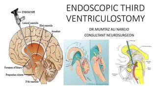
Endoscopic 3rd ventriculostomy is procedure of choice in obstructive hydrocephalus in which stoma is formed in floor of 3rd ventricle .
Report
Share
Report
Share

Recommended
More Related Content
Similar to Endoscopic 3rd Ventriculostomy-DR.MUMTAZ ALI NAREJO.pptx
Similar to Endoscopic 3rd Ventriculostomy-DR.MUMTAZ ALI NAREJO.pptx (20)
Know how to thoracocentesis and thoracostomy tubes

Know how to thoracocentesis and thoracostomy tubes
Diagnosis and radiological management of varicose vein

Diagnosis and radiological management of varicose vein
Thoracoscopy vs laparoscopy technical feasibility and complications in cong...

Thoracoscopy vs laparoscopy technical feasibility and complications in cong...
More from Neurosurgeon Mumtaz Ali Narejo
More from Neurosurgeon Mumtaz Ali Narejo (20)
head injury according to ATLS BY DR MUMTAZ ALI.pptx

head injury according to ATLS BY DR MUMTAZ ALI.pptx
Spine injury according to atls by dr mumtaz ali narejo.pptx

Spine injury according to atls by dr mumtaz ali narejo.pptx
Statistical software packages ,their layout & applications

Statistical software packages ,their layout & applications
Spine lecture for Final year MBBS by Dr.Mumtaz Ali.pptx

Spine lecture for Final year MBBS by Dr.Mumtaz Ali.pptx
Vestibular scwanomma (case pressentation)dr.mumtaz ali

Vestibular scwanomma (case pressentation)dr.mumtaz ali
Spinal schwannoma(case presentation) dr.mumtaz ali

Spinal schwannoma(case presentation) dr.mumtaz ali
Spinal ependymoma (case presentation)dr.mumtaz ali

Spinal ependymoma (case presentation)dr.mumtaz ali
Spinal meningioma (case presentation)dr.mumtaz ali

Spinal meningioma (case presentation)dr.mumtaz ali
Right sphenoid wing meningioma (case presentation)dr.mumtaz a li

Right sphenoid wing meningioma (case presentation)dr.mumtaz a li
Olfactory groove meningioma(case presentation) dr.mumtaz ali

Olfactory groove meningioma(case presentation) dr.mumtaz ali
Recently uploaded
A rare case of double-diverticulae of the Gallbladder found during a routine elective cholecystectomy is presented including intra operative and specimen images.Gallbladder Double-Diverticular: A Case Report المرارة مزدوجة التج: تقرير حالة

Gallbladder Double-Diverticular: A Case Report المرارة مزدوجة التج: تقرير حالةMohamad محمد Al-Gailani الكيلاني
Young & Hot ℂall Girls Salem 8250077686 WhatsApp Number Best Rates of Surat ℂall Girl Serviℂes Available 24x7x365 Young & Hot ℂall Girls Salem 8250077686 WhatsApp Number Best Rates of Surat ℂ...

Young & Hot ℂall Girls Salem 8250077686 WhatsApp Number Best Rates of Surat ℂ...Call Girls in Nagpur High Profile Call Girls
Saudi Arabia [ Abortion pills) Jeddah/riaydh/dammam/++918133066128☎️] cytotec tablets uses abortion pills 💊💊 How effective is the abortion pill? 💊💊 +918133066128) "Abortion pills in Jeddah" how to get cytotec tablets in Riyadh " Abortion pills in dammam*💊💊 The abortion pill is very effective. If you’re taking mifepristone and misoprostol, it depends on how far along the pregnancy is, and how many doses of medicine you take:💊💊 +918133066128) how to buy cytotec pills
At 8 weeks pregnant or less, it works about 94-98% of the time. +918133066128[ 💊💊💊 At 8-9 weeks pregnant, it works about 94-96% of the time. +918133066128) At 9-10 weeks pregnant, it works about 91-93% of the time. +918133066128)💊💊 If you take an extra dose of misoprostol, it works about 99% of the time. At 10-11 weeks pregnant, it works about 87% of the time. +918133066128) If you take an extra dose of misoprostol, it works about 98% of the time. In general, taking both mifepristone and+918133066128 misoprostol works a bit better than taking misoprostol only. +918133066128 Taking misoprostol alone works to end the+918133066128 pregnancy about 85-95% of the time — depending on how far along the+918133066128 pregnancy is and how you take the medicine. +918133066128 The abortion pill usually works, but if it doesn’t, you can take more medicine or have an in-clinic abortion. +918133066128 When can I take the abortion pill?+918133066128 In general, you can have a medication abortion up to 77 days (11 weeks)+918133066128 after the first day of your last period. If it’s been 78 days or more since the first day of your last+918133066128 period, you can have an in-clinic abortion to end your pregnancy.+918133066128
Why do people choose the abortion pill? Which kind of abortion you choose all depends on your personal+918133066128 preference and situation. With+918133066128 medication+918133066128 abortion, some people like that you don’t need to have a procedure in a doctor’s office. You can have your medication abortion on your own+918133066128 schedule, at home or in another comfortable place that you choose.+918133066128 You get to decide who you want to be with during your abortion, or you can go it alone. Because+918133066128 medication abortion is similar to a miscarriage, many people feel like it’s more “natural” and less invasive. And some+918133066128 people may not have an in-clinic abortion provider close by, so abortion pills are more available to+918133066128 them. +918133066128 Your doctor, nurse, or health center staff can help you decide which kind of abortion is best for you. +918133066128 More questions from patients: Saudi Arabia+918133066128 CYTOTEC Misoprostol Tablets. Misoprostol is a medication that can prevent stomach ulcers if you also take NSAID medications. It reduces the amount of acid in your stomach, which protects your stomach lining. The brand name of this medication is Cytotec®.+918133066128) Unwanted Kit is a combination of two medicines, which iBest medicine 100% Effective&Safe Mifepristion ௵+918133066128௹Abortion pills ...

Best medicine 100% Effective&Safe Mifepristion ௵+918133066128௹Abortion pills ...Abortion pills in Kuwait Cytotec pills in Kuwait
Recently uploaded (20)
NDCT Rules, 2019: An Overview | New Drugs and Clinical Trial Rules 2019

NDCT Rules, 2019: An Overview | New Drugs and Clinical Trial Rules 2019
Bhimrad + ℂall Girls Serviℂe Surat (Adult Only) 8849756361 Esℂort Serviℂe 24x...

Bhimrad + ℂall Girls Serviℂe Surat (Adult Only) 8849756361 Esℂort Serviℂe 24x...
Gallbladder Double-Diverticular: A Case Report المرارة مزدوجة التج: تقرير حالة

Gallbladder Double-Diverticular: A Case Report المرارة مزدوجة التج: تقرير حالة
Charbagh { ℂall Girls Serviℂe Lucknow ₹7.5k Pick Up & Drop With Cash Payment ...

Charbagh { ℂall Girls Serviℂe Lucknow ₹7.5k Pick Up & Drop With Cash Payment ...
Hemodialysis: Chapter 1, Physiological Principles of Hemodialysis - Dr.Gawad

Hemodialysis: Chapter 1, Physiological Principles of Hemodialysis - Dr.Gawad
Young & Hot ℂall Girls Salem 8250077686 WhatsApp Number Best Rates of Surat ℂ...

Young & Hot ℂall Girls Salem 8250077686 WhatsApp Number Best Rates of Surat ℂ...
CAD CAM DENTURES IN PROSTHODONTICS : Dental advancements

CAD CAM DENTURES IN PROSTHODONTICS : Dental advancements
CONGENITAL HYPERTROPHIC PYLORIC STENOSIS by Dr M.KARTHIK EMMANUEL

CONGENITAL HYPERTROPHIC PYLORIC STENOSIS by Dr M.KARTHIK EMMANUEL
Best medicine 100% Effective&Safe Mifepristion ௵+918133066128௹Abortion pills ...

Best medicine 100% Effective&Safe Mifepristion ௵+918133066128௹Abortion pills ...
Report Back from SGO: What’s the Latest in Ovarian Cancer?

Report Back from SGO: What’s the Latest in Ovarian Cancer?
Failure to thrive in neonates and infants + pediatric case.pptx

Failure to thrive in neonates and infants + pediatric case.pptx
SEMESTER-V CHILD HEALTH NURSING-UNIT-1-INTRODUCTION.pdf

SEMESTER-V CHILD HEALTH NURSING-UNIT-1-INTRODUCTION.pdf
The Clean Living Project Episode 24 - Subconscious

The Clean Living Project Episode 24 - Subconscious
Treatment Choices for Slip Disc at Gokuldas Hospital

Treatment Choices for Slip Disc at Gokuldas Hospital
Endoscopic 3rd Ventriculostomy-DR.MUMTAZ ALI NAREJO.pptx
- 1. ENDOSCOPIC THIRD VENTRICULOSTOMY DR.MUMTAZ ALI NAREJO CONSULTANT NEUROSURGEON
- 2. OUTLINES • Neuroendoscopic Anatomy of 3rd V • Indications • Contraindications • Complications • Ventriculoscope set & O.T • Ventricular irrigation • Procedure • Postop care • Pearls
- 3. NEUROENDOSCOPIC ANATOMY 3rd VENTRICLE
- 5. INDICATIONS • Obstuctive HCP • Shunt infections • Post-shunting subdural hematoma • Slit Ventricle Syndrom • NPH • DESH • Pineal gland tectal & PF tumors • DWM • Chiari type 1
- 6. CONTRAINDICATIONS • Communicating HCP • Low ETV success rate • Extensive scarring of Prepontine cistern • SAH • Agenesis of corpus callosum • Fused fornices with isolated 3rd V • Thickened 3rd ventricle floor • Pinpoint/Faint foramen monro • Dec space between BA & DS
- 7. COMPLICATIONS • Hypothalamic injury • Injury to pituitary stalk or gland • Transient 3rd and 6th nerve palsies • Injury to basilar A, PComA, or PCA • Uncontrollable bleeding • Cardiac arrest • Traumatic basilar artery aneurysm • Injury to Fornix • Injury to thalamostriate & septal vein • Injury to Thalamus • ETV failure 10-50%
- 9. ETV SUCCESS SCORE • <40% =Low Chance • 50-70%=Intermediate • >80%=Highest • 1 from each category=3 • 76% (72 of 95 patients) 6 Patient = 2nd ETV 3patients Partially functioning shunts
- 11. SUCCESS RATE • OverAll =56% • Non-tumoral Aqs=60-90% • Tectal tumors: 88% • Untreated Acquired Aqs = Highest • Infants = Poor • Low success rate 20%: Tumor Previous shunt Previous SAH Previous whole brain radiation Significant adhesion
- 13. VENTRICULAR IRRIGATION • an intermittent /continuous • operative field • maintain ventricular volumes & working space • stop low-pressure venous bleeding • 20- to 50-mL syringe • Ringer’s lactate heated to 37.5°C • Hypothalmus injury
- 14. PROCEDURE • Rigid endoscope • Position :15 degree • Incision :Curvlinear • Kochers Point:Burr • Durotomy : cruciate form • LV=>Foramen monro=>3rd V • Fixate sheath • Visualization = Floor of 3rd V • BA & Mamillary bodies • If not seen=Abort
- 15. PROCEDURE • Location Of opening : Midline: Avoids PCOM & PCA In Tuberum cinerum Posterior : Infundibular recess Anterior : Mamillary bodies Anterior : Tip of BA • Rubbing through floor 3rd V Probe or decq forcep • Hydrodissection/Bipolar • Avoid Laser
- 16. PROCEDURE • Opening : enlarged Decq forcep 3F fogarty balloon 4-9mm • Surety • Web membrane/liliquist Certain vessels Diluted iohexol intrathecal contrast • CT scan after 1 hour • T2WI sagittal thin slice sequence drop-out of T2 signal at stoma of ETV CISS/FIESTA:floor of 3rd V , thickness , bowing ,clivius & BA proximity
- 18. POSTOP • Seizures • Blood pressure lability • Bradycardia • Hyperthermia • Diabetes insipidus • Antiobiotics : 24 hour • AEM:24-48 hours • High dose dexamethasone prevent inflammation & stoma closure
- 19. • Neuroendoscopy provides monocular vision inside small structures. Slow and gentle movements are safe and allow for depth perception. • The etiology of hydrocephalus, clinical status, and results of imaging can all help determine which patients are ideal candidates for ETV. The ETVSS is a valuable tool for properly selecting patients for the procedure. • Excessive manipulation while in the foramen can lead to damage of the anterior column of the fornix, which can result in a significant loss of quality of life. • A key principle in neuroendoscopy is to always maintain the instrument in your field of view. To accomplish this goal, keep the endoscope behind the instrument tip, and advance the instrument and endoscope in small successive steps. • Identify the relevant anatomy initially. Do not assume that the endoscope has entered the ipsilateral ventricle. • Both the tuber cinereum and the membrane of Liliequist, when present, must be perforated. The best way to ensure entry into the prepontine cistern is to have a clear view of the basilar artery. • Intraoperative bleeding generally stops with gentle irrigation. Aggressive cautery should be avoided. An EVD should be left in place if there is significant hemorrhaging. • Unfavorable anatomic characteristics include small ventricles, a large massa intermedia, and a small space between the dorsum sella and basilar artery.
- 20. REFRENCES • Greenberg Handbook of Neurosurgery 10th edition • Youmans & winn text book of Neurological surgery 8th edition • Neurosurgical Atlas • Google
- 21. THANKS