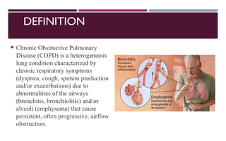
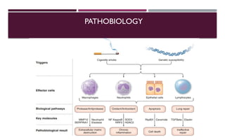
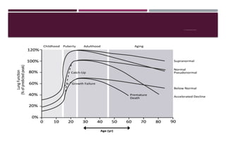
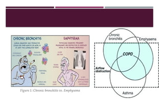
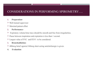
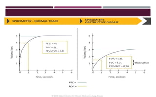
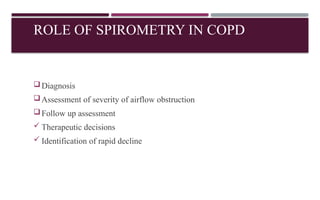
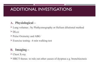
DR. AMMAR SABIR SIDDIQUI,
ASSISTANT PROFESSOR,
DEPARTMENT OF MEDICINE,
IIMSR, LUCKNOW
CHRONIC
OBSTRUCTIVE
PULMONARY DISEASE
DEFINITION
 Chronic Obstructive Pulmonary
Disease (COPD) is a heterogeneous
lung condition characterized by
chronic respiratory symptoms
(dyspnea, cough, sputum production
and/or exacerbations) due to
abnormalities of the airways
(bronchitis, bronchiolitis) and/or
alveoli (emphysema) that cause
persistent, often progressive, airflow
obstruction.
BURDEN OF COPD
 Global prevalence is 10.8% (11.8% in men, 8.5 % in women) .
 Prevalence of COPD increased steeply with age, with the highest
prevalence among those > 60 years.
 Globally, 3 million deaths each year due to COPD .
 3rd
leading cause of death
 5th
leading cause of reduced DALY (disability-adjusted life years) (2013) .
 In India NCDs were estimated to have accounted for 53% of all deaths and
44% of disability-adjusted life-years (DALYs) lost in 2005. Of these
chronic respiratory disease accounted for 7% deaths and 3% DALYs lost
PATHOGENESIS
GETomics :
Complex and dynamic interaction between Genes and Environment over the
lifeTime
Genes
Environment Lifetime
ENVIRONMENTAL RISK FACTORS
Cigarette
smoking
Biomass
exposure
Occupational
exposure
Air pollution
Respiratory
infections
GENETIC FACTORS
 To date, hundred of genetic variants are discovered with a link to COPD
(including genes encoding matrix metalloproteinase 12 (MMP-12),
glutathione S-transferase, the alpha-nicotinic acetylcholine receptor, and
the hedgehog interacting protein (HHIP)
 Most important genetic mutation is SERPINA1 which encodes for alfa-
1 antitrypsin (AATD)
 PiZZ (homozygous) genotype in 0.14% patients of COPD.
 There is controversy in link of heterozygotes (MZ & SZ) with the
development of COPD
PATHOBIOLOGY
Inflammatory changes
Inflammation within large and
small airways and in terminal
space of lung
Proteinases released by
inflammatory cells damage ECM
(extracellular matrix) around
airways, vasculature and alveoli.
Structural changes
Structural cell death leading to
extensive loss of small airways,
vascular pruning ,and alveolar
destruction
Disordered repair of elastin and other
ECM leads to air space enlargement
and emphysema.
PATHOBIOLOGY
COPD NORMAL LUNG
TISSUE
(A) COPD lung tissue with severe emphysema showing a small
airway with extensive loss of alveolar attachments.
(B) normal lung tissue with a small airway with normal
parenchyma
A B
PATHOPHYSIOLOGY
 Airflow obstruction and gas trapping
caused by a mixture of small airways disease (which increases airway resistance)
and parenchymal destruction (emphysema, that reduces the normal elastic recoil
of the lung parenchyma).
 Hyperinflation
Static hyperinflation: due to the loss of elastic lung recoil (as a consequence of
emphysema)
Dynamic hyperinflation: as a consequence of airflow obstruction when
ventilatory demands are increased and expiratory times are reduced
 Gas exchange abnormalities
 resulting in different degrees of arterial hypoxemia, without or with hypercapnia.
 also leads to decreased lung diffusing capacity (Dlco)
 Pulmonary hypertension
Centrilobular emphysema in
chronic smoker (70 pack year) Panlobular emphysema with severe Paraseptal emphysema in 56 yr female
alfa-1 AT deficiency. (37 pack year smoking history)
FEV1 TRAJECTORIES OVER LIFETIME
HOW DOES EMPHYSEMA LEAD TO
OBSTRUCTION
• Elastic recoil - generates the
pressure responsible for flow
under conditions of dynamic
compression.
• Loss of tethering caused by
alveolar wall destruction in
emphysema - airway collapse
• Pressure-volume curve shifts
upwards and leftwards.
West, J. B., Luks, A., & West, J. B. (2017). West's pulmonary pathophysiology: The essentials. Chapter 4 : Obstructive diseases
 The posteroanterior (A) and
lateral (B) chest x-rays of a 71-
year-old female with emphysema
show increased lung volumes
with flattened hemidiaphragms
on the lateral examination
(arrow) and increase in the
retrosternal space (arrowhead).
The normal retrosternal airspace
is less than 2.5 cm. A prominent
pulmonary artery on the
posteroanterior view (dashed
arrow) reflects secondary
pulmonary hypertension.
ETIOTYPES OF COPD
 COPD G- genetically determined COPD (AATD)
 COPD D- COPD due to abnormal lung development (e.g.premature birth)
 Environmental COPD –
COPD C -cigarette smoking
COPD P-pollution and biomass exposure
 COPD I-COPD due to childhood infections,tuberculosis
 COPD A –COPD and asthma
 COPD U –COPD of unknown cause
MEDICAL HISTORY
1. History of exposure to risk factors, such as smoking and environmental
exposures (household/outdoor).
2. Past medical history, including early life events (prematurity, low
birthweight, maternal smoking during pregnancy, passive smoking exposure
during infancy), asthma, allergy, sinusitis, or nasal polyps; respiratory infections
in childhood; HIV; tuberculosis.
3. Family history of COPD or other chronic respiratory disease.
4. History of more frequent or prolonged “winter colds,” .
5. History of exacerbations or previous hospitalizations for respiratory disorder.
6. Impact of disease on patient’s life.
SPIROMETRY
Forced spirometry measures the –
1) the volume of air forcibly exhaled
from the point of maximal
inspiration (forced vital capacity,
FVC);
2) the volume of air exhaled during
the first second of this maneuver
(forced expiratory volume in one
second, FEV1); and
3) the ratio of these two
measurements (FEV1/FVC).
CONSIDERATIONS IN PERFORMING SPIROMETRY….
1. Preparation-
 Well trained supervisor
 Maximal patient effort
2. Performance-
 Expiratory volume/time tracs should be smooth and free from irregularities.
 Pause between inspiration and expiration is less than 1 second.
 Largest value of FVC and FEV1 to be considered.
3. Bronchodilation-
 400mcg beta2 agonist/160mcg short acting anticholinergic is given.
4. Evaluation
ROLE OF SPIROMETRY IN COPD
Diagnosis
Assessment of severity of airflow obstruction
Follow up assessment
 Therapeutic decisions
 Identification of rapid decline
ADDITIONAL INVESTIGATIONS
A. Physiological –
 Lung volumes : by Plethysmography or Helium dilutional method
 DLco
 Pulse Oximetry and ABG
 Exercise testing : 6 min walking test
B. Imaging –
 Chest X-ray
 HRCT thorax: to rule out other causes of dyspnea e.g. bronchiectasis
CONTD…..
C. Alpha-1 antitrypsin levels:
 <20% of normal concentration suggests homozygous deficiency.
D. Biomarkers :
 e.g. absolute eosinophil count .
E. Composite scores :
 e.g. BODE index for COPD survival (BMI, Obstruction, Dyspnea
And Exercise )
IMPULSE OSCILLOMETRY
• An advanced spirometry technique, patient friendly and does not require any tedious
and exhaustive breathing maneuver.
• Simple, noninvasive method requiring only passive patient cooperation.
• It allows for the evaluation of lung function through the measurement of both airway
resistance and airway reactance.
• It has much higher sensitivity than FEV1 and peak expiratory flow.
 IOS can detect subtle changes in the small airway function even in the setting of
normal spirometry
 The frequencies of the waves delivered in IOS ranges from 5 to 30 Hz.
* While frequencies higher than 30 Hz can cause discomfort to the patient, the
parameters measured at <5 Hz are influenced by breath dynamics.
STEPS OF IOS INTERPRETATION
Check X5 Insp or X5 Exp
Check Reversibility Values
Check Value Of R5-R20
Check Value Of R20
Check Value Of R5
Check CoV, if < 10% → Valid
IF INCREASED→ TOTAL AIRWAY RESISTANCE
INCREASED
IF INCREASED→ LARGER AIRWAY
RESISTANCE INCREASED
SMALLER AIRWAY RESISTANCE
INCREASED
If X5insp Is More Negative → Restrictive Pathology
If X5exp Is More Negative → Obstructive Pathology
INTERPRETATION WITH PARAMETERS
 Increase R5,increase R19/20, Normal R5 - R19/20 -> Central airway obstruction
 Increase R5, Normal R19/20, Increase R5,R19/20-> Peripheral airway obstruction
 Increase R5, increase R19/20, Increase R5 - R19/20-> Total airway obstruction
 More negative X5 and increase Fres with normal R -> Restrictive disease
 Response to bronchodilator - Significant change in R5 (40%), X5 (50%), Ax (80%) &
Fres (leftward shift) -> Reversible obstruction
 Increase R5, more negative Xs and increase Fres - Peripheral airway obstruction
 Normal R5, more negative X5 and 1 Fres - Restrictive lung disease
 Increase R5, & increase R19/20 with normal Xs & Fres - Central airway obstruction
INITIALASSESSMENT OF COPD
Severity of airflow obstruction
Nature and magnitude of current symptoms
Previous history of moderate and severe exacerbations
Blood eosinophil count
Presence and type of other diseases (multimorbidity)
CLASSIFICATION OF AIRFLOW SEVERITY IN COPD
MODIFIED mMRC DYSPNEA SCALE
CAT (COPD ASSESSMENT TEST)
COMBINED COPD ASSESSMENT (GOLD)
In 2011, GOLD proposed combined assessment of COPD Based on –
 Levels of symptoms (mMRC or CAT)
Severity of airflow obstruction (GOLD grades 1-4)
Frequency of exacerbation
EARLIER GOLD 2023
Earlier
C D
A B
E
B
A
1or more severe
exacerbation leading
to hospitalization or
2 or more moderate
exacerbation
0 or1 moderate
exacerbation (not leading
to hospitalization)
WHAT’S NEW IN GOLD 2023 AND 2024?
 Proposed taxonomy / etio-types of COPD
 The ABCD assessment tool has been revised to ABE assessment tool
 Classification of severity of COPD exacerbation
 Vaccine recommendations for people with COPD .
 Therapeutic intervention to reduce COPD mortality
MANAGEMENT
• STABLE COPD
 EXACERBATION OF COPD
MANAGEMENT OFSTABLE COPD
Reduce Risk
factors
Pharmacological
Non
pharmacological
Maintenance
and follow
up
Smoking
cessation Reduces exposure
to household and
outdoor pollution
• Education
• Self
management
• Nutritional
supplement
• Palliative care
Oxygen therapy
Ventilatory
support
Interventional
bronchoscopy and
surgery
INITIAL PHARMACOLOGICAL TREATMENT
Group B
LABA + LAMA
Group A
BRONCHO-
DIALATOR
Group E
LABA + LAMA
Consider LABA + LAMA+ICS WHEN BLOOD EOS
≥300
mMRC-0-1,
CAT SCORE <10
mMRC-≥2
CAT SCORE >10
≥2 Moderate
Exacerbations
or
≥1 leading to
Hospitalization
0-1 moderate
Exacerbation
Not leading to
hospitalization
PHARMACOLOGICAL
THERAPY
Anti
Inflammatory
drugs
Bronchodilator
Antibiotics
Azithromycin [250mg/day or
500 mg three times per week
Erythromycin 250 mg / day
X 1 year
Mucolytics agents
Other pharmacological agents – alpha 1 antitrypsin augmentation therapy
USE OF ANTI-INFLAMMATORY AGENTS
Long term mono-therapy with ICS is not recommended
(Evidence A)
 Long term treatment with ICS may be considered with LABA
for patients with history of exacerbations despite appropriate
treatment with long acting bronchodilators (Evidence A)
 Long term therapy with oral corticosteroids not recommended
(Evidence A)
 In patients with severe airflow limitation, chronic bronchitis,
and exacerbations addition of PDE-4 inhibitor (Roflumilast) to
long acting bronchodilators with or without ICS (Evidence B)
CLASSIFICATION OF EXACERBATION
Mild
[No respiratory failure]
Moderate
[Acute respiratory failure
- Non life threatening]
Severe
[Acute respiratory failure-
Life threatening]
Respiratory rate ≤24/min[20-30 /min] >24/min[>30 /min] >24/min[>30 /min]
Accessory muscle use Absent Present Present
Mental status Normal Normal Altered
Hypoxemia Improved with supplemental
oxygen (FiO2: 24-35 % )
Improved with supplemental
oxygen
(FiO2 > 35 % )
Not improved even with
supplemental oxygen
(FiO2 > 40 % )
PaCO2 Normal 50-60 mmHg >60 mmHg (pH < 7.2)
MANAGEMENT OF EXACERBATION
SABA with or without Short acting anti cholinergic are recommended as initial
bronchodilator (Evidence C)
Systemic corticosteroid {40 mg prednisone per day for 5 days } (Evidence A)
Antibiotics when indicated shorten recovery time and hospital stay.
Methylxanthines are not recommended due to increased side effect profile (Evidence B)
NIV should be the first mode of ventilation provided no contraindication to its use is present
(Evidence A)
FACTORS TO CONSIDER WHEN USE ICS
Strongly favour use-History of hospitalization for exacerbation
• ≥2moderate exacerbation of COPD per year
• Blood eosinophils count ≥300 cells/micro L
• History of, or concomitant asthma
Favours use-1 moderate exacerbation of COPD per year
• Blood eosinophils count 100 to <300 cells/micro L
Against use -repeated pneumonia
Blood eosinophils count <100 cells/micro L
History of mycobacterial infection
IF LABA+LAMA+ICS FAILS…..
 Add roflumilast. This may be considered in patients with an
FEV1 < 50% predicted and chronic bronchitis.
 Add a macrolide. The best available evidence exists for the use of
azithromycin, especially in those who are not current smokers .
 Withdrawing ICS can be considered if pneumonia or other
considerable side-effects develop .
COMMONLY USED MEDICATIONS IN COPD
OVERVIEW OF MANAGEMENT
DIAGNOSIS
On the basis of
• Symptoms
• Risk factors
• Spirometry
INITIALASSESSMENT
• Categorise into GOLD 1- 4 (FEV1)
and ABE(CAT, mMRC,
exacerbations)
• Smoking status
• Comorbidities
ADJUST
• Pharmacotherapy
• Non-pharmacological
therapy
INITIAL MANAGEMENT
• Smoking cessation
• Vaccination
• Lifestyle and exercise
• Initial pharmacotherapy
• Self management education
• Management of comorbidities
REVIEW
• Symptoms
• Exacerbations
• Smoking status & other risk factors
• Inhaler techniques and adherence
• Physical activity
• Need for pulmonary rehabilitation
• Need for oxygen, NIV, intervention, palliation
• Vaccination
• Spirometry (at least annually)
FOLLOW UPPHARMACOLOGICALTREATMENT
If appropriate
Maintain it
Assess response to initial treatment
If not appropriate
 Assess inhaler technique and adherence
 Consider predominant treatable trait to
target (Dyspnoea or exacerbation) and
treat accordingly
 Assess response, adjust and review
INHALATION OF DRUGS
 Choice of inhaler device has to be individually tailored and depends on
access, cost, patient’s ability and preference
 It is essential to demonstrate proper inhalational technique when
prescribing a device
 Inhaler technique (and adherence) should be assessed before changing
present therapy
INDICATIONS OF HOSPITALISATION
 Symptoms like worsening of resting dyspnoea, high respiratory rate,
decrease oxygen saturation, confusion, drowsiness
 Presence of cyanosis, peripheral edema
 Failure to respond to initial medication
 Serious comorbidities like heart failure, arrhythmias etc.
 Insufficient home support
Supplemental oxygen to COPD Patient
Arterial hypoxemia –Pao2 ≤55 mm hg or
Sao2 <88% Or
Pao2 > 55 but <60 mm hg with right heart
failure
Prescribe supplemental oxygen
and titrate to keep Sao2 ≥ 90 %
Recheck in 60 to 90 days to assess
 If supplemental oxygen still needed
 If prescribed supplemental oxygen is
effective
INDICATIONS OF
MECHANICAL VENTILATION
 At least one of the following
1. Respiratory acidosis (PaCO2 >45
mmHg and arterial pH )
2. Severe dyspnoea with clinical signs
like use of accessory muscles,
retraction of intercostal spaces,
paradoxical motion of abdomen
3. Persistent hypoxemia despite
supplemental oxygen therapy
 Unable to tolerate NIV or NIV failure
 Diminished consciousness, psychomotor
agitation
 Massive aspiration or persistent vomiting
 Inability to remove respiratory secretions
 Hemodynamic instability
 Severe ventricular or supraventricular
arrhythmias
INDICATIONS OF NIV
VACCINATION IN COPD
INTERVENTIONAL THERAPY IN STABLE COPD
 Bullectomy
 Bronchoscopic interventions-endobronchial valves, lung
coils
 Lung volume reduction surgery
 Bronchoscopic intervention under study-
cryospray,targeted lung denervation technology
 Lung transplantation
THANK
YOU

COPD - DEFINITION, PATHOPHYSIOLOGY AND MANAGEMENT

  • 1.
    DR. AMMAR SABIRSIDDIQUI, ASSISTANT PROFESSOR, DEPARTMENT OF MEDICINE, IIMSR, LUCKNOW CHRONIC OBSTRUCTIVE PULMONARY DISEASE
  • 2.
    DEFINITION  Chronic ObstructivePulmonary Disease (COPD) is a heterogeneous lung condition characterized by chronic respiratory symptoms (dyspnea, cough, sputum production and/or exacerbations) due to abnormalities of the airways (bronchitis, bronchiolitis) and/or alveoli (emphysema) that cause persistent, often progressive, airflow obstruction.
  • 3.
    BURDEN OF COPD Global prevalence is 10.8% (11.8% in men, 8.5 % in women) .  Prevalence of COPD increased steeply with age, with the highest prevalence among those > 60 years.  Globally, 3 million deaths each year due to COPD .  3rd leading cause of death  5th leading cause of reduced DALY (disability-adjusted life years) (2013) .  In India NCDs were estimated to have accounted for 53% of all deaths and 44% of disability-adjusted life-years (DALYs) lost in 2005. Of these chronic respiratory disease accounted for 7% deaths and 3% DALYs lost
  • 4.
    PATHOGENESIS GETomics : Complex anddynamic interaction between Genes and Environment over the lifeTime Genes Environment Lifetime
  • 5.
  • 6.
    GENETIC FACTORS  Todate, hundred of genetic variants are discovered with a link to COPD (including genes encoding matrix metalloproteinase 12 (MMP-12), glutathione S-transferase, the alpha-nicotinic acetylcholine receptor, and the hedgehog interacting protein (HHIP)  Most important genetic mutation is SERPINA1 which encodes for alfa- 1 antitrypsin (AATD)  PiZZ (homozygous) genotype in 0.14% patients of COPD.  There is controversy in link of heterozygotes (MZ & SZ) with the development of COPD
  • 7.
    PATHOBIOLOGY Inflammatory changes Inflammation withinlarge and small airways and in terminal space of lung Proteinases released by inflammatory cells damage ECM (extracellular matrix) around airways, vasculature and alveoli. Structural changes Structural cell death leading to extensive loss of small airways, vascular pruning ,and alveolar destruction Disordered repair of elastin and other ECM leads to air space enlargement and emphysema.
  • 8.
  • 9.
    COPD NORMAL LUNG TISSUE (A)COPD lung tissue with severe emphysema showing a small airway with extensive loss of alveolar attachments. (B) normal lung tissue with a small airway with normal parenchyma A B
  • 10.
    PATHOPHYSIOLOGY  Airflow obstructionand gas trapping caused by a mixture of small airways disease (which increases airway resistance) and parenchymal destruction (emphysema, that reduces the normal elastic recoil of the lung parenchyma).  Hyperinflation Static hyperinflation: due to the loss of elastic lung recoil (as a consequence of emphysema) Dynamic hyperinflation: as a consequence of airflow obstruction when ventilatory demands are increased and expiratory times are reduced
  • 11.
     Gas exchangeabnormalities  resulting in different degrees of arterial hypoxemia, without or with hypercapnia.  also leads to decreased lung diffusing capacity (Dlco)  Pulmonary hypertension Centrilobular emphysema in chronic smoker (70 pack year) Panlobular emphysema with severe Paraseptal emphysema in 56 yr female alfa-1 AT deficiency. (37 pack year smoking history)
  • 12.
  • 13.
    HOW DOES EMPHYSEMALEAD TO OBSTRUCTION • Elastic recoil - generates the pressure responsible for flow under conditions of dynamic compression. • Loss of tethering caused by alveolar wall destruction in emphysema - airway collapse • Pressure-volume curve shifts upwards and leftwards. West, J. B., Luks, A., & West, J. B. (2017). West's pulmonary pathophysiology: The essentials. Chapter 4 : Obstructive diseases
  • 15.
     The posteroanterior(A) and lateral (B) chest x-rays of a 71- year-old female with emphysema show increased lung volumes with flattened hemidiaphragms on the lateral examination (arrow) and increase in the retrosternal space (arrowhead). The normal retrosternal airspace is less than 2.5 cm. A prominent pulmonary artery on the posteroanterior view (dashed arrow) reflects secondary pulmonary hypertension.
  • 17.
    ETIOTYPES OF COPD COPD G- genetically determined COPD (AATD)  COPD D- COPD due to abnormal lung development (e.g.premature birth)  Environmental COPD – COPD C -cigarette smoking COPD P-pollution and biomass exposure  COPD I-COPD due to childhood infections,tuberculosis  COPD A –COPD and asthma  COPD U –COPD of unknown cause
  • 18.
    MEDICAL HISTORY 1. Historyof exposure to risk factors, such as smoking and environmental exposures (household/outdoor). 2. Past medical history, including early life events (prematurity, low birthweight, maternal smoking during pregnancy, passive smoking exposure during infancy), asthma, allergy, sinusitis, or nasal polyps; respiratory infections in childhood; HIV; tuberculosis. 3. Family history of COPD or other chronic respiratory disease. 4. History of more frequent or prolonged “winter colds,” . 5. History of exacerbations or previous hospitalizations for respiratory disorder. 6. Impact of disease on patient’s life.
  • 19.
    SPIROMETRY Forced spirometry measuresthe – 1) the volume of air forcibly exhaled from the point of maximal inspiration (forced vital capacity, FVC); 2) the volume of air exhaled during the first second of this maneuver (forced expiratory volume in one second, FEV1); and 3) the ratio of these two measurements (FEV1/FVC).
  • 20.
    CONSIDERATIONS IN PERFORMINGSPIROMETRY…. 1. Preparation-  Well trained supervisor  Maximal patient effort 2. Performance-  Expiratory volume/time tracs should be smooth and free from irregularities.  Pause between inspiration and expiration is less than 1 second.  Largest value of FVC and FEV1 to be considered. 3. Bronchodilation-  400mcg beta2 agonist/160mcg short acting anticholinergic is given. 4. Evaluation
  • 22.
    ROLE OF SPIROMETRYIN COPD Diagnosis Assessment of severity of airflow obstruction Follow up assessment  Therapeutic decisions  Identification of rapid decline
  • 23.
    ADDITIONAL INVESTIGATIONS A. Physiological–  Lung volumes : by Plethysmography or Helium dilutional method  DLco  Pulse Oximetry and ABG  Exercise testing : 6 min walking test B. Imaging –  Chest X-ray  HRCT thorax: to rule out other causes of dyspnea e.g. bronchiectasis
  • 24.
    CONTD….. C. Alpha-1 antitrypsinlevels:  <20% of normal concentration suggests homozygous deficiency. D. Biomarkers :  e.g. absolute eosinophil count . E. Composite scores :  e.g. BODE index for COPD survival (BMI, Obstruction, Dyspnea And Exercise )
  • 25.
    IMPULSE OSCILLOMETRY • Anadvanced spirometry technique, patient friendly and does not require any tedious and exhaustive breathing maneuver. • Simple, noninvasive method requiring only passive patient cooperation. • It allows for the evaluation of lung function through the measurement of both airway resistance and airway reactance. • It has much higher sensitivity than FEV1 and peak expiratory flow.  IOS can detect subtle changes in the small airway function even in the setting of normal spirometry  The frequencies of the waves delivered in IOS ranges from 5 to 30 Hz. * While frequencies higher than 30 Hz can cause discomfort to the patient, the parameters measured at <5 Hz are influenced by breath dynamics.
  • 27.
    STEPS OF IOSINTERPRETATION Check X5 Insp or X5 Exp Check Reversibility Values Check Value Of R5-R20 Check Value Of R20 Check Value Of R5 Check CoV, if < 10% → Valid IF INCREASED→ TOTAL AIRWAY RESISTANCE INCREASED IF INCREASED→ LARGER AIRWAY RESISTANCE INCREASED SMALLER AIRWAY RESISTANCE INCREASED If X5insp Is More Negative → Restrictive Pathology If X5exp Is More Negative → Obstructive Pathology
  • 28.
    INTERPRETATION WITH PARAMETERS Increase R5,increase R19/20, Normal R5 - R19/20 -> Central airway obstruction  Increase R5, Normal R19/20, Increase R5,R19/20-> Peripheral airway obstruction  Increase R5, increase R19/20, Increase R5 - R19/20-> Total airway obstruction  More negative X5 and increase Fres with normal R -> Restrictive disease  Response to bronchodilator - Significant change in R5 (40%), X5 (50%), Ax (80%) & Fres (leftward shift) -> Reversible obstruction  Increase R5, more negative Xs and increase Fres - Peripheral airway obstruction  Normal R5, more negative X5 and 1 Fres - Restrictive lung disease  Increase R5, & increase R19/20 with normal Xs & Fres - Central airway obstruction
  • 29.
    INITIALASSESSMENT OF COPD Severityof airflow obstruction Nature and magnitude of current symptoms Previous history of moderate and severe exacerbations Blood eosinophil count Presence and type of other diseases (multimorbidity)
  • 30.
    CLASSIFICATION OF AIRFLOWSEVERITY IN COPD
  • 31.
  • 32.
  • 33.
    COMBINED COPD ASSESSMENT(GOLD) In 2011, GOLD proposed combined assessment of COPD Based on –  Levels of symptoms (mMRC or CAT) Severity of airflow obstruction (GOLD grades 1-4) Frequency of exacerbation EARLIER GOLD 2023 Earlier C D A B E B A 1or more severe exacerbation leading to hospitalization or 2 or more moderate exacerbation 0 or1 moderate exacerbation (not leading to hospitalization)
  • 34.
    WHAT’S NEW INGOLD 2023 AND 2024?  Proposed taxonomy / etio-types of COPD  The ABCD assessment tool has been revised to ABE assessment tool  Classification of severity of COPD exacerbation  Vaccine recommendations for people with COPD .  Therapeutic intervention to reduce COPD mortality
  • 35.
    MANAGEMENT • STABLE COPD EXACERBATION OF COPD
  • 36.
    MANAGEMENT OFSTABLE COPD ReduceRisk factors Pharmacological Non pharmacological Maintenance and follow up Smoking cessation Reduces exposure to household and outdoor pollution • Education • Self management • Nutritional supplement • Palliative care Oxygen therapy Ventilatory support Interventional bronchoscopy and surgery
  • 37.
    INITIAL PHARMACOLOGICAL TREATMENT GroupB LABA + LAMA Group A BRONCHO- DIALATOR Group E LABA + LAMA Consider LABA + LAMA+ICS WHEN BLOOD EOS ≥300 mMRC-0-1, CAT SCORE <10 mMRC-≥2 CAT SCORE >10 ≥2 Moderate Exacerbations or ≥1 leading to Hospitalization 0-1 moderate Exacerbation Not leading to hospitalization
  • 38.
    PHARMACOLOGICAL THERAPY Anti Inflammatory drugs Bronchodilator Antibiotics Azithromycin [250mg/day or 500mg three times per week Erythromycin 250 mg / day X 1 year Mucolytics agents Other pharmacological agents – alpha 1 antitrypsin augmentation therapy
  • 39.
    USE OF ANTI-INFLAMMATORYAGENTS Long term mono-therapy with ICS is not recommended (Evidence A)  Long term treatment with ICS may be considered with LABA for patients with history of exacerbations despite appropriate treatment with long acting bronchodilators (Evidence A)  Long term therapy with oral corticosteroids not recommended (Evidence A)  In patients with severe airflow limitation, chronic bronchitis, and exacerbations addition of PDE-4 inhibitor (Roflumilast) to long acting bronchodilators with or without ICS (Evidence B)
  • 40.
    CLASSIFICATION OF EXACERBATION Mild [Norespiratory failure] Moderate [Acute respiratory failure - Non life threatening] Severe [Acute respiratory failure- Life threatening] Respiratory rate ≤24/min[20-30 /min] >24/min[>30 /min] >24/min[>30 /min] Accessory muscle use Absent Present Present Mental status Normal Normal Altered Hypoxemia Improved with supplemental oxygen (FiO2: 24-35 % ) Improved with supplemental oxygen (FiO2 > 35 % ) Not improved even with supplemental oxygen (FiO2 > 40 % ) PaCO2 Normal 50-60 mmHg >60 mmHg (pH < 7.2)
  • 41.
    MANAGEMENT OF EXACERBATION SABAwith or without Short acting anti cholinergic are recommended as initial bronchodilator (Evidence C) Systemic corticosteroid {40 mg prednisone per day for 5 days } (Evidence A) Antibiotics when indicated shorten recovery time and hospital stay. Methylxanthines are not recommended due to increased side effect profile (Evidence B) NIV should be the first mode of ventilation provided no contraindication to its use is present (Evidence A)
  • 42.
    FACTORS TO CONSIDERWHEN USE ICS Strongly favour use-History of hospitalization for exacerbation • ≥2moderate exacerbation of COPD per year • Blood eosinophils count ≥300 cells/micro L • History of, or concomitant asthma Favours use-1 moderate exacerbation of COPD per year • Blood eosinophils count 100 to <300 cells/micro L Against use -repeated pneumonia Blood eosinophils count <100 cells/micro L History of mycobacterial infection
  • 43.
    IF LABA+LAMA+ICS FAILS….. Add roflumilast. This may be considered in patients with an FEV1 < 50% predicted and chronic bronchitis.  Add a macrolide. The best available evidence exists for the use of azithromycin, especially in those who are not current smokers .  Withdrawing ICS can be considered if pneumonia or other considerable side-effects develop .
  • 44.
  • 45.
  • 46.
    OVERVIEW OF MANAGEMENT DIAGNOSIS Onthe basis of • Symptoms • Risk factors • Spirometry INITIALASSESSMENT • Categorise into GOLD 1- 4 (FEV1) and ABE(CAT, mMRC, exacerbations) • Smoking status • Comorbidities ADJUST • Pharmacotherapy • Non-pharmacological therapy INITIAL MANAGEMENT • Smoking cessation • Vaccination • Lifestyle and exercise • Initial pharmacotherapy • Self management education • Management of comorbidities REVIEW • Symptoms • Exacerbations • Smoking status & other risk factors • Inhaler techniques and adherence • Physical activity • Need for pulmonary rehabilitation • Need for oxygen, NIV, intervention, palliation • Vaccination • Spirometry (at least annually)
  • 47.
    FOLLOW UPPHARMACOLOGICALTREATMENT If appropriate Maintainit Assess response to initial treatment If not appropriate  Assess inhaler technique and adherence  Consider predominant treatable trait to target (Dyspnoea or exacerbation) and treat accordingly  Assess response, adjust and review
  • 48.
    INHALATION OF DRUGS Choice of inhaler device has to be individually tailored and depends on access, cost, patient’s ability and preference  It is essential to demonstrate proper inhalational technique when prescribing a device  Inhaler technique (and adherence) should be assessed before changing present therapy
  • 49.
    INDICATIONS OF HOSPITALISATION Symptoms like worsening of resting dyspnoea, high respiratory rate, decrease oxygen saturation, confusion, drowsiness  Presence of cyanosis, peripheral edema  Failure to respond to initial medication  Serious comorbidities like heart failure, arrhythmias etc.  Insufficient home support
  • 50.
    Supplemental oxygen toCOPD Patient Arterial hypoxemia –Pao2 ≤55 mm hg or Sao2 <88% Or Pao2 > 55 but <60 mm hg with right heart failure Prescribe supplemental oxygen and titrate to keep Sao2 ≥ 90 % Recheck in 60 to 90 days to assess  If supplemental oxygen still needed  If prescribed supplemental oxygen is effective
  • 51.
    INDICATIONS OF MECHANICAL VENTILATION At least one of the following 1. Respiratory acidosis (PaCO2 >45 mmHg and arterial pH ) 2. Severe dyspnoea with clinical signs like use of accessory muscles, retraction of intercostal spaces, paradoxical motion of abdomen 3. Persistent hypoxemia despite supplemental oxygen therapy  Unable to tolerate NIV or NIV failure  Diminished consciousness, psychomotor agitation  Massive aspiration or persistent vomiting  Inability to remove respiratory secretions  Hemodynamic instability  Severe ventricular or supraventricular arrhythmias INDICATIONS OF NIV
  • 52.
  • 53.
    INTERVENTIONAL THERAPY INSTABLE COPD  Bullectomy  Bronchoscopic interventions-endobronchial valves, lung coils  Lung volume reduction surgery  Bronchoscopic intervention under study- cryospray,targeted lung denervation technology  Lung transplantation
  • 54.