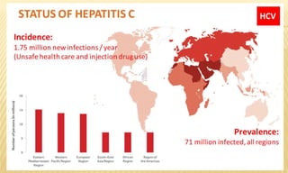
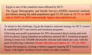
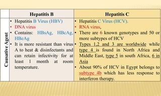
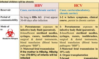
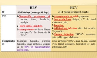
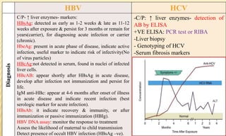
CONTACT
INFECTIONS
By
Noha Hesham
Under supervision of
Prof Dr Mona Aboserea
Zagazig university
 Invasion of the skin or mucous membranes by a pathogenic
organism or parasite.
 Infection in which entrance of the pathogenic organism (or the
parasite) occurs through the skin or mucus membranes.
 Some infectious agents can invade the intact (undamaged)
skin or mucous membranes, but the majority needs injured
surfaces in the form of abrasions, scratches, wounds or ulcers.
Viral
AIDS
Hepatitis
“B&C”
Rabies
cytomegalovir
us
Bacterial
Anthrax
Gas gangrene
Gonorrhea
Leprosy
Tetanus
Parasitic
Ancylostomiasis
Schistosomiasis
Spirochetes
Syphilis
HBV, HCV and HDV are transmitted mainly percutaneous “contact infections”.
HAV is transmitted mainly by faeco-oral transmission )food borne disease(.
HDV & HFV are similar to HBV.
HEV is similar to HAV.
HGV is similar to HCV.
Other viruses cause acute hepatitis e.g. cytomegalovirus, Epstein-Barr virus.
EPIDEMIOLOGY OF VIRAL HEPATITIS
• Usually the disease is self-limited, however some cases show
massive or chronic hepatic necrosis & chronic hepatitis.
• Virus B and C may be complicated by liver cirrhosis &
hepatocellular carcinoma.
• An estimated 248 million people have chronic HBV infection
globally. In Egypt the prevalence is < 2%.
Viral hepatitis C is a major health problem in Egypt.
• Egypt is one of the countries most affected by HCV.
• The Egypt Demographic and Health Surveys (EDHS) measured antibody
prevalence among the adult population aged 15–59 years at 14.7% in 2009
and at 10.0% in 2015 substantially higher than global levels.
• To attend to this challenge, Egypt developed a national strategy for HCV control
& established HCV prevention and treatment programs.
• Following successful negotiations for 99% discounted direct-acting antivirals
(DAAs) prices, Egypt launched an ambitious national HCV treatment program
aiming to treat over 250,000 chronically infected individuals per year, with the
goal of achieving a national chronic infection prevalence of < 2% by 2025.
• Despite this progress, existing evidence suggests ongoing HCV transmission in
Egypt, with higher incidence levels relative to other countries.
Hepatitis B Hepatitis C
CausativeAgent
• Hepatitis B Virus (HBV)
• DNA virus
• Contains: HBsAg, HBcAg,
HBeAg
• It is more resistant than virus
A to heat & disinfectants and
can retain infectivity for at
least 1 month at room
temperature.
• Hepatitis C Virus (HCV).
• RNA virus,
• There are 6 known genotypes and 50 or
more subtypes of HCV
• Types 1,2 and 3 are worldwide while
type 4 is found in North Africa and
Middle East, type 5 in south Africa, 6 in
Asia
• About 90% of HCV in Egypt belongs to
subtype 4b which has less response to
interferon therapy.
HCVHBV
Cases, carriers(incubatory,
chronic carrier)
Cases, carriers(chronic carrier)Reservoir
1-2 w before symptoms, clinical
course, persist in chronic carriers
So long is HBs AG (+ve) appear
30-60 days after infection
Period of
communicability
1- Percutaneous exposure to
infective body fluid (parentral).
2-Insufficient sterilized needles,
syringes, razors, toothbrushes,
surgical & dental instruments,
blood transfusion (Blood borne
pathogens “BBP”).
3-Maternal fetal transmission in
10% only
4- Organ transplantation
5-Sexual transmission
1- Percutaneous exposure to
infective body fluid (parentral).
2-Insufficient sterilized needles,
syringes, razors, toothbrushes,
surgical & dental instruments,
blood transfusion (Blood borne
pathogens “BBP”).
3- Maternal fetal transmission
If the mother is HBsAg, HBeAg
+ve: (70-90%) of infants will be
infected
Mode of
transmission
infected children will be chronic
HCVHBV
2-12 weeks (average 6 weeks)60-150 days (average 90 days)IP
1-Asymptomatic or mild symptoms.
2.Low grade fever, fatigue, N,V , Rt. sided
abdominal pain,
3.Jaundice.
4.Self-limiting infection after 3-6 months.
No jaundice.
5.Chronic infection “80%”: weakness,
pain in Rt. upper abdomen.
1-Nonspecific prodroma of
malaise, fever, headache,
myalgia.
2- Dark urine, jaundice.
3-Asymptomatic or have illness
not specific for hepatitis B
“50%”.
C/P
Liver cirrhosis “25%”, liver failure, Cancer
liver, Renal disorders, formation of auto-
antibodies, DM
Fulminant hepatitis, Chronic
hepatitis, Liver cirrhosis, Causes
up to 80% of hepatocellular
carcinoma.
Complicatio
ns
HCVHBV
-C/P: ↑ liver enzymes- detection of
AB by ELISA
+VE ELISA: PCR test or RIBA
-Liver biopsy
- Genotyping of HCV
-Serum fibrosis markers
C/P- ↑ liver enzymes- markers:
HBsAg: detected as early as 1-2 weeks & late as 11-12
weeks after exposure & persist for 3 months or remain for
years(carrier), for diagnosing acute infection or carrier
(chronic).
HbeAg: present in acute phase of disease, indicate active
infection, useful marker to indicate risk of infectivity(No
of virus particles)
HBcAg:not detected in serum, found in nuclei of infected
liver cells.
HBcAB: appear shortly after HBsAg in acute disease,
develop after infection not immunization and persist for
life.
IgM anti-HBc: appear at 4-6 months after onset of illness
in acute disease and indicate recent infection (best
serologic marker for acute infection).
HBsAb: it indicate recovery & immunity, or after
immunization or passive immunization (HBIg).
HBV DNA assay: monitor the response to treatment
Assess the likelihood of maternal to child transmission
Detect presence of occult HBV infection (HBsAg –ve).
Diagnosis
Test Result Interpretation
HBsAg
Anti-HBc
Anti-HBs
-ve
-ve
-ve
susceptible
HBsAg
Anti-HBc
Anti-HBs
-ve
-ve
+ve >10m IU/mL
Immune due to vaccination
Post vaccination testing after 1-2 month of vaccination
HBsAg
Anti-HBc
Anti-HBs
-ve
+ve
+ve
Immune due to natural infection
HBsAg
Anti-HBc
IgM anti-HBc
Anti-HBs
+ve
+ve
+ve
-ve
Acute infection
HBsAg
Anti-HBc
IgM anti-HBc
Anti-HBs
+ve
+ve
-ve
-ve
Chronic infection
HBsAg
Anti-HBc
Anti-HBs
-ve
+ve
-ve
1-Recovering from acute infection.
2-distantly immune & test is not sensitive to detect very low level
of anti-HBs.
3-Susceptibel with false +ve anti-HBc.
4-chronically infected & have undetectable level of HBsAg.
N.B: Window period: the period of infection when neither HBsAg nor it’s Ab can be
detected in the serum of the patient
Serologic tests conducted during the window period will be +ve HBcAb & HbeAb.
HCVHBV
No
specific
preventiv
e
measures
Active immunization:
1- Plasma derived HB: purified inactivated HBsAg prepared from healthy
carrier (not well accepted due to fear of transmission of live HBV & other
blood born pathogens)
2-Recombinant vaccine:recombinant HBsAg, 95% protection for 20 years
Formulations:
Recombivax Hb, Engerix-B: 3 doses, (0.5 ml each in pediatrics & 1 ml in
adults) at 0,1,6, booster doesn’t routinely recommended.
Heplisav-B: >18 years, 0.5 ml, 2 doses at 0, 1.
Indications: 1- compulsory to all infants, 1st dose is taken at birth, 2nd (1-2
months) & 3rd (6-18) but if the mother is HBsAg +ve must completed in 1st 6
months.
2- all children & adolescents <19 years who haven’t the vaccine should be
vaccinated.
3- at risk adults ( medical & paramedical, repeated blood transfusion &
heamodialysis, drug abusers, people whose sex parteners have HB, household
contacts of HBsAg +ve person, traveler to regions with high rates if Hb, people
with chronic liver diseases, kidney diseases, HIV or diabetes).
specifi
c
prevention
HBV
Combined vaccine:
1- COMVAX: HB-Hib combination.
2- Pediarix: DTaP- HB- IPV combination.
3- Twinirx : Hepatistis A & B.
Contraindications: to vaccinations: sever allergic reaction to
vaccine component or following 1st dose, moderate of sever acute
illness.
Adverse reaction: anaphylaxis.
Passive immunization: seroprophylaxis
Immunoglobilins, 0.1 ml/kg, 2 doses 1 month apart given at once
after exposure not after 48 hrs.
Indications: 1- high risk persons 2- exposure to infected blood
3- after sexual exposure to infected person
4- infants born to infected mothers: after birth by 12hrs then after
3 and 6 months.
prevention
TREATMENT
• HCV TTT continues to evolve rapidly.
• Since 2014, several new all-oral direct-acting
antiviral agents have been approved for use.
• The Egyptian MOH proposed a new national
strategy to control the HCV epidemic in
Egypt with a greater capital fund and with
support from the WHO as well as other
institutes.
• This scheme was entitled “The Plan of Action
for the Prevention, Care and Treatment of
Viral Hepatitis 2014–2018” & promoted
sofosbuvir (Sovaldi™) as its 1ry treatment.
Patients with hepatocellular carcinoma awaiting liver transplantation
•For prevention of post-transplant HCV reinfection
•400 mg, once daily + ribavirin for up to 48 weeks or until the time of liver
transplantation, whichever occurs first.
Treatment regimen and duration depends on both viral
genotype and patient population:
• Genotype 1, 4: 400mg, tablet orally, taken once a day + ribavirin
& peginterferon alpha for 12 weeks.
For patients who should not take interferon: sofosbuvir + ribavirin
for 24 weeks.
• Genotype 2: 400 mg, orally, once daily + ribavirin for 12 weeks.
• Genotype 3: 400 mg orally once daily + ribavirin for 24 weeks.
The scheme aims to treat 300,000 patients annually
MOH
38%
HIO
51%
Private
payments
3% Patients
8%
TTT Cost
RABIES
Reservoir: Domestic & wild animals “dog, cat, fox, wolf, rats, bat,
etc”.
Exit: in saliva of rabid animals.
Modes of transmission: Infection is transmitted with saliva through
the bite of an infected animal usually or when saliva gets on an
injured skin by lick, also organ transplantation from infected person
as corneal transplant. No man to man infection.
Causative organism: Rabies virus, RNA rhabdovirus with 5
genetically related viruses.
1 antigenic type but 2 biological forms:
1-Street virus: propagated in nature, infects by peripheral inoculation lead to encephalitis.
2- fixed virus: the virus lost its ability to infect by peripheral inoculation but cause
encephalitis if injected by intracerebral inoculation (attenuated), used in preparation of
different vaccines.
IP:
- Depends on the distance between the site of bite & brain.
- Usually 2-8 weeks, but up to 1 year.
C/P:
1- History of animal bite.
2- Pain & paresthesia at site of exposure.
3- Nonspecific manifestations: fever, anorexia.
4- Specific manifestations: paresthesia, aerophobia, hydrophobia, difficult swallowing,
convulsions.
5- Death within 1-2 weeks during convulsive state or due to respiratory paralysis.
Diagnosis:
1- C/P
2- Detect virus particles in saliva & CSF
3- Detect viral antigen in skin biopsy using fluorescent antibody technique
4- Rising antibody titer.
Pre-exposure vaccination Control:
RISK CATEGORY TYPICAL POPULATIONS PREEXPOSURE REGIMEN
Continuous Rabies research laboratory
workers
Primary course; serologic testing
every 6 months; booster
vaccination if antibody titter is
below acceptable level
Frequent Rabies diagnostic laboratory
workers, cavers, veterinarians and
staff, and animal control and
wildlife workers in areas where
rabies is enzootic. All people who
frequently handle bats.
Primary course; serologic testing
every 2 years; booster vaccination
if antibody titer is below
acceptable level
2
Infrequent (greater than general
population)
Veterinarians and animal control
staff working in areas where rabies
is uncommon to rare; veterinary
students; and travellers visiting
areas where rabies is enzootic and
immediate access to medical care
is limited
Primary course; no serologic
testing or booster vaccination
Rare (general population) population at large No preexposure immunization
necessary
N.B: Patients who are immunosuppressed by disease or medications should postpone
preexposure vaccinations and consider avoiding activities for which rabies preexposure
prophylaxis is indicated during the period of expected immunosuppression. If this is not
possible, immunosuppressed people who are at risk for rabies should have their antibody
titers checked after vaccination.
Types of vaccine:
1- human diploid cell vaccine
2- rabies vaccine adsorped
3- duck embryo vaccine
4- purified chick embryo vaccine
Dose: 1 ml IM in deltoid region “ never in gluteal” 3 doses at 0, 7, 21 or 28.
Post-exposure
IMMUNIZA-
TION STATUS
VACCINE/
PRODUCT
DOSE NUMBER OF
DOSES
SCHEDULE
(DAYS)
ROUTE
Not previously
vaccinated
RIG plus 20 IU/kg body
weight
1 0 Infiltrated at bite
site (if possible);
remainder IM
HDCV or PCEC 1.0 mL 4 0, 3, 7, 14 (28 if
immuno-
compromised)
IM
Previously
vaccinated
HDCV or PCEC 1.0 mL 2
3
0, 3
0,3,7
IM
Recommended post-exposure prophylaxis for rabies infection
Category of exposure to suspect rabid animal Post-exposure measures
Category I – touching or feeding animals, licks on intact skin
(i.e. no exposure)
None
Category II – nibbling of uncovered skin, minor scratches or
abrasions without bleeding
Immediate vaccination and local treatment of the
wound
Category III – single or multiple transdermal bites or scratches,
licks on broken skin; contamination of mucous membrane with
saliva from licks, exposures to bats.
Immediate vaccination and administration of rabies
immunoglobulin; local treatment of the wound
MEASURES TO THE WOUND (BITE)
• 1-Immediate local treatment of animal bite & scratches through repeated flushing
& cleaning of wound with soap & water.
• 2-A rabies immunoglobulin (20 IU/kg) or antiserum (40 IU/kg) is infiltrated
locally around the wound (half the dose). The other half is given I.M.
• 3-Wound should not be sutured then wound is dressed.
• 4-Give tetanus prophylaxis & antibacterial treatment.
Acquired Immunodeficiency Syndrome (AIDS)
Causative agent:
 Human immune deficiency virus (HIV): DNA retrovirus.
 Two serologically & geographically distinct types with similar
epidemiological characteristics:HIV-1 & HIV-2.
 Reservoir: man
 Exit: in blood and body fluids e.g. semen, vaginal secretion,
saliva and tears.
 Period of communicability: so long the infected person is alive.
 IP: variable, but 50% of those infected develop AIDS about 10
years after infection.
Mode of
transmission
C/P:
Non-specific manifestations:
 Lymphadenopathy, anorexia, weight loss, fever. Fatigue, chronic diarrhea.
Specific manifestations:
 1- opportunistic infections: pneumocystis carnii pneumonia,chronic
cryptosporidiosis, toxoplasmosis of CNS.
 2- Neurologic Diseases: dementia, sensory neuropathy
 3-cancers: Kaposi sarcoma, hodgikin lymphoma
 4- others: pulmonary, extrapulmonary T.B
HIV CONTROL
Preventive measures:
1ry (HE - ↑ religious roots - disposable syringe -
testing blood donors - no tattooing or acupuncture)
2ry
Measures for cases: Case finding – Notification - Isolation is
unnecessary - Disinfection - ttt of opportunistic infections,
Antiretroviral ttt.
Measures for contacts: Notification – Screening – HE - No vaccination
or chemoprophylaxis).
Pre-exposure prophylaxis (PrEP)
• Highly effective in preventing HIV infection (90%).
• 2 oral antiretroviral “ARV” medications (tenofovir & emtricitabine)
• Co-formulated as a single pill (Truvada) that is taken once daily.
No vaccination is available yet.
Post-exposure prophylaxis (PEP)
• As soon as possible to be effective
• Within 72 hours (3 days) after a possible exposure, reduce infection by 80%
• 3 or more ARV medicines every day for 28 days.
TETANUS
Causative agent: Clostridium tetani
Reservoir of infection: herbivorous animals
Period of communicability: It is the only vaccine-preventable
disease that is infectious but not contagious from person to
person.
Mode of
infection
Surgical
(postoperative)
tetanus
Puerperal
tetanus
Contamination
of wounds
Neonatal
tetanus
- Tetanic seizures (painful, powerful bursts of
ms. contraction)
- Ms. spasms of larynx or chest wall:
asphyxiation
-Stiffness of abdominal & back ms.: resus
sardonicus
-Lock jaw
IP: 6-8 days C/P
The back muscles are more powerful, thus
creating the arc backward “Oposthotonus”
TYPES OF TETANUS:
A- MOST COMMON TYPES
Generalized tetanus
- descending pattern: lockjaw  stiffness of neck  difficulty swallowing
 rigidity of abdominal and back muscles.
- Spasms continue for 3-4 weeks, and recovery can last for months
- Death occurs when spasms interfere with respiration.
Neonatal tetanus:
- Form of generalized tetanus that occurs in newborn infants born without
protective passive immunity because the mother is not immune.
- Usually occurs through infection of the unhealed umbilical stump,
particularly when the stump is cut with an unsterile instrument.
B-Uncommon types:
• Local tetanus: persistent muscle contractions in the same
anatomic area as the injury, which will however subside after
many weeks; very rarely fatal; milder than generalized tetanus,
although it could precede it.
• Cephalic tetanus: occurs with ear infections or following
injuries of the head; facial muscles contractions.
DIAGNOSIS OF TEATANUS
History
C/P
Lab diagnosis:
Difficult
MEASURES FOR SPECIAL FORMS OF TETANUS
2. Surgical infection:
• Proper sterilization of catgut &
instruments and asepsis in care
of surgical wounds.
• Clean hospital environment &
surrounding.
1. Neonatal tetanus (tetanus
neonatorum):
Aseptic cutting & dressing of
the umbilical cord
Training & health education of
birth attendants for sound health
behavior.
Preconceptional or prenatal
active immunization of mothers
in high risk areas.
3. Puerperal infection:
• Active immunization of pregnant in high risk areas.
• Proper asepsis & sterilization in labor or abortion.
• Chemoprophylaxis after labor or abortion.
SPECIFIC MEASURES:
A-ACTIVE IMMUNIZATION BY TETANUS TOXOID VACCINE
1. Infants:
DPT (Diphtheria, Pertussis and Tetanus), compulsory, at 2, 4 & 6 months,
0.5 ml, I.M., then booster dose at 18 months. DT is used instead of DPT
after 4 years.
SPECIFIC MEASURES:
A-ACTIVE IMMUNIZATION BY TETANUS TOXOID VACCINE
1st
• 1st contact with the health service (in 1st pregnancy).
2nd
• At least 4 weeks after 1st dose.
3rd
• 6-12 months after 2nd dose or during subsequent pregnancy.
4th
• 5 years after 3rd dose or during subsequent pregnancy.
5th
• 10 years after 4th dose or during subsequent pregnancy.
2. Pregnant mothers: to prevent neonatal tetanus. 5 doses of tetanus toxoid:
3 doses are given as
follows
1st dose (0.5ml)
2nd dose (0.5 ml)
3rd dose (1 ml)
3-High risk groups: e.g. Military
forces, soldiers, farmers, night
guards and pregnant.
It gives immunity for 10 years, so
booster dose is given every 10
years.
4 ws
6 months
B-MANAGEMENT OF INJURED PERSONS
Prevent further toxin production by:
Cleaning & local debridement of
the wound by removing foreign matters
& necrotic tissues, then application of
local antiseptic solution.
Suturing & dressing of the wound.
Chemoprophylaxis: Antibiotic (e.g.
penicillin or tetracycline) &
metronidazole.
TetanusImmunization
No record of
immunization or less
than 3 doses
1st dose TT + HIG
2nd dose after 4 weaks
3rd dose after 6 months
vaccinated
<5 years: no further
>5 years: DT
II-CONTROL MEASURES FOR CASES:
Notification to the LHO.
Isolation in a quiet room.
Treatment: Human immunoglobulin (3000-6000 I.U.) or equine antitoxin
(30,000 I.U.) I.M. with precautions for serum reaction.
Surgical care of the wound.
Chemoprophylaxis (e.g. penicillin & metronidazole).
Sedatives to avoid stimuli.
Tracheostomy is performed if needed.
Active immunization should be initiated concurrently with therapy.
GAS GANGRENE
Causative organisms:
Two groups of anaerobic spore-forming clostridia:
2ry Invaders
Proteolytic & Facilitate growth
of the 1ry organisms.
Cl. histolyticum, Cl.
sporogenes & others.
1ry Organisms
Saccharolytic & Toxigenic.
Cl. welchii, Cl. oedematients,
& Cl. septicum.
Modes of transmission
Contamination of deep lacerated wounds with spores of causative organisms.
Infection of the uterus following septic abortion.
Postoperative infection may occur due to contamination and deficient sterilization of
skin.
SYMPTOMS
High fever Shock
Massive tissue
destruction
Blackening of
skin
Severe pain
around a skin of
wound
Blisters with gas
bubbles near the
infected area
Tachycardia &
tachypnea
Toxemia &
Death
I/P: 1-7 days
CONTROL OF GAS GANGRENE
Prevention
• Surgical care of injuries, removal of any foreign matter and excision of
damaged tissues.
• Aseptic techniques in surgery and chemoprophylaxis with antibiotics.
• Emergency care of abortion to prevent puerperal infection.
• Sero-prophylaxis by polyvalent antitoxin I.M.
Measures
for cases
• Early diagnosis and surgical management.
• Penicillin in massive doses
• Sero-therapy in proper dose.
• Sterilization of any used object or material.
ANTHRAX
Source of infectionAgent
- Domestic animals: Cattle, goat, horse, pigs, sheep.
- In blood, hair, stool, meat
- No man to man infection except in pneumonic anthrax
Bacillus anthracis
spore forming bacilli
(zoonotic disease)
IP: 2-7 days
Anthrax
(Wool sorter’s disease)
Malignant pustule
Modes of transmission
Cutaneous
anthrax
• Contamination of skin abrasions through contact with diseased animals or
handling infected tissues or organs.
• Using unsterilized shaving brushes made of natural bristles.
Intestinal
anthrax
• Ingestion of contaminated milk or insufficiently cooked meat of diseased
animals.
Pneumonic
anthrax
• Inhalation of spores with dusty air of raw wool during the process of wool
sorting (wool sorter's disease).
• Droplet infection from pneumonic anthrax case to a contact.
Cutaneous: “most common, benign”
pruritic vesicle at contact site as face,
neck. Ulcer, black scar.
Gastrointestinal: GE, intest. oedma,
obstruction, death
Inhalation (pneumonic): “most serious
form” cough, fever, massive oedma of
neck & chest, death within 48hs
Diagnosis
History &
clinical
picture
Gram stained
smears from
skin lesions.
Blood
cultures.
PREVENTION
1-Protection of people at
risk
• Vaccination with anthrax
vaccine for occupational
exposure AVA- Anthrax
Vaccine Adsorbed
(BioThrax): IM in deltoid at
0,4 weeks with 3 booster at 6,
12,18 then anually
2-Control of reservoir
of infection
3- measures for
cases
4- measures for
contact
LEPROSY
(Hansen's disease)
 Causative agent:
Mycobacterium leprae, acid-fast, gram-positive bacillus.
 Reservoir:
1 - Humans (the main reservoir).
2 - Wild armadillos, mangabey monkeys & Chimpanzee: naturally
infected with M.leprae.
Modes of transmission:
• Humans are the only significant reservoirs.
• The disease is in all likelihood transmitted from the nasal
mucosa of a patient to the skin and respiratory tract of another
person.
Transmission requires close contact.
IP: 9 months to 20 years (average 4-8 years).
Indeterminate leprosy
• Hypo-pigmented maculae with ill-defined borders; if untreated, it may progress
to tuberculoid, borderline or lepromatous disease.
Lepromatous (multibacillary) leprosy
• Symmetrical & bilateral nodules, papules, macules & diffuse infiltrations “numerous &
extensive”.
• Involvement of the nasal mucosa: crusting, obstructed breathing & epistaxis.
• Ocular involvement: iritis & keratitis.
Tuberculoid (paucibacillary) leprosy
• Skin lesions: single or few, sharply demarcated, anesthetic or hypo-anesthetic.
• Bilateral asymmetrical involvement of peripheral nerves tends to be severe.
Borderline leprosy
• Features of both polar forms and is more labile.
DIAGNOSIS OF LEPROSY:
Characteristic skin lesions (↓or lost sensation, thickened peripheral nerves).
Skin smear stained with Ziel-Neelsen stain: most important method.
Nasal scrapping: assessing the infectiousness of a patient.
Nerve biopsy: when there is no skin lesion.
PCR to detect M. leprae DNA.
Lepromin test
PREVENTIVE MEASURES:
General measures
• Improvement in general hygiene, better
housing & prevent overcrowding.
• Health education.
Specific measures
• BCG immunization: ↓ incidence of
tuberculoid leprosy.
CONTROL MEASURES
Measures for cases
• Case finding e.g. surveys in endemic areas.
• Notification to LHO.
• Isolation: “stigmatization”. No restrictions in
work or school, only for lepromatos type.
• Disinfection.
• Specific treatment(MDT):
• Combined chemotherapy “avoid resistance”
• for MB leprosy: Rifampicin 600mg once
monthly + dapsone 100 mg/ day + clofazimine
50 mg once a day and 300 mg once a month.
• Min. duration of ttt is 12 months or more
until skin smears are negative.
• For PB leprosy: rifampicin 600mg once
monthly+dapsone 100mg daily for 6 months.
• Single skin lesion PB: single dose of
rifampicine 600+ ofloxacine 400+
minocycline 100
• Release: after becoming bacteriological free.
Measures for contacts
• Enlistment
• Initial examination then periodically at 12
months intervals for at least 5 years after last
contact with an infectious case.
• Chemoprophylaxis with dapsone is not
recommended “limited effectiveness & danger
of resistance”.
Schistosoma hematobium
• Urinary tract
• Bullinus trancutus snail
• Egypt
Schistosoma mansoni
• Large intestine
• Biomphilaria alexandrina
snail
• Egypt
Schistosoma japonicum
• Large intestine
• Oncomelania snails e.g.
Oncomelaniahupensis
• China
 Mode of transmission:
Cercaria in polluted water penetrates skin of new host
during bathing or irrigating lands.
 IP.:
5-8 ws (1-2 ms) from penetration of skin by cercaria till
appearance of eggs in stool or urine.
 Infective stage:
Cercaria “lives 24-72 hs (1-3 ds) in water then dies”.
Itching
Dermatitis
“Cercaria
penetration
”
Terminal
hematuria
or blood in
faeces
General
weakness
Anemia
Cystitis
Urethritis
Stone
formation
Colitis
Liver
cirrhosis
Portal HPN
Esophageal
varices &
bleeding
Cancer
Clinical picture:
DIAGNOSIS
• Suggestive in endemic areas.
• e.g. terminal haematuria or dysentery
C/P
• (a) Microscopic exam.: ova in urine & stools.
• (b) Immunologic tests:
• Complement fixation test: +ve few ws after infection & persists for
many ys.
• Intradermal test: injection of Ag. prepared from adult worms. Reading is
taken within 15 min. +ve reaction signifies infection (past or present).
Lab examination
CONTROL
1ry prevention
• 1. Sanitary water supply &waste
disposal e.g. sanitary latrines.
• 2. Mechanization of agriculture,
irrigation & drainage system.
• 4. Provision of recreation places in
rural areas.
• 5. Snail control:
• Periodic drying of canals.
• Clearance from vegetation.
• Trapping snails.
• Manual collection of snails.
• Molluscicides to kill snails.
Protection of susceptible host
• Health education for:
• 1. Mode of transmission.
• 2. Role of defecation & urination in
canal water.
• 3.Drying of skin after water contact
to kill cercaria before entering skin.
• 4.Wearing PPE on water contact e.g.
gloves & boots.
• 5. Seek medical ttt early when
infected.
Secondary prevention:
Measures for cases
• Early case finding: routine stool &
urine examination for S. eggs:
• (a)Health appraisal of school
children.
• (b) Examination of army recruits.
• (c) Periodic checkup.
• (d) Pre-placement examination.
• (e) All attendants of health services.
• ttt of discovered cases.
• Re-examination to be sure of cure.
• Health education.
Measures for contacts
• No special measures as there is no
man to man infection; but the
following are recommended:
• Stool & urine examination for
early detection of cases.
• Health education.
• Mass ttt: controls reservoir of
infection & limits spread of disease.
Praziquantel (non-antimonial drug)
“orally in a single dose, 30 mg/Kg
BW with a max. of 2400 mg (4
tablets)”.
ANCYLOSTOMIASIS
(Hookworm disease)
Period of communicability: untreated cases remain
infective for years.
IP: 6 ws from penetration of skin to appearance of eggs
in stool.
Reservoir of infection: Man: infected person who
discharges eggs in faeces
Infective stage: filariform larva
Clinical picture:
May be asymptomatic.
Ground
itch
• Local
dermatitis at
site of entry
of larvae.
Respiratory
manifestations
• Migrating
larvae
• Patchy lung
consolidation,
Upper resp.
catarrh.
Anaemia
• Microcytic
hypochromic
“chronic blood
loss by worm
sucking
blood”.
General
manifestations
• Retarded
physical
growth &
mental
development
of children.
CONTROL
I- Prevention:
Community development: safe water supply & sanitary latrines.
Health education: Avoid promiscuous defecation & contamination of soil. Not to
walk bare footed or sit on the soil.
Upgrading of health services.
II. Measures for cases: as bilharzioasis
III. Measures for contacts: non as there is no man to man transmission.
SEXUALLY TRANSMITTED DISEASES (STDS)
(VENEREALDISEASES)
Group of communicable diseases in which sexual contact is the
most important mode of transmission.
Increasing incidence worldwide.
Cost & Difficulties in ttt of diseases & complications.
Socioeconomic & behavioral problem “linked to addiction, low level of
religious values, increase age of marriage, etc”.
Importance:
AIDS
Genital
herpes
simplex
Genital warts
(HPV)
Molluscum
contagiosum
Syphilis
Gonorrhea
Non-gonorrheal
urethritis
Chancroid
Non-specific
vaginitis
Granuloma
inguinale
chlamydia
Trichomonus
vaginitis &
urethritis.
Scabies
Pediculosis
pubis.
Vaginal
thrush
Valvovaginitis.
Balanitis
Gonorrhea
Bacterial
• C.O: Spirochaete, treponema
pallidum.
Delicate & is rapidly killed by: temp, drying
Syphilis
C.O: Neisseria gonorrhea (Gonococcus)
Delicate Gram -ve, intracellular
diplococcus that perishes rapidly outside
the body.
Reservoir: Man: untreated case is
infectious during the 1ry & 2ry stages
of disease, usually for 2-4 years.
Exit:
• Exudates of skin & mucous
membranes.
• Blood & body fluids (semen, saliva,
vaginal & cervical discharge).
IP: About 3 weeks
Gonorrhea
Reservoir: Man: case “infectious for
months or years if not treated, while
ttt eliminates infection within days”.
Exit:
Discharges of infected mucous membranes.
IP: About 3 -4 days
Syphilis Gonorrhea
Contact with open lesion
Sexuel contact (Most important mode).
Kissing.
Contact with baby having congenital syphilis.
Contact with contaminated articles.
Congenital infection
Trans-placental from 4th month till delivery (not
before as treponema can’t pass BPB).
Inoculation infection
Contaminated blood & body fluids
(contaminated syringes & needles & blood
transfusion).
Direct sexual contact only.
Transmission
Primary syphilis(2-10 weaks after exposure)
• Chancre at portal of entry: firm, indurate, painless &
highly infectious ulcer.
• Enlarged lymph nodes.
• Spontaneously disappears without treatment after 4-6
weeks.
Secondary(1-3 months later)
• Generalized skin rash “Patchy lesions of mucous
membranes especially mouth”.(maculopapular rash).
• Condyloma lata.
• Spontaneously disappears within weeks or months
followed after a latent period (years) by the 3rd stage.
Late symptomatic syphilis(years later in 30% of
untreated)
 Reappearance of symptoms( gummas).
 Characterized by occurrence of neuro & cardiovascular
syphilis & characteristic lesions involving different parts of
body.
Starts as acute infection &
if not properly treated it
becomes chronic.
• In males: urethritis with
purulent discharges.
• In females: urethritis
and/or cervicitis with
discharges.
Arthritis, pharyngitis, rectal
infection, septicaemia,
endocarditis or meningitis
may occur in both sexes.
Syphilis
Gonorrhea
Latent(none)
Diagnosis
History & C/P
Lab investigations:
Dark field microscopic exam
Serologic testing
1-Non-treponemal test (non-specific): for
screening e.g. Wassermann Reaction (WR)
& Venereal Disease Research Laboratory
test (VDRL) “↑false +ve”.
•2-Treponemal tests (specific test): Use
treponema Ag. e.g. fluorescent
treponema antibody absorption test.
Lab investigations:
Acute case: demonstration of causative
organism from film of pus taken from cervix or
urethra.
Chronic case: serologic test such as
complement fixation test
History & C/p
Syphilis Gonorrhea
PREVENTION
A. General measures:
Avoidance of sexual promiscuity.
Health education to increase awareness.
Religious & social guidance especially of youth.
Convenient family life & supervision of youth.
Suitable places for leisure time & development of hobbies.
Socioeconomic development & provide facility for marriage.
Chemoprophylaxis: 1 dose of 2.4
million units of long acting penicillin
I.M. soon after exposure for syphilis
Oral penicillin 400,000 I.U.
just before or after sexual
exposure for gonorrhoea
B. Specific:
A. Cases:
1. Early case finding: during survey & on health appraisal:
• Premarital & prenatal examination & Suspected attendants of medical services.
• Exam of food handlers, blood donors, army recruits, child nurses.
• Diagnosis of congenital syphilis when mother is syphilitic.
2. Measures for cases:
• Notification: LHO.
• Isolation: not needed but avoid sexual contact till elimination of infectivity.
• Disinfection: non but precautions with blood & body fluids.
CONTROL
TREATMENT
Cases:
 Amoxicillin: 3 gm orally as a
single dose.
 Penicillin resistant strains:
Ceftrioxone 250 mg as a single
dose.
 Re-examination after treatment.
•Specific ttt: Long acting penicillin 2.4
million units in a single dose I.M.
Penicillin sensitive patients:
doxycycline 100 mg twice daily for 14
days.
•Re-examination after treatment.
For Congenital syphilis:
• Serologic testing & ttt.
• Proper handling of baby with
congenital syphilis with caution to
avoid infection.
Syphilis Gonorrhea
B. Contacts:
• Tracing & Enlistment.
• Examination.
• Health education.
• Surveillance.
• .
Syphilis: 1 dose of 2.4 million
units of long acting penicillin
I.M
Gonorrhea:
Oral penicillin 400,000 I.U.
Chemoprophylaxis:
Chancroid:
causative agent: Haemophilus ducreyi
c/p: chancroid start as erythematous popular lesion that breaks into painful bleeding ulce
associated with regional lymphadenitis.
Diagnosis: sample: swab , gram stain& Culture & DNA
Chlamydia trachomatis:
causative agent: chlamydia trachomatis serotypes D through K& L1, L2,L3
cause lymphogranuloma venereum (LGV).
C/p: painless papule or ulceration of external genitalia
Inguinal lymphadenopathy: enlargement, tenderness, suppuration.
Heal by scar cause lymphatic obstruction & elephantiasis in late stage.
Viral
Genital herpes
Causative agent::
is an STD caused by two types of viruses. The viruses are called herpes simplex virus type 1
(HSV-1) and herpes simplex virus type 2 (HSV-2)
Incubation period: for an initial herpes infection is 4 days (range, 2 to 12) after
exposure.
Mode of transmission:
contact with HSV in herpes lesions, mucosal surfaces, genital secretions, or oral
secretions. HSV-1 and HSV-2 can be shed from normal-appearing oral or genital mucosa
or skin.
Symptoms
-Mostly asymptomatic or very mild symptoms that go unnoticed or are mistaken for another skin condition.
-When symptoms occur, herpes lesions typically appear as one or more vesicles, or small blisters,
on or around the genitals, rectum or mouth.
-The vesicles break and leave painful ulcers that may take two to four weeks to heal after the initial herpes
infection. Experiencing these symptoms is referred to as having a first herpes “outbreak” or episode.
-Clinical manifestations of genital herpes differ between the first and recurrent (i.e., subsequent)
outbreaks.
-The first outbreak of herpes is often associated with a longer duration of herpetic lesions, increased viral
shedding (making HSV transmission more likely) and systemic symptoms including fever, body aches, swollen
lymph nodes, or headache.
Recurrent outbreaks: common, and many patients who recognize recurrences have prodromal symptoms, either
localized genital pain, or tingling or shooting pains in the legs, hips or buttocks, which occur hours to days
before the eruption of herpetic lesions. typically, shorter in duration and less severe than the first outbreak of
genital herpes.
Complications:
painful genital ulcers that can be severe and persistent in persons with suppressed
immune systems, such as HIV-infected persons.
rare: aseptic meningitis (inflammation of the linings of the brain). Development
of extragenital lesions (e.g. buttocks, groin, thigh, finger, or eye)
increased risk of acquiring HIV, if individuals with genital herpes infection are
genitally exposed to HIV
Neonatal herpes is one of the most serious complications of genital herpes.
Diagnosis:
detection of HSV DNA by nucleic acid amplification tests such as (PCR).
isolation by viral culture and ElIsa identification
Herpes serologic tests are blood tests that detect antibodies to the herpes virus.
Detection of the virus antigen by immunoflouresence or DNA probe.
Prevention:
General:
1-Correct and consistent use of latex condoms can reduce the risk of transmitting or
acquiring genital herpes
2- Persons with herpes should avoid sexual activity with partners when herpes lesions
or other symptoms of herpes are present. It is important to know that even if a person
does not have any symptoms, he or she can still infect sex partners.
Specific:
1-There is currently no commercially available vaccine that is protective against genital
herpes infection. Candidate vaccines are in clinical trials.
2-Daily treatment with valacyclovir decreases the rate of HSV-2 transmission in
discordant, heterosexual couples in which the source partner has a history of genital
HSV-2 infection.
Case:
Treatment:
There is no cure for herpes.
Antiviral medications can prevent or shorten outbreaks during the period of time
the person takes the medication. In addition, daily suppressive therapy (i.e., daily
use of antiviral medication) for herpes can reduce the likelihood of transmission
to partners.
Acyclovir for encephalitis early in disease.
Contact:
Sex partners of infected persons should be advised that they may become
infected and they should use condoms to reduce the risk.
Sex partners can seek testing to determine if they are infected with HSV.

Contact infections

  • 1.
    CONTACT INFECTIONS By Noha Hesham Under supervisionof Prof Dr Mona Aboserea Zagazig university
  • 2.
     Invasion ofthe skin or mucous membranes by a pathogenic organism or parasite.  Infection in which entrance of the pathogenic organism (or the parasite) occurs through the skin or mucus membranes.  Some infectious agents can invade the intact (undamaged) skin or mucous membranes, but the majority needs injured surfaces in the form of abrasions, scratches, wounds or ulcers.
  • 3.
  • 4.
    HBV, HCV andHDV are transmitted mainly percutaneous “contact infections”. HAV is transmitted mainly by faeco-oral transmission )food borne disease(. HDV & HFV are similar to HBV. HEV is similar to HAV. HGV is similar to HCV. Other viruses cause acute hepatitis e.g. cytomegalovirus, Epstein-Barr virus.
  • 5.
    EPIDEMIOLOGY OF VIRALHEPATITIS • Usually the disease is self-limited, however some cases show massive or chronic hepatic necrosis & chronic hepatitis. • Virus B and C may be complicated by liver cirrhosis & hepatocellular carcinoma. • An estimated 248 million people have chronic HBV infection globally. In Egypt the prevalence is < 2%.
  • 7.
    Viral hepatitis Cis a major health problem in Egypt.
  • 8.
    • Egypt isone of the countries most affected by HCV. • The Egypt Demographic and Health Surveys (EDHS) measured antibody prevalence among the adult population aged 15–59 years at 14.7% in 2009 and at 10.0% in 2015 substantially higher than global levels. • To attend to this challenge, Egypt developed a national strategy for HCV control & established HCV prevention and treatment programs. • Following successful negotiations for 99% discounted direct-acting antivirals (DAAs) prices, Egypt launched an ambitious national HCV treatment program aiming to treat over 250,000 chronically infected individuals per year, with the goal of achieving a national chronic infection prevalence of < 2% by 2025. • Despite this progress, existing evidence suggests ongoing HCV transmission in Egypt, with higher incidence levels relative to other countries.
  • 10.
    Hepatitis B HepatitisC CausativeAgent • Hepatitis B Virus (HBV) • DNA virus • Contains: HBsAg, HBcAg, HBeAg • It is more resistant than virus A to heat & disinfectants and can retain infectivity for at least 1 month at room temperature. • Hepatitis C Virus (HCV). • RNA virus, • There are 6 known genotypes and 50 or more subtypes of HCV • Types 1,2 and 3 are worldwide while type 4 is found in North Africa and Middle East, type 5 in south Africa, 6 in Asia • About 90% of HCV in Egypt belongs to subtype 4b which has less response to interferon therapy.
  • 11.
    HCVHBV Cases, carriers(incubatory, chronic carrier) Cases,carriers(chronic carrier)Reservoir 1-2 w before symptoms, clinical course, persist in chronic carriers So long is HBs AG (+ve) appear 30-60 days after infection Period of communicability 1- Percutaneous exposure to infective body fluid (parentral). 2-Insufficient sterilized needles, syringes, razors, toothbrushes, surgical & dental instruments, blood transfusion (Blood borne pathogens “BBP”). 3-Maternal fetal transmission in 10% only 4- Organ transplantation 5-Sexual transmission 1- Percutaneous exposure to infective body fluid (parentral). 2-Insufficient sterilized needles, syringes, razors, toothbrushes, surgical & dental instruments, blood transfusion (Blood borne pathogens “BBP”). 3- Maternal fetal transmission If the mother is HBsAg, HBeAg +ve: (70-90%) of infants will be infected Mode of transmission infected children will be chronic
  • 12.
    HCVHBV 2-12 weeks (average6 weeks)60-150 days (average 90 days)IP 1-Asymptomatic or mild symptoms. 2.Low grade fever, fatigue, N,V , Rt. sided abdominal pain, 3.Jaundice. 4.Self-limiting infection after 3-6 months. No jaundice. 5.Chronic infection “80%”: weakness, pain in Rt. upper abdomen. 1-Nonspecific prodroma of malaise, fever, headache, myalgia. 2- Dark urine, jaundice. 3-Asymptomatic or have illness not specific for hepatitis B “50%”. C/P Liver cirrhosis “25%”, liver failure, Cancer liver, Renal disorders, formation of auto- antibodies, DM Fulminant hepatitis, Chronic hepatitis, Liver cirrhosis, Causes up to 80% of hepatocellular carcinoma. Complicatio ns
  • 13.
    HCVHBV -C/P: ↑ liverenzymes- detection of AB by ELISA +VE ELISA: PCR test or RIBA -Liver biopsy - Genotyping of HCV -Serum fibrosis markers C/P- ↑ liver enzymes- markers: HBsAg: detected as early as 1-2 weeks & late as 11-12 weeks after exposure & persist for 3 months or remain for years(carrier), for diagnosing acute infection or carrier (chronic). HbeAg: present in acute phase of disease, indicate active infection, useful marker to indicate risk of infectivity(No of virus particles) HBcAg:not detected in serum, found in nuclei of infected liver cells. HBcAB: appear shortly after HBsAg in acute disease, develop after infection not immunization and persist for life. IgM anti-HBc: appear at 4-6 months after onset of illness in acute disease and indicate recent infection (best serologic marker for acute infection). HBsAb: it indicate recovery & immunity, or after immunization or passive immunization (HBIg). HBV DNA assay: monitor the response to treatment Assess the likelihood of maternal to child transmission Detect presence of occult HBV infection (HBsAg –ve). Diagnosis
  • 14.
    Test Result Interpretation HBsAg Anti-HBc Anti-HBs -ve -ve -ve susceptible HBsAg Anti-HBc Anti-HBs -ve -ve +ve>10m IU/mL Immune due to vaccination Post vaccination testing after 1-2 month of vaccination HBsAg Anti-HBc Anti-HBs -ve +ve +ve Immune due to natural infection HBsAg Anti-HBc IgM anti-HBc Anti-HBs +ve +ve +ve -ve Acute infection HBsAg Anti-HBc IgM anti-HBc Anti-HBs +ve +ve -ve -ve Chronic infection HBsAg Anti-HBc Anti-HBs -ve +ve -ve 1-Recovering from acute infection. 2-distantly immune & test is not sensitive to detect very low level of anti-HBs. 3-Susceptibel with false +ve anti-HBc. 4-chronically infected & have undetectable level of HBsAg.
  • 15.
    N.B: Window period:the period of infection when neither HBsAg nor it’s Ab can be detected in the serum of the patient Serologic tests conducted during the window period will be +ve HBcAb & HbeAb.
  • 16.
    HCVHBV No specific preventiv e measures Active immunization: 1- Plasmaderived HB: purified inactivated HBsAg prepared from healthy carrier (not well accepted due to fear of transmission of live HBV & other blood born pathogens) 2-Recombinant vaccine:recombinant HBsAg, 95% protection for 20 years Formulations: Recombivax Hb, Engerix-B: 3 doses, (0.5 ml each in pediatrics & 1 ml in adults) at 0,1,6, booster doesn’t routinely recommended. Heplisav-B: >18 years, 0.5 ml, 2 doses at 0, 1. Indications: 1- compulsory to all infants, 1st dose is taken at birth, 2nd (1-2 months) & 3rd (6-18) but if the mother is HBsAg +ve must completed in 1st 6 months. 2- all children & adolescents <19 years who haven’t the vaccine should be vaccinated. 3- at risk adults ( medical & paramedical, repeated blood transfusion & heamodialysis, drug abusers, people whose sex parteners have HB, household contacts of HBsAg +ve person, traveler to regions with high rates if Hb, people with chronic liver diseases, kidney diseases, HIV or diabetes). specifi c prevention
  • 17.
    HBV Combined vaccine: 1- COMVAX:HB-Hib combination. 2- Pediarix: DTaP- HB- IPV combination. 3- Twinirx : Hepatistis A & B. Contraindications: to vaccinations: sever allergic reaction to vaccine component or following 1st dose, moderate of sever acute illness. Adverse reaction: anaphylaxis. Passive immunization: seroprophylaxis Immunoglobilins, 0.1 ml/kg, 2 doses 1 month apart given at once after exposure not after 48 hrs. Indications: 1- high risk persons 2- exposure to infected blood 3- after sexual exposure to infected person 4- infants born to infected mothers: after birth by 12hrs then after 3 and 6 months. prevention
  • 18.
    TREATMENT • HCV TTTcontinues to evolve rapidly. • Since 2014, several new all-oral direct-acting antiviral agents have been approved for use. • The Egyptian MOH proposed a new national strategy to control the HCV epidemic in Egypt with a greater capital fund and with support from the WHO as well as other institutes. • This scheme was entitled “The Plan of Action for the Prevention, Care and Treatment of Viral Hepatitis 2014–2018” & promoted sofosbuvir (Sovaldi™) as its 1ry treatment.
  • 19.
    Patients with hepatocellularcarcinoma awaiting liver transplantation •For prevention of post-transplant HCV reinfection •400 mg, once daily + ribavirin for up to 48 weeks or until the time of liver transplantation, whichever occurs first. Treatment regimen and duration depends on both viral genotype and patient population: • Genotype 1, 4: 400mg, tablet orally, taken once a day + ribavirin & peginterferon alpha for 12 weeks. For patients who should not take interferon: sofosbuvir + ribavirin for 24 weeks. • Genotype 2: 400 mg, orally, once daily + ribavirin for 12 weeks. • Genotype 3: 400 mg orally once daily + ribavirin for 24 weeks.
  • 20.
    The scheme aimsto treat 300,000 patients annually MOH 38% HIO 51% Private payments 3% Patients 8% TTT Cost
  • 21.
  • 22.
    Reservoir: Domestic &wild animals “dog, cat, fox, wolf, rats, bat, etc”. Exit: in saliva of rabid animals. Modes of transmission: Infection is transmitted with saliva through the bite of an infected animal usually or when saliva gets on an injured skin by lick, also organ transplantation from infected person as corneal transplant. No man to man infection. Causative organism: Rabies virus, RNA rhabdovirus with 5 genetically related viruses. 1 antigenic type but 2 biological forms: 1-Street virus: propagated in nature, infects by peripheral inoculation lead to encephalitis. 2- fixed virus: the virus lost its ability to infect by peripheral inoculation but cause encephalitis if injected by intracerebral inoculation (attenuated), used in preparation of different vaccines.
  • 23.
    IP: - Depends onthe distance between the site of bite & brain. - Usually 2-8 weeks, but up to 1 year. C/P: 1- History of animal bite. 2- Pain & paresthesia at site of exposure. 3- Nonspecific manifestations: fever, anorexia. 4- Specific manifestations: paresthesia, aerophobia, hydrophobia, difficult swallowing, convulsions. 5- Death within 1-2 weeks during convulsive state or due to respiratory paralysis. Diagnosis: 1- C/P 2- Detect virus particles in saliva & CSF 3- Detect viral antigen in skin biopsy using fluorescent antibody technique 4- Rising antibody titer.
  • 24.
    Pre-exposure vaccination Control: RISKCATEGORY TYPICAL POPULATIONS PREEXPOSURE REGIMEN Continuous Rabies research laboratory workers Primary course; serologic testing every 6 months; booster vaccination if antibody titter is below acceptable level Frequent Rabies diagnostic laboratory workers, cavers, veterinarians and staff, and animal control and wildlife workers in areas where rabies is enzootic. All people who frequently handle bats. Primary course; serologic testing every 2 years; booster vaccination if antibody titer is below acceptable level 2 Infrequent (greater than general population) Veterinarians and animal control staff working in areas where rabies is uncommon to rare; veterinary students; and travellers visiting areas where rabies is enzootic and immediate access to medical care is limited Primary course; no serologic testing or booster vaccination Rare (general population) population at large No preexposure immunization necessary
  • 25.
    N.B: Patients whoare immunosuppressed by disease or medications should postpone preexposure vaccinations and consider avoiding activities for which rabies preexposure prophylaxis is indicated during the period of expected immunosuppression. If this is not possible, immunosuppressed people who are at risk for rabies should have their antibody titers checked after vaccination. Types of vaccine: 1- human diploid cell vaccine 2- rabies vaccine adsorped 3- duck embryo vaccine 4- purified chick embryo vaccine Dose: 1 ml IM in deltoid region “ never in gluteal” 3 doses at 0, 7, 21 or 28.
  • 26.
    Post-exposure IMMUNIZA- TION STATUS VACCINE/ PRODUCT DOSE NUMBEROF DOSES SCHEDULE (DAYS) ROUTE Not previously vaccinated RIG plus 20 IU/kg body weight 1 0 Infiltrated at bite site (if possible); remainder IM HDCV or PCEC 1.0 mL 4 0, 3, 7, 14 (28 if immuno- compromised) IM Previously vaccinated HDCV or PCEC 1.0 mL 2 3 0, 3 0,3,7 IM Recommended post-exposure prophylaxis for rabies infection Category of exposure to suspect rabid animal Post-exposure measures Category I – touching or feeding animals, licks on intact skin (i.e. no exposure) None Category II – nibbling of uncovered skin, minor scratches or abrasions without bleeding Immediate vaccination and local treatment of the wound Category III – single or multiple transdermal bites or scratches, licks on broken skin; contamination of mucous membrane with saliva from licks, exposures to bats. Immediate vaccination and administration of rabies immunoglobulin; local treatment of the wound
  • 27.
    MEASURES TO THEWOUND (BITE) • 1-Immediate local treatment of animal bite & scratches through repeated flushing & cleaning of wound with soap & water. • 2-A rabies immunoglobulin (20 IU/kg) or antiserum (40 IU/kg) is infiltrated locally around the wound (half the dose). The other half is given I.M. • 3-Wound should not be sutured then wound is dressed. • 4-Give tetanus prophylaxis & antibacterial treatment.
  • 29.
    Acquired Immunodeficiency Syndrome(AIDS) Causative agent:  Human immune deficiency virus (HIV): DNA retrovirus.  Two serologically & geographically distinct types with similar epidemiological characteristics:HIV-1 & HIV-2.  Reservoir: man  Exit: in blood and body fluids e.g. semen, vaginal secretion, saliva and tears.  Period of communicability: so long the infected person is alive.  IP: variable, but 50% of those infected develop AIDS about 10 years after infection.
  • 31.
  • 32.
    C/P: Non-specific manifestations:  Lymphadenopathy,anorexia, weight loss, fever. Fatigue, chronic diarrhea. Specific manifestations:  1- opportunistic infections: pneumocystis carnii pneumonia,chronic cryptosporidiosis, toxoplasmosis of CNS.  2- Neurologic Diseases: dementia, sensory neuropathy  3-cancers: Kaposi sarcoma, hodgikin lymphoma  4- others: pulmonary, extrapulmonary T.B
  • 34.
    HIV CONTROL Preventive measures: 1ry(HE - ↑ religious roots - disposable syringe - testing blood donors - no tattooing or acupuncture) 2ry Measures for cases: Case finding – Notification - Isolation is unnecessary - Disinfection - ttt of opportunistic infections, Antiretroviral ttt. Measures for contacts: Notification – Screening – HE - No vaccination or chemoprophylaxis).
  • 35.
    Pre-exposure prophylaxis (PrEP) •Highly effective in preventing HIV infection (90%). • 2 oral antiretroviral “ARV” medications (tenofovir & emtricitabine) • Co-formulated as a single pill (Truvada) that is taken once daily. No vaccination is available yet. Post-exposure prophylaxis (PEP) • As soon as possible to be effective • Within 72 hours (3 days) after a possible exposure, reduce infection by 80% • 3 or more ARV medicines every day for 28 days.
  • 36.
    TETANUS Causative agent: Clostridiumtetani Reservoir of infection: herbivorous animals Period of communicability: It is the only vaccine-preventable disease that is infectious but not contagious from person to person. Mode of infection Surgical (postoperative) tetanus Puerperal tetanus Contamination of wounds Neonatal tetanus
  • 37.
    - Tetanic seizures(painful, powerful bursts of ms. contraction) - Ms. spasms of larynx or chest wall: asphyxiation -Stiffness of abdominal & back ms.: resus sardonicus -Lock jaw IP: 6-8 days C/P The back muscles are more powerful, thus creating the arc backward “Oposthotonus”
  • 38.
    TYPES OF TETANUS: A-MOST COMMON TYPES Generalized tetanus - descending pattern: lockjaw  stiffness of neck  difficulty swallowing  rigidity of abdominal and back muscles. - Spasms continue for 3-4 weeks, and recovery can last for months - Death occurs when spasms interfere with respiration. Neonatal tetanus: - Form of generalized tetanus that occurs in newborn infants born without protective passive immunity because the mother is not immune. - Usually occurs through infection of the unhealed umbilical stump, particularly when the stump is cut with an unsterile instrument.
  • 39.
    B-Uncommon types: • Localtetanus: persistent muscle contractions in the same anatomic area as the injury, which will however subside after many weeks; very rarely fatal; milder than generalized tetanus, although it could precede it. • Cephalic tetanus: occurs with ear infections or following injuries of the head; facial muscles contractions.
  • 40.
  • 41.
    MEASURES FOR SPECIALFORMS OF TETANUS 2. Surgical infection: • Proper sterilization of catgut & instruments and asepsis in care of surgical wounds. • Clean hospital environment & surrounding. 1. Neonatal tetanus (tetanus neonatorum): Aseptic cutting & dressing of the umbilical cord Training & health education of birth attendants for sound health behavior. Preconceptional or prenatal active immunization of mothers in high risk areas. 3. Puerperal infection: • Active immunization of pregnant in high risk areas. • Proper asepsis & sterilization in labor or abortion. • Chemoprophylaxis after labor or abortion.
  • 42.
    SPECIFIC MEASURES: A-ACTIVE IMMUNIZATIONBY TETANUS TOXOID VACCINE 1. Infants: DPT (Diphtheria, Pertussis and Tetanus), compulsory, at 2, 4 & 6 months, 0.5 ml, I.M., then booster dose at 18 months. DT is used instead of DPT after 4 years.
  • 43.
    SPECIFIC MEASURES: A-ACTIVE IMMUNIZATIONBY TETANUS TOXOID VACCINE 1st • 1st contact with the health service (in 1st pregnancy). 2nd • At least 4 weeks after 1st dose. 3rd • 6-12 months after 2nd dose or during subsequent pregnancy. 4th • 5 years after 3rd dose or during subsequent pregnancy. 5th • 10 years after 4th dose or during subsequent pregnancy. 2. Pregnant mothers: to prevent neonatal tetanus. 5 doses of tetanus toxoid:
  • 44.
    3 doses aregiven as follows 1st dose (0.5ml) 2nd dose (0.5 ml) 3rd dose (1 ml) 3-High risk groups: e.g. Military forces, soldiers, farmers, night guards and pregnant. It gives immunity for 10 years, so booster dose is given every 10 years. 4 ws 6 months
  • 45.
    B-MANAGEMENT OF INJUREDPERSONS Prevent further toxin production by: Cleaning & local debridement of the wound by removing foreign matters & necrotic tissues, then application of local antiseptic solution. Suturing & dressing of the wound. Chemoprophylaxis: Antibiotic (e.g. penicillin or tetracycline) & metronidazole.
  • 46.
    TetanusImmunization No record of immunizationor less than 3 doses 1st dose TT + HIG 2nd dose after 4 weaks 3rd dose after 6 months vaccinated <5 years: no further >5 years: DT
  • 47.
    II-CONTROL MEASURES FORCASES: Notification to the LHO. Isolation in a quiet room. Treatment: Human immunoglobulin (3000-6000 I.U.) or equine antitoxin (30,000 I.U.) I.M. with precautions for serum reaction. Surgical care of the wound. Chemoprophylaxis (e.g. penicillin & metronidazole). Sedatives to avoid stimuli. Tracheostomy is performed if needed. Active immunization should be initiated concurrently with therapy.
  • 48.
  • 49.
    Causative organisms: Two groupsof anaerobic spore-forming clostridia: 2ry Invaders Proteolytic & Facilitate growth of the 1ry organisms. Cl. histolyticum, Cl. sporogenes & others. 1ry Organisms Saccharolytic & Toxigenic. Cl. welchii, Cl. oedematients, & Cl. septicum.
  • 50.
    Modes of transmission Contaminationof deep lacerated wounds with spores of causative organisms. Infection of the uterus following septic abortion. Postoperative infection may occur due to contamination and deficient sterilization of skin.
  • 51.
    SYMPTOMS High fever Shock Massivetissue destruction Blackening of skin Severe pain around a skin of wound Blisters with gas bubbles near the infected area Tachycardia & tachypnea Toxemia & Death I/P: 1-7 days
  • 52.
    CONTROL OF GASGANGRENE Prevention • Surgical care of injuries, removal of any foreign matter and excision of damaged tissues. • Aseptic techniques in surgery and chemoprophylaxis with antibiotics. • Emergency care of abortion to prevent puerperal infection. • Sero-prophylaxis by polyvalent antitoxin I.M. Measures for cases • Early diagnosis and surgical management. • Penicillin in massive doses • Sero-therapy in proper dose. • Sterilization of any used object or material.
  • 53.
  • 54.
    Source of infectionAgent -Domestic animals: Cattle, goat, horse, pigs, sheep. - In blood, hair, stool, meat - No man to man infection except in pneumonic anthrax Bacillus anthracis spore forming bacilli (zoonotic disease) IP: 2-7 days Anthrax (Wool sorter’s disease) Malignant pustule
  • 55.
    Modes of transmission Cutaneous anthrax •Contamination of skin abrasions through contact with diseased animals or handling infected tissues or organs. • Using unsterilized shaving brushes made of natural bristles. Intestinal anthrax • Ingestion of contaminated milk or insufficiently cooked meat of diseased animals. Pneumonic anthrax • Inhalation of spores with dusty air of raw wool during the process of wool sorting (wool sorter's disease). • Droplet infection from pneumonic anthrax case to a contact.
  • 56.
    Cutaneous: “most common,benign” pruritic vesicle at contact site as face, neck. Ulcer, black scar. Gastrointestinal: GE, intest. oedma, obstruction, death Inhalation (pneumonic): “most serious form” cough, fever, massive oedma of neck & chest, death within 48hs
  • 57.
  • 58.
    PREVENTION 1-Protection of peopleat risk • Vaccination with anthrax vaccine for occupational exposure AVA- Anthrax Vaccine Adsorbed (BioThrax): IM in deltoid at 0,4 weeks with 3 booster at 6, 12,18 then anually 2-Control of reservoir of infection 3- measures for cases 4- measures for contact
  • 59.
  • 60.
     Causative agent: Mycobacteriumleprae, acid-fast, gram-positive bacillus.  Reservoir: 1 - Humans (the main reservoir). 2 - Wild armadillos, mangabey monkeys & Chimpanzee: naturally infected with M.leprae.
  • 61.
    Modes of transmission: •Humans are the only significant reservoirs. • The disease is in all likelihood transmitted from the nasal mucosa of a patient to the skin and respiratory tract of another person. Transmission requires close contact.
  • 62.
    IP: 9 monthsto 20 years (average 4-8 years).
  • 63.
    Indeterminate leprosy • Hypo-pigmentedmaculae with ill-defined borders; if untreated, it may progress to tuberculoid, borderline or lepromatous disease. Lepromatous (multibacillary) leprosy • Symmetrical & bilateral nodules, papules, macules & diffuse infiltrations “numerous & extensive”. • Involvement of the nasal mucosa: crusting, obstructed breathing & epistaxis. • Ocular involvement: iritis & keratitis. Tuberculoid (paucibacillary) leprosy • Skin lesions: single or few, sharply demarcated, anesthetic or hypo-anesthetic. • Bilateral asymmetrical involvement of peripheral nerves tends to be severe. Borderline leprosy • Features of both polar forms and is more labile.
  • 64.
    DIAGNOSIS OF LEPROSY: Characteristicskin lesions (↓or lost sensation, thickened peripheral nerves). Skin smear stained with Ziel-Neelsen stain: most important method. Nasal scrapping: assessing the infectiousness of a patient. Nerve biopsy: when there is no skin lesion. PCR to detect M. leprae DNA. Lepromin test
  • 65.
    PREVENTIVE MEASURES: General measures •Improvement in general hygiene, better housing & prevent overcrowding. • Health education. Specific measures • BCG immunization: ↓ incidence of tuberculoid leprosy.
  • 66.
    CONTROL MEASURES Measures forcases • Case finding e.g. surveys in endemic areas. • Notification to LHO. • Isolation: “stigmatization”. No restrictions in work or school, only for lepromatos type. • Disinfection. • Specific treatment(MDT): • Combined chemotherapy “avoid resistance” • for MB leprosy: Rifampicin 600mg once monthly + dapsone 100 mg/ day + clofazimine 50 mg once a day and 300 mg once a month. • Min. duration of ttt is 12 months or more until skin smears are negative. • For PB leprosy: rifampicin 600mg once monthly+dapsone 100mg daily for 6 months. • Single skin lesion PB: single dose of rifampicine 600+ ofloxacine 400+ minocycline 100 • Release: after becoming bacteriological free. Measures for contacts • Enlistment • Initial examination then periodically at 12 months intervals for at least 5 years after last contact with an infectious case. • Chemoprophylaxis with dapsone is not recommended “limited effectiveness & danger of resistance”.
  • 68.
    Schistosoma hematobium • Urinarytract • Bullinus trancutus snail • Egypt Schistosoma mansoni • Large intestine • Biomphilaria alexandrina snail • Egypt Schistosoma japonicum • Large intestine • Oncomelania snails e.g. Oncomelaniahupensis • China
  • 70.
     Mode oftransmission: Cercaria in polluted water penetrates skin of new host during bathing or irrigating lands.  IP.: 5-8 ws (1-2 ms) from penetration of skin by cercaria till appearance of eggs in stool or urine.  Infective stage: Cercaria “lives 24-72 hs (1-3 ds) in water then dies”.
  • 71.
  • 72.
    DIAGNOSIS • Suggestive inendemic areas. • e.g. terminal haematuria or dysentery C/P • (a) Microscopic exam.: ova in urine & stools. • (b) Immunologic tests: • Complement fixation test: +ve few ws after infection & persists for many ys. • Intradermal test: injection of Ag. prepared from adult worms. Reading is taken within 15 min. +ve reaction signifies infection (past or present). Lab examination
  • 73.
    CONTROL 1ry prevention • 1.Sanitary water supply &waste disposal e.g. sanitary latrines. • 2. Mechanization of agriculture, irrigation & drainage system. • 4. Provision of recreation places in rural areas. • 5. Snail control: • Periodic drying of canals. • Clearance from vegetation. • Trapping snails. • Manual collection of snails. • Molluscicides to kill snails. Protection of susceptible host • Health education for: • 1. Mode of transmission. • 2. Role of defecation & urination in canal water. • 3.Drying of skin after water contact to kill cercaria before entering skin. • 4.Wearing PPE on water contact e.g. gloves & boots. • 5. Seek medical ttt early when infected.
  • 74.
    Secondary prevention: Measures forcases • Early case finding: routine stool & urine examination for S. eggs: • (a)Health appraisal of school children. • (b) Examination of army recruits. • (c) Periodic checkup. • (d) Pre-placement examination. • (e) All attendants of health services. • ttt of discovered cases. • Re-examination to be sure of cure. • Health education. Measures for contacts • No special measures as there is no man to man infection; but the following are recommended: • Stool & urine examination for early detection of cases. • Health education. • Mass ttt: controls reservoir of infection & limits spread of disease. Praziquantel (non-antimonial drug) “orally in a single dose, 30 mg/Kg BW with a max. of 2400 mg (4 tablets)”.
  • 75.
  • 76.
    Period of communicability:untreated cases remain infective for years. IP: 6 ws from penetration of skin to appearance of eggs in stool. Reservoir of infection: Man: infected person who discharges eggs in faeces Infective stage: filariform larva
  • 77.
    Clinical picture: May beasymptomatic. Ground itch • Local dermatitis at site of entry of larvae. Respiratory manifestations • Migrating larvae • Patchy lung consolidation, Upper resp. catarrh. Anaemia • Microcytic hypochromic “chronic blood loss by worm sucking blood”. General manifestations • Retarded physical growth & mental development of children.
  • 78.
    CONTROL I- Prevention: Community development:safe water supply & sanitary latrines. Health education: Avoid promiscuous defecation & contamination of soil. Not to walk bare footed or sit on the soil. Upgrading of health services. II. Measures for cases: as bilharzioasis III. Measures for contacts: non as there is no man to man transmission.
  • 79.
    SEXUALLY TRANSMITTED DISEASES(STDS) (VENEREALDISEASES)
  • 80.
    Group of communicablediseases in which sexual contact is the most important mode of transmission. Increasing incidence worldwide. Cost & Difficulties in ttt of diseases & complications. Socioeconomic & behavioral problem “linked to addiction, low level of religious values, increase age of marriage, etc”. Importance:
  • 81.
  • 82.
  • 83.
    • C.O: Spirochaete,treponema pallidum. Delicate & is rapidly killed by: temp, drying Syphilis C.O: Neisseria gonorrhea (Gonococcus) Delicate Gram -ve, intracellular diplococcus that perishes rapidly outside the body. Reservoir: Man: untreated case is infectious during the 1ry & 2ry stages of disease, usually for 2-4 years. Exit: • Exudates of skin & mucous membranes. • Blood & body fluids (semen, saliva, vaginal & cervical discharge). IP: About 3 weeks Gonorrhea Reservoir: Man: case “infectious for months or years if not treated, while ttt eliminates infection within days”. Exit: Discharges of infected mucous membranes. IP: About 3 -4 days
  • 84.
    Syphilis Gonorrhea Contact withopen lesion Sexuel contact (Most important mode). Kissing. Contact with baby having congenital syphilis. Contact with contaminated articles. Congenital infection Trans-placental from 4th month till delivery (not before as treponema can’t pass BPB). Inoculation infection Contaminated blood & body fluids (contaminated syringes & needles & blood transfusion). Direct sexual contact only. Transmission
  • 85.
    Primary syphilis(2-10 weaksafter exposure) • Chancre at portal of entry: firm, indurate, painless & highly infectious ulcer. • Enlarged lymph nodes. • Spontaneously disappears without treatment after 4-6 weeks. Secondary(1-3 months later) • Generalized skin rash “Patchy lesions of mucous membranes especially mouth”.(maculopapular rash). • Condyloma lata. • Spontaneously disappears within weeks or months followed after a latent period (years) by the 3rd stage. Late symptomatic syphilis(years later in 30% of untreated)  Reappearance of symptoms( gummas).  Characterized by occurrence of neuro & cardiovascular syphilis & characteristic lesions involving different parts of body. Starts as acute infection & if not properly treated it becomes chronic. • In males: urethritis with purulent discharges. • In females: urethritis and/or cervicitis with discharges. Arthritis, pharyngitis, rectal infection, septicaemia, endocarditis or meningitis may occur in both sexes. Syphilis Gonorrhea Latent(none)
  • 86.
    Diagnosis History & C/P Labinvestigations: Dark field microscopic exam Serologic testing 1-Non-treponemal test (non-specific): for screening e.g. Wassermann Reaction (WR) & Venereal Disease Research Laboratory test (VDRL) “↑false +ve”. •2-Treponemal tests (specific test): Use treponema Ag. e.g. fluorescent treponema antibody absorption test. Lab investigations: Acute case: demonstration of causative organism from film of pus taken from cervix or urethra. Chronic case: serologic test such as complement fixation test History & C/p Syphilis Gonorrhea
  • 87.
    PREVENTION A. General measures: Avoidanceof sexual promiscuity. Health education to increase awareness. Religious & social guidance especially of youth. Convenient family life & supervision of youth. Suitable places for leisure time & development of hobbies. Socioeconomic development & provide facility for marriage. Chemoprophylaxis: 1 dose of 2.4 million units of long acting penicillin I.M. soon after exposure for syphilis Oral penicillin 400,000 I.U. just before or after sexual exposure for gonorrhoea B. Specific:
  • 88.
    A. Cases: 1. Earlycase finding: during survey & on health appraisal: • Premarital & prenatal examination & Suspected attendants of medical services. • Exam of food handlers, blood donors, army recruits, child nurses. • Diagnosis of congenital syphilis when mother is syphilitic. 2. Measures for cases: • Notification: LHO. • Isolation: not needed but avoid sexual contact till elimination of infectivity. • Disinfection: non but precautions with blood & body fluids. CONTROL
  • 89.
    TREATMENT Cases:  Amoxicillin: 3gm orally as a single dose.  Penicillin resistant strains: Ceftrioxone 250 mg as a single dose.  Re-examination after treatment. •Specific ttt: Long acting penicillin 2.4 million units in a single dose I.M. Penicillin sensitive patients: doxycycline 100 mg twice daily for 14 days. •Re-examination after treatment. For Congenital syphilis: • Serologic testing & ttt. • Proper handling of baby with congenital syphilis with caution to avoid infection. Syphilis Gonorrhea
  • 90.
    B. Contacts: • Tracing& Enlistment. • Examination. • Health education. • Surveillance. • . Syphilis: 1 dose of 2.4 million units of long acting penicillin I.M Gonorrhea: Oral penicillin 400,000 I.U. Chemoprophylaxis:
  • 91.
    Chancroid: causative agent: Haemophilusducreyi c/p: chancroid start as erythematous popular lesion that breaks into painful bleeding ulce associated with regional lymphadenitis. Diagnosis: sample: swab , gram stain& Culture & DNA Chlamydia trachomatis: causative agent: chlamydia trachomatis serotypes D through K& L1, L2,L3 cause lymphogranuloma venereum (LGV). C/p: painless papule or ulceration of external genitalia Inguinal lymphadenopathy: enlargement, tenderness, suppuration. Heal by scar cause lymphatic obstruction & elephantiasis in late stage.
  • 92.
  • 93.
    Genital herpes Causative agent:: isan STD caused by two types of viruses. The viruses are called herpes simplex virus type 1 (HSV-1) and herpes simplex virus type 2 (HSV-2) Incubation period: for an initial herpes infection is 4 days (range, 2 to 12) after exposure. Mode of transmission: contact with HSV in herpes lesions, mucosal surfaces, genital secretions, or oral secretions. HSV-1 and HSV-2 can be shed from normal-appearing oral or genital mucosa or skin.
  • 94.
    Symptoms -Mostly asymptomatic orvery mild symptoms that go unnoticed or are mistaken for another skin condition. -When symptoms occur, herpes lesions typically appear as one or more vesicles, or small blisters, on or around the genitals, rectum or mouth. -The vesicles break and leave painful ulcers that may take two to four weeks to heal after the initial herpes infection. Experiencing these symptoms is referred to as having a first herpes “outbreak” or episode. -Clinical manifestations of genital herpes differ between the first and recurrent (i.e., subsequent) outbreaks. -The first outbreak of herpes is often associated with a longer duration of herpetic lesions, increased viral shedding (making HSV transmission more likely) and systemic symptoms including fever, body aches, swollen lymph nodes, or headache. Recurrent outbreaks: common, and many patients who recognize recurrences have prodromal symptoms, either localized genital pain, or tingling or shooting pains in the legs, hips or buttocks, which occur hours to days before the eruption of herpetic lesions. typically, shorter in duration and less severe than the first outbreak of genital herpes.
  • 95.
    Complications: painful genital ulcersthat can be severe and persistent in persons with suppressed immune systems, such as HIV-infected persons. rare: aseptic meningitis (inflammation of the linings of the brain). Development of extragenital lesions (e.g. buttocks, groin, thigh, finger, or eye) increased risk of acquiring HIV, if individuals with genital herpes infection are genitally exposed to HIV Neonatal herpes is one of the most serious complications of genital herpes. Diagnosis: detection of HSV DNA by nucleic acid amplification tests such as (PCR). isolation by viral culture and ElIsa identification Herpes serologic tests are blood tests that detect antibodies to the herpes virus. Detection of the virus antigen by immunoflouresence or DNA probe.
  • 96.
    Prevention: General: 1-Correct and consistentuse of latex condoms can reduce the risk of transmitting or acquiring genital herpes 2- Persons with herpes should avoid sexual activity with partners when herpes lesions or other symptoms of herpes are present. It is important to know that even if a person does not have any symptoms, he or she can still infect sex partners. Specific: 1-There is currently no commercially available vaccine that is protective against genital herpes infection. Candidate vaccines are in clinical trials. 2-Daily treatment with valacyclovir decreases the rate of HSV-2 transmission in discordant, heterosexual couples in which the source partner has a history of genital HSV-2 infection.
  • 97.
    Case: Treatment: There is nocure for herpes. Antiviral medications can prevent or shorten outbreaks during the period of time the person takes the medication. In addition, daily suppressive therapy (i.e., daily use of antiviral medication) for herpes can reduce the likelihood of transmission to partners. Acyclovir for encephalitis early in disease. Contact: Sex partners of infected persons should be advised that they may become infected and they should use condoms to reduce the risk. Sex partners can seek testing to determine if they are infected with HSV.