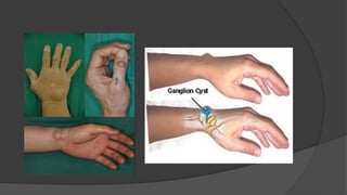
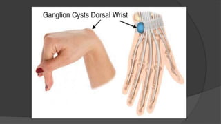
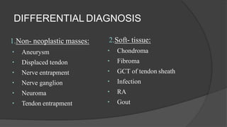
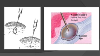
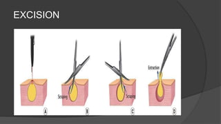
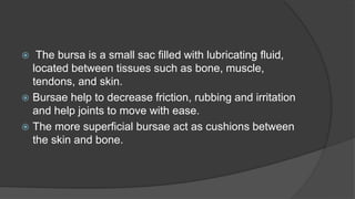
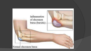
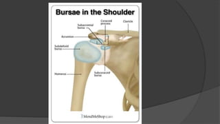
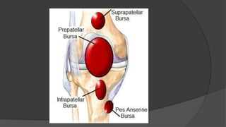
GANGLION
Dr. Anshu Sharma
Assistant Prof.
Dept. of Orthopaediscs, GMC&H.
INTRODUCTION
Most common swelling in the wrists.
Arises from leakage of synovial fluid from a joint or tendon sheath.
Contains a glairy, viscous fluid.
Can appear anywhere around the carpus.
Usually develops on the dorsal surface of the scapho-lunate
ligament.
Common sites are:
a.Dosrsally:
Dorsal carpus
Second and third CMC joint
Intratendon area
DIP (muscous cyst)
b.Volarlly:
Flexor sheath
Guyon’s canal
Volar carpus ganglion
ETIOLOGY
Unknown
Traumatic
Mucous cysts associated with OA of DIPjoint
PATHOLOGY
Multi- loculated
Outer wall: several layers of collagen layers, few fibroblasts
and
mesenchymal cells
No synovial lining: not a cyst in true sense
Content: clear, viscous, mucin- glucosamine, albumin, globulin,
hyaluronic acid
PATHOGENESIS
Unclear,
Communication of fluid from wrist joint: herniation,
Rent in capsule and tendon sheath→ escape of synovial
fluid→ irritation of surrounding tissues→ pseudo capsule
formation
Enlarge when fluid is pumped in and reduced when water
content is absorbed.
Mucoid degeneration of connective tissue and breakdown
products coalesce.
CLINICAL FEATURES
Often young adults.
More common in woman.
Older patients: mucous cyst.
Painless lump.
Occasionally slight ache and weakness.
Well defined swelling.
Cystic and not tender.
Trans- illuminated.
DORSAL CARPALGANGLION
Most common location
Originates from the scapho- lunate interval
May decompress into EPL or common extensor tendon sheath
Asymptomatic, decreasing and increasing in size
May be associated with aching pain, tenderness and interference
with activity atthe time of presentation
Occult ganglion: pressure on branches of posterior inter- osseous
nerve
Large ganglion: pressure on radial nerve branches
VOLAR CARPAL GANGLION
Second most common location.
Originates from the FCR tendon sheath or from the
articulation between the radius and the scaphoid (2/3),
the scaphoid and the trapezium (1/3).
Located between radial artery and FCR.
May present with sensory or motor nerve palsy involving
palmar cutaneous branch of median nerve, ulnar nerve, deep
ulnar motor branches.
 Aspiration is not recommended.
VOLAR RETINACULAR GANGLION
7- 12 %.
Arise from A1 or A2pulley.
Firm, minimally mobile mass near proximal
digital crease or MCP joint.
Excision if pressure symptoms.
MUCOUS
CYSTS
Middle age or later.
Females.
Dorsal DIP joint.
Observation, aspiration or excision.
Pain is the indication for excision.
DIFFERENTIAL DIAGNOSIS
1.Non- neoplastic masses:
Aneurysm
Displaced tendon
Nerve entrapment
Nerve ganglion
Neuroma
Tendon entrapment
2.Soft- tissue:
Chondroma
Fibroma
GCT of tendon sheath
Infection
RA
Gout
TREATMENT
Observation
Closed rupture
Cyst puncture
Aspiration
Excision
Spontaneous resolution in 50% cases.
EXCISION
REHABILITATION
 2 weeks:
 Remove the short arm splint and sutures
 AROM and AAROM of wrist flexion and extension
 Continue interval splint wear during the day between exercises
and at night.
 2-4 weeks:
 RROM and gradual strengthening exercises
 Discontinue the splint at 4 weeks
 4-6 weeks: normal activities to tolerance.
 6 weeks: allow full activity.
COMPLICATIONS
Due to ganglion is rare.
Due to surgery: infection, decreased motion, instability
and neurovascular injury.
Bursa & Bursitis
 The bursa is a small sac filled with lubricating fluid,
located between tissues such as bone, muscle,
tendons, and skin.
 Bursae help to decrease friction, rubbing and irritation
and help joints to move with ease.
 The more superficial bursae act as cushions between
the skin and bone.
 Bursitis is the inflammation or irritation of the bursa
which results in pain and discomfort.
 The hallmark symptom of bursitis is localized swelling
at the joint.
 A fever, localized skin warmth and redness, tenderness
and joint pain, may be signs of septic bursitis
 Bursitis can be treated in a number of ways,including:
-Avoiding activities that aggravate the problem
-Resting the injured area
-Ice pack application,
-NSAIDs.
-Surgical excision of bursa.
THANK YOU…..!!!!

Ganglion & Bursitis.

  • 1.
    GANGLION Dr. Anshu Sharma AssistantProf. Dept. of Orthopaediscs, GMC&H.
  • 2.
    INTRODUCTION Most common swellingin the wrists. Arises from leakage of synovial fluid from a joint or tendon sheath. Contains a glairy, viscous fluid. Can appear anywhere around the carpus. Usually develops on the dorsal surface of the scapho-lunate ligament.
  • 3.
    Common sites are: a.Dosrsally: Dorsalcarpus Second and third CMC joint Intratendon area DIP (muscous cyst) b.Volarlly: Flexor sheath Guyon’s canal Volar carpus ganglion
  • 5.
  • 6.
    PATHOLOGY Multi- loculated Outer wall:several layers of collagen layers, few fibroblasts and mesenchymal cells No synovial lining: not a cyst in true sense Content: clear, viscous, mucin- glucosamine, albumin, globulin, hyaluronic acid
  • 7.
    PATHOGENESIS Unclear, Communication of fluidfrom wrist joint: herniation, Rent in capsule and tendon sheath→ escape of synovial fluid→ irritation of surrounding tissues→ pseudo capsule formation Enlarge when fluid is pumped in and reduced when water content is absorbed. Mucoid degeneration of connective tissue and breakdown products coalesce.
  • 8.
    CLINICAL FEATURES Often youngadults. More common in woman. Older patients: mucous cyst. Painless lump. Occasionally slight ache and weakness. Well defined swelling. Cystic and not tender. Trans- illuminated.
  • 9.
    DORSAL CARPALGANGLION Most commonlocation Originates from the scapho- lunate interval May decompress into EPL or common extensor tendon sheath Asymptomatic, decreasing and increasing in size May be associated with aching pain, tenderness and interference with activity atthe time of presentation Occult ganglion: pressure on branches of posterior inter- osseous nerve Large ganglion: pressure on radial nerve branches
  • 11.
    VOLAR CARPAL GANGLION Secondmost common location. Originates from the FCR tendon sheath or from the articulation between the radius and the scaphoid (2/3), the scaphoid and the trapezium (1/3). Located between radial artery and FCR. May present with sensory or motor nerve palsy involving palmar cutaneous branch of median nerve, ulnar nerve, deep ulnar motor branches.  Aspiration is not recommended.
  • 13.
    VOLAR RETINACULAR GANGLION 7-12 %. Arise from A1 or A2pulley. Firm, minimally mobile mass near proximal digital crease or MCP joint. Excision if pressure symptoms.
  • 14.
    MUCOUS CYSTS Middle age orlater. Females. Dorsal DIP joint. Observation, aspiration or excision. Pain is the indication for excision.
  • 16.
    DIFFERENTIAL DIAGNOSIS 1.Non- neoplasticmasses: Aneurysm Displaced tendon Nerve entrapment Nerve ganglion Neuroma Tendon entrapment 2.Soft- tissue: Chondroma Fibroma GCT of tendon sheath Infection RA Gout
  • 17.
  • 19.
  • 20.
    REHABILITATION  2 weeks: Remove the short arm splint and sutures  AROM and AAROM of wrist flexion and extension  Continue interval splint wear during the day between exercises and at night.  2-4 weeks:  RROM and gradual strengthening exercises  Discontinue the splint at 4 weeks  4-6 weeks: normal activities to tolerance.  6 weeks: allow full activity.
  • 22.
    COMPLICATIONS Due to ganglionis rare. Due to surgery: infection, decreased motion, instability and neurovascular injury.
  • 23.
  • 24.
     The bursais a small sac filled with lubricating fluid, located between tissues such as bone, muscle, tendons, and skin.  Bursae help to decrease friction, rubbing and irritation and help joints to move with ease.  The more superficial bursae act as cushions between the skin and bone.
  • 25.
     Bursitis isthe inflammation or irritation of the bursa which results in pain and discomfort.  The hallmark symptom of bursitis is localized swelling at the joint.  A fever, localized skin warmth and redness, tenderness and joint pain, may be signs of septic bursitis
  • 29.
     Bursitis canbe treated in a number of ways,including: -Avoiding activities that aggravate the problem -Resting the injured area -Ice pack application, -NSAIDs. -Surgical excision of bursa.
  • 30.