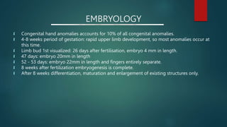
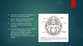
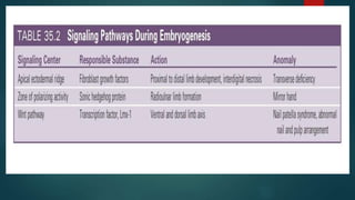
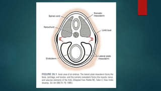
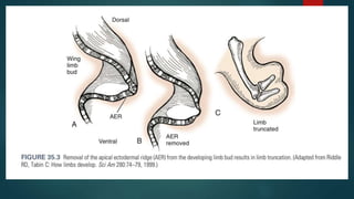
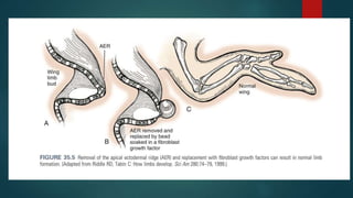
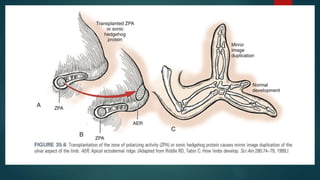
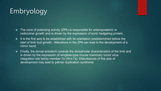
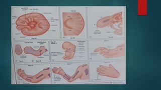
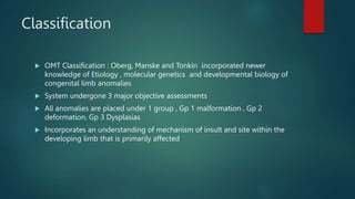
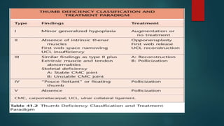
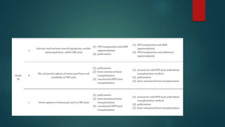
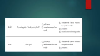
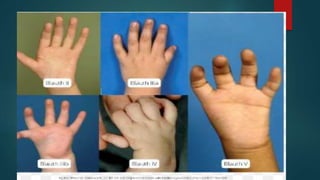
Seminar on Thumb
Hypoplasia
BY DR SATISH SINGH
EMBRYOLOGY
 Congenital hand anomalies accounts for 10% of all congenital anomalies.
 4-8 weeks period of gestation: rapid upper limb development, so most anomalies occur at
this time.
 Limb bud 1st visualized: 26 days after fertilisation, embryo 4 mm in length.
 47 days: embryo 20mm in length
 52 - 53 days: embryo 22mm in length and fingers entirely separate.
 8 weeks after fertilization embryogenesis is complete.
 After 8 weeks differentiation, maturation and enlargement of existing structures only.
 Limb bud: Outgrowth of mesoderm
into the overlying ectoderm.
 Lateral plate mesoderm: Becomes
bones, cartilage and tendon.
 Somatic mesoderm: Forms the
muscular, nervous and vascular
elements.
 Bone Morphogenetic Proteins (BMPs)
- role in chondrogenesis and
osteogenesis, trigger apoptotic
pathways of the interdigital
mesenchyme to produce separated
fingers.
EMBROYOLOGY
 Upper extremity development takes place between the fifth and eighth weeks of
intrauterine growth.
 It begins with the outgrowth of the arm bud at approximately 30 days gestation, and
by 37 days gestation the hand plate is well developed.
 Concentration gradients of a variety of growth factors influence growth in three
spatial axes: proximodistal, anteroposterior (radial-ulnar), and dorsoventral.
 Proximodistal limb growth is controlled by a thickened ridge of ectodermal tissue
known as the apical ectodermal ridge (AER).
 It is the last axis to develop and its function is dependent on the secretion of
several fibroblastic growth factors that control the AER differentiation and growth.
 The delineation of digital rays and finger separation occurs through apoptosis of
specific portions of the AER between 47 and 53 days.
EMBRYOLOGY
 Muscular blastema forms muscles, chondrogenic blastema forms bones.
 Muscles form sequentially from proximal to distal.
 Joints form in interzones between the ends of two blastemas.
 Intrauterine motion is necessary to form a mobile functioning joint.
 In the absence of movement, such as arthrogryposis, the joints space is infiltrated by fibrous
tissue leading to immobile joint.
GENETIC ABNORMALITIES
 The HOX and T-box genes encode transcription factors crucial for limb formation.
 Synpolydactyly and hand-foot-genital syndrome have mutations within the HOX genes.
 Holt-Oram syndrome: T-box genes affected.
Embryology
 The zone of polarizing activity (ZPA) is responsible for anteroposterior or
radio/ulnar growth and is driven by the expression of sonic hedgehog protein.
 It is the first axis to be established with its orientation predetermined before the
start of limb bud growth. Alterations in the ZPA can lead to the development of a
mirror hand.
 Finally, the dorsal ectoderm controls the dorsal/volar characteristics of the limb and
is driven by the expression of wingless-type mouse mammary tumor virus
integration site family member 7a (Wnt-7a). Disturbances of this axis of
development may lead to palmar duplication syndrome
Classification
 OMT Classification : Oberg, Manske and Tonkin incorporated newer
knowledge of Etiology , molecular genetics and developmental biology of
congenital limb anomalies
 System undergone 3 major objective assessments
 All anomalies are placed under 1 group , Gp 1 malformation , Gp 2
deformation, Gp 3 Dysplasias
 Incorporates an understanding of mechanism of insult and site within the
developing limb that is primarily affected
 Involvement of family doctor and pediatrician , therapist and and
psychologist in overall management of child and family is necessary
 Cardiac, hematological, neurological, gastroenterological and urological
systems must be investigated
Various congenital malformations
 Syndactyly
 Camptodactyly
 Clinodactyly
 Symbrachydactyly
 Polydactyly
 Radial Column Abnormalities
 Constriction Ring Syndrome
 Radial Longitudinal deficiencies
SYNDACTYLY
 Variable fusion of soft tissue or skeletal elements or both of adjacent digits, occurring when
the normal processes of digital separation and webspace formation fail to some degree.
 Normal webspace slopes 45 deg in dorsal to palmar direction from metacarpal heads to the
mid proximal phalanx.
 Second and fourth webs are wider than 3rd web.
 First webspace is broader diamond shaped, with glabrous skin on the palm and thinner
mobile skin dorsally.
 Radial longitudinal deficiency (RLD), or radial club hand, is a result of a congenital
failure of formation of the radial column. The severity of presentation can vary from
mild hypoplasia to complete absence of the radius, carpal bones, and radial digits.
 The radial musculotendinous units may be absent or hypoplastic and often form a
fibrous tether at the wrist, contributing to the angled position of the hand.
 It has an incidence of approximately 1 in 5400 live births. RLD occurs because of
an insult to the apical ectodermal ridge during the fourth to seventh weeks of
intrauterine development. The condition gained widespread attention in the 1960s
with the thalidomide induced epidemic of phocomelia. 45
 One-third of patients have an associated syndrome such as Holt-Oram, Fanconi anemia,
TAR (thrombocytopenia-absent radius), or VACTERL (vertebral abnormalities, anal atresia,
cardiac
defects, tracheoesophageal fistula, renal agenesis, limb anomalies).
Two-thirds of patients have an associated medical or musculoskeletal anomaly ranging from
congenital heart malformations,
 blood dyscrasias, and renal or gastrointestinal dysfunction. 45 Often the limb abnormality
may be the only
 outward manifestation of the underlying syndrome, necessitating the surgeon to perform an
appropriate
 work-up including a renal ultrasound, echocardiogram, and complete blood count. 46 The
Bayne and
 Klug classification divides the radial dysplasias into four types, based on the radiographic
appearance 47
Two-thirds of patients have an associated medical or musculoskeletal
anomaly ranging from congenital heart malformations, blood dyscrasias, and
renal or gastrointestinal dysfunction.
Often the limb abnormality may be the only outward manifestation of the
underlying syndrome, necessitating the surgeon to perform an appropriate
work-up including a renal ultrasound, echocardiogram, and complete blood
count.
The Bayne and Klug classification divides the radial dysplasias into four
types, based on the radiographic appearance
 Type I: short distal radius
 Type II: hypoplastic radius
 Type III: partial absence of the radius
 Type IV: total absence of the radius
 This classification system was further modified by James to include the presence of
thumb hypoplasia, carpal anomalies, and radioulnar synostosis.
.
 Treatment for radial longitudinal deficiency initially involves passive stretching and
serial splinting. In severe cases, some advocate distraction of the tight radial
tissues with an external fixator device for at least 6-8 weeks before surgical
reconstruction, a procedure we advocate.
 The goals of surgical treatment include the following :
straightening of the radial bowing of the forearm,
correction of radial and volar subluxation of the carpusl
improvement of the limb length to optimize function,
improvement of aesthetic appearance, and
treatment of the thumb hypoplasia.
 All cases of radial longitudinal deficiency have an associated thumb hypoplasia, though
thumb hypoplasia may exist without an abnormality of the radius.
 Blauth developed a classification for thumb hypoplasia based on the size of the thumb,
the thenar musculature, first webspace contracture, and stability of the carpometacarpal
joint
 Type I: minor thumb hypoplasia,
 Type II: first webspace narrowing, hypoplastic or absent thenar intrinsic muscles, MCP
joint instability,
 Type III: first webspace narrowing, hypoplastic or absent thenar intrinsic muscles, MCP
joint instability, extrinsic tendon abnormalities, skeletal abnormalities:
 Type IIIA: hypoplastic metacarpal, stable CMC joint and
 Type IIIB: partial metacarpal aplasia, unstable CMC joint,
 Type IV: floating thumb (pouce flottant), and
 Type V: absent thumb
 Cleft Hand (Central Longitudinal Deficiency)
 Cleft hand or central longitudinal deficiency is characterized by varying degrees of growth
suppression of the bones and soft tissue of the central hand, most frequently involving the central
rays.
 The defect does not extend proximal to the wrist but may have associated carpal coalition or
radioulnar synostosis.
 It is frequently associated with central polydactyly or osseous syndactyly. Although the cleft itself
causes little functional limitation, the main deficit is secondary to a narrowed thumb-index
webspace.
 Surgical repair involves: (1)
 preservation of a stable and mobile thumb; (2) transposition of the index ray to the ulnar side of the
cleft;
 (3) creation of a wide and functional first webspace; (4) correction of any index finger malrotation or
 deviation; (5) preservation of an adductor pollicis muscle; and (6) creation of a satisfactory aesthetic
 appearance.
 Surgical repair involves:
(1)preservation of a stable and mobile thumb
(2) transposition of the index ray to the ulnar side of the cleft;
(3) creation of a wide and functional first webspace
(4) correction of any index finger malrotation or deviation
(5) preservation of an adductor pollicis muscle and
(6) creation of a satisfactory aesthetic appearance.
Thumb Hypoplasia
 Part of Radial deficiency even with normal forearm architecture
 Associated anomalies need to be evaluated on initial visit
 Work up must include Vacterl association, thrombocytopenia- absent
radius syndrome, Holt Oram Syndrome , Charge syndrome and Fanconi s
syndrome
 Work up include Renal USG , ECHO, Spine imaging and chromosal
challenge test
Thumb Hypolasia
 Blauth classification
 Type I : Minor generalised hypoplasia usually affecting thenar muscles
 May present later in life as hand function becomes intricate
 May warrant treatment
 If the opposition is causing functional difficulties a tendon transfer is
recommended
 Type II hypoplasia : absence of thenar muscle innervated by recurrent
branch of the median nerve(APB, OP and superficial head of flexor policis
brevis ), Deep head innervated by ulnar provides MCP Joint Flexion. MCP is
unstable wit laxity or incompetency of UCL
 Type III hypoplasia: similar characterstics to Type II + extrinsic muscle and
tendon deficiencies
 Absence or Hypoplasia of extrinsic extensors or flexor policis longus
requires observant examination at MCP and IP joint
 Type IIIA : stable CMC , worthy of Reconstruction and can provide a
worthwhile prehensile thumb
 Type IIIB Hypoplasia : Unstable CMC Joint
 Type IV Hypoplasia : Represents a profound deficiency known as pouce
flotant , Thumb attached to hand solely by a narrow bridge, Thumb is
floppy and entirely ignored
 Type V hypoplasia: complete absence of thumb, Small skin Nubbin can be
present
 Thumb hypoplasia can also be found in children with ulnar deficiency
 Treatment for hypoplasia in ulnar of radial deficiency remains the same
Treatment
 An experienced hand therapist is valuable for opinion
 Type I doesn’t require surgical intervention because functional impairment
is negligible
 Thumb Reconstruction for Type II and II A requires addressing narrowed
thumb /Index finger web space, MCP instability and thenar muscle absence
require treatment
Policization

Thumb hypoplasia(4).pptx

  • 1.
  • 2.
    EMBRYOLOGY  Congenital handanomalies accounts for 10% of all congenital anomalies.  4-8 weeks period of gestation: rapid upper limb development, so most anomalies occur at this time.  Limb bud 1st visualized: 26 days after fertilisation, embryo 4 mm in length.  47 days: embryo 20mm in length  52 - 53 days: embryo 22mm in length and fingers entirely separate.  8 weeks after fertilization embryogenesis is complete.  After 8 weeks differentiation, maturation and enlargement of existing structures only.
  • 3.
     Limb bud:Outgrowth of mesoderm into the overlying ectoderm.  Lateral plate mesoderm: Becomes bones, cartilage and tendon.  Somatic mesoderm: Forms the muscular, nervous and vascular elements.  Bone Morphogenetic Proteins (BMPs) - role in chondrogenesis and osteogenesis, trigger apoptotic pathways of the interdigital mesenchyme to produce separated fingers.
  • 5.
    EMBROYOLOGY  Upper extremitydevelopment takes place between the fifth and eighth weeks of intrauterine growth.  It begins with the outgrowth of the arm bud at approximately 30 days gestation, and by 37 days gestation the hand plate is well developed.  Concentration gradients of a variety of growth factors influence growth in three spatial axes: proximodistal, anteroposterior (radial-ulnar), and dorsoventral.  Proximodistal limb growth is controlled by a thickened ridge of ectodermal tissue known as the apical ectodermal ridge (AER).  It is the last axis to develop and its function is dependent on the secretion of several fibroblastic growth factors that control the AER differentiation and growth.  The delineation of digital rays and finger separation occurs through apoptosis of specific portions of the AER between 47 and 53 days.
  • 11.
    EMBRYOLOGY  Muscular blastemaforms muscles, chondrogenic blastema forms bones.  Muscles form sequentially from proximal to distal.  Joints form in interzones between the ends of two blastemas.  Intrauterine motion is necessary to form a mobile functioning joint.  In the absence of movement, such as arthrogryposis, the joints space is infiltrated by fibrous tissue leading to immobile joint.
  • 12.
    GENETIC ABNORMALITIES  TheHOX and T-box genes encode transcription factors crucial for limb formation.  Synpolydactyly and hand-foot-genital syndrome have mutations within the HOX genes.  Holt-Oram syndrome: T-box genes affected.
  • 13.
    Embryology  The zoneof polarizing activity (ZPA) is responsible for anteroposterior or radio/ulnar growth and is driven by the expression of sonic hedgehog protein.  It is the first axis to be established with its orientation predetermined before the start of limb bud growth. Alterations in the ZPA can lead to the development of a mirror hand.  Finally, the dorsal ectoderm controls the dorsal/volar characteristics of the limb and is driven by the expression of wingless-type mouse mammary tumor virus integration site family member 7a (Wnt-7a). Disturbances of this axis of development may lead to palmar duplication syndrome
  • 15.
    Classification  OMT Classification: Oberg, Manske and Tonkin incorporated newer knowledge of Etiology , molecular genetics and developmental biology of congenital limb anomalies  System undergone 3 major objective assessments  All anomalies are placed under 1 group , Gp 1 malformation , Gp 2 deformation, Gp 3 Dysplasias  Incorporates an understanding of mechanism of insult and site within the developing limb that is primarily affected
  • 17.
     Involvement offamily doctor and pediatrician , therapist and and psychologist in overall management of child and family is necessary  Cardiac, hematological, neurological, gastroenterological and urological systems must be investigated
  • 19.
    Various congenital malformations Syndactyly  Camptodactyly  Clinodactyly  Symbrachydactyly  Polydactyly  Radial Column Abnormalities  Constriction Ring Syndrome  Radial Longitudinal deficiencies
  • 20.
    SYNDACTYLY  Variable fusionof soft tissue or skeletal elements or both of adjacent digits, occurring when the normal processes of digital separation and webspace formation fail to some degree.  Normal webspace slopes 45 deg in dorsal to palmar direction from metacarpal heads to the mid proximal phalanx.  Second and fourth webs are wider than 3rd web.  First webspace is broader diamond shaped, with glabrous skin on the palm and thinner mobile skin dorsally.
  • 21.
     Radial longitudinaldeficiency (RLD), or radial club hand, is a result of a congenital failure of formation of the radial column. The severity of presentation can vary from mild hypoplasia to complete absence of the radius, carpal bones, and radial digits.  The radial musculotendinous units may be absent or hypoplastic and often form a fibrous tether at the wrist, contributing to the angled position of the hand.  It has an incidence of approximately 1 in 5400 live births. RLD occurs because of an insult to the apical ectodermal ridge during the fourth to seventh weeks of intrauterine development. The condition gained widespread attention in the 1960s with the thalidomide induced epidemic of phocomelia. 45
  • 22.
     One-third ofpatients have an associated syndrome such as Holt-Oram, Fanconi anemia, TAR (thrombocytopenia-absent radius), or VACTERL (vertebral abnormalities, anal atresia, cardiac defects, tracheoesophageal fistula, renal agenesis, limb anomalies). Two-thirds of patients have an associated medical or musculoskeletal anomaly ranging from congenital heart malformations,  blood dyscrasias, and renal or gastrointestinal dysfunction. 45 Often the limb abnormality may be the only  outward manifestation of the underlying syndrome, necessitating the surgeon to perform an appropriate  work-up including a renal ultrasound, echocardiogram, and complete blood count. 46 The Bayne and  Klug classification divides the radial dysplasias into four types, based on the radiographic appearance 47
  • 23.
    Two-thirds of patientshave an associated medical or musculoskeletal anomaly ranging from congenital heart malformations, blood dyscrasias, and renal or gastrointestinal dysfunction. Often the limb abnormality may be the only outward manifestation of the underlying syndrome, necessitating the surgeon to perform an appropriate work-up including a renal ultrasound, echocardiogram, and complete blood count. The Bayne and Klug classification divides the radial dysplasias into four types, based on the radiographic appearance
  • 24.
     Type I:short distal radius  Type II: hypoplastic radius  Type III: partial absence of the radius  Type IV: total absence of the radius  This classification system was further modified by James to include the presence of thumb hypoplasia, carpal anomalies, and radioulnar synostosis.
  • 25.
    .  Treatment forradial longitudinal deficiency initially involves passive stretching and serial splinting. In severe cases, some advocate distraction of the tight radial tissues with an external fixator device for at least 6-8 weeks before surgical reconstruction, a procedure we advocate.  The goals of surgical treatment include the following : straightening of the radial bowing of the forearm, correction of radial and volar subluxation of the carpusl improvement of the limb length to optimize function, improvement of aesthetic appearance, and treatment of the thumb hypoplasia.
  • 26.
     All casesof radial longitudinal deficiency have an associated thumb hypoplasia, though thumb hypoplasia may exist without an abnormality of the radius.  Blauth developed a classification for thumb hypoplasia based on the size of the thumb, the thenar musculature, first webspace contracture, and stability of the carpometacarpal joint  Type I: minor thumb hypoplasia,  Type II: first webspace narrowing, hypoplastic or absent thenar intrinsic muscles, MCP joint instability,  Type III: first webspace narrowing, hypoplastic or absent thenar intrinsic muscles, MCP joint instability, extrinsic tendon abnormalities, skeletal abnormalities:  Type IIIA: hypoplastic metacarpal, stable CMC joint and  Type IIIB: partial metacarpal aplasia, unstable CMC joint,  Type IV: floating thumb (pouce flottant), and  Type V: absent thumb
  • 27.
     Cleft Hand(Central Longitudinal Deficiency)  Cleft hand or central longitudinal deficiency is characterized by varying degrees of growth suppression of the bones and soft tissue of the central hand, most frequently involving the central rays.  The defect does not extend proximal to the wrist but may have associated carpal coalition or radioulnar synostosis.  It is frequently associated with central polydactyly or osseous syndactyly. Although the cleft itself causes little functional limitation, the main deficit is secondary to a narrowed thumb-index webspace.  Surgical repair involves: (1)  preservation of a stable and mobile thumb; (2) transposition of the index ray to the ulnar side of the cleft;  (3) creation of a wide and functional first webspace; (4) correction of any index finger malrotation or  deviation; (5) preservation of an adductor pollicis muscle; and (6) creation of a satisfactory aesthetic  appearance.
  • 28.
     Surgical repairinvolves: (1)preservation of a stable and mobile thumb (2) transposition of the index ray to the ulnar side of the cleft; (3) creation of a wide and functional first webspace (4) correction of any index finger malrotation or deviation (5) preservation of an adductor pollicis muscle and (6) creation of a satisfactory aesthetic appearance.
  • 29.
    Thumb Hypoplasia  Partof Radial deficiency even with normal forearm architecture  Associated anomalies need to be evaluated on initial visit  Work up must include Vacterl association, thrombocytopenia- absent radius syndrome, Holt Oram Syndrome , Charge syndrome and Fanconi s syndrome  Work up include Renal USG , ECHO, Spine imaging and chromosal challenge test
  • 30.
    Thumb Hypolasia  Blauthclassification  Type I : Minor generalised hypoplasia usually affecting thenar muscles  May present later in life as hand function becomes intricate  May warrant treatment  If the opposition is causing functional difficulties a tendon transfer is recommended
  • 31.
     Type IIhypoplasia : absence of thenar muscle innervated by recurrent branch of the median nerve(APB, OP and superficial head of flexor policis brevis ), Deep head innervated by ulnar provides MCP Joint Flexion. MCP is unstable wit laxity or incompetency of UCL  Type III hypoplasia: similar characterstics to Type II + extrinsic muscle and tendon deficiencies  Absence or Hypoplasia of extrinsic extensors or flexor policis longus requires observant examination at MCP and IP joint  Type IIIA : stable CMC , worthy of Reconstruction and can provide a worthwhile prehensile thumb
  • 32.
     Type IIIBHypoplasia : Unstable CMC Joint  Type IV Hypoplasia : Represents a profound deficiency known as pouce flotant , Thumb attached to hand solely by a narrow bridge, Thumb is floppy and entirely ignored  Type V hypoplasia: complete absence of thumb, Small skin Nubbin can be present
  • 33.
     Thumb hypoplasiacan also be found in children with ulnar deficiency  Treatment for hypoplasia in ulnar of radial deficiency remains the same
  • 34.
    Treatment  An experiencedhand therapist is valuable for opinion  Type I doesn’t require surgical intervention because functional impairment is negligible  Thumb Reconstruction for Type II and II A requires addressing narrowed thumb /Index finger web space, MCP instability and thenar muscle absence require treatment
  • 42.