
Downloaded 945 times


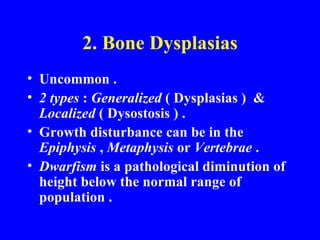


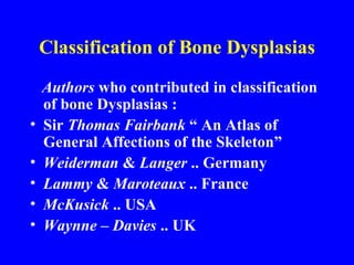


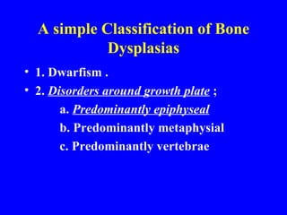


This document provides an overview of congenital bone and joint diseases. It begins with an introduction and outline. The first section discusses genetic aspects, including basic genetic principles like chromosomes, genes, alleles, and patterns of inheritance. It describes numerical and structural chromosomal abnormalities. The second section covers bone dysplasias, including classification systems and a simplified classification covering disorders like dwarfism and abnormalities around the growth plate.


