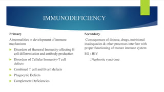
This document summarizes primary defects of antibody production and immunodeficiency. It discusses various types of primary immunodeficiencies including disorders of humoral immunity affecting B cell differentiation and antibody production. Specific disorders covered include X-linked agammaglobulinemia, common variable immunodeficiency, selective IgA deficiency, transient hypogammaglobulinemia of infancy, immunodeficiency with hyper-IgM, and X-linked lymphoproliferative disease. Treatment options for humoral defects such as immunoglobulin administration are also summarized.
![Subcutaneous Immunoglobulin Replacement Therapy in Patients with Primary
Immunodeficiency in Routine Clinical Practice: The VISPO Prospective Multicenter Study
Alessandra Vultaggio, Chiara Azzari, Cinzia Milito, Andrea Finocchi, Claudia Toppino, Giuseppe Spadaro, Antonino Trizzino, Martire Baldassarr
Aim
To evaluate the shifting from intravenous immunoglobulins (IVIGs) replacement therapy to SCIG in patients with
primary immunodeficiency (PID) in a routine real-life situation.
Methods
In a multicenter prospective observational study, we enrolled 50 patients suffering from PID who were monitored for
24 months; 44 patients switched from IVIG and six from different SCIG preparations. The study preparation (human
IgG 16 %, Vivaglobin®, CSL Behring GmbH, Germany) was subcutaneously infused weekly (maximum volume
15 mL/site; maximum infusion rate 22 mL/h). The study endpoints were: annual rate of severe bacterial infections
(SBIs), local adverse reactions, quality of life, days off school/work, and days of hospitalization
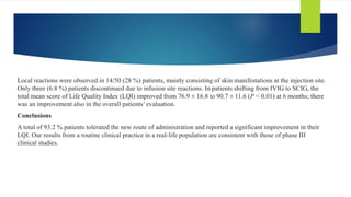
Results
Thirty-three of 39 (84.6 %) patients who completed the study experienced an infection or signs thereof. Only five
SBIs were observed, corresponding to an annual rate of 0.056 episodes per patient in 44 subjects [intention-to-treat
(ITT) population]. A significant decrease in both days of hospitalization (1.93 ± 4.08 vs. 0.64 ± 2.94) and days off
school/work (15.27 ± 23.17 vs. 2.26 ± 4.45) was recorded at 24 months.](https://image.slidesharecdn.com/primarydefectinantibodyproduction-231221122708-2e035f7c/85/primary-defect-in-antibody-production-pptx-26-320.jpg)