Download to read offline

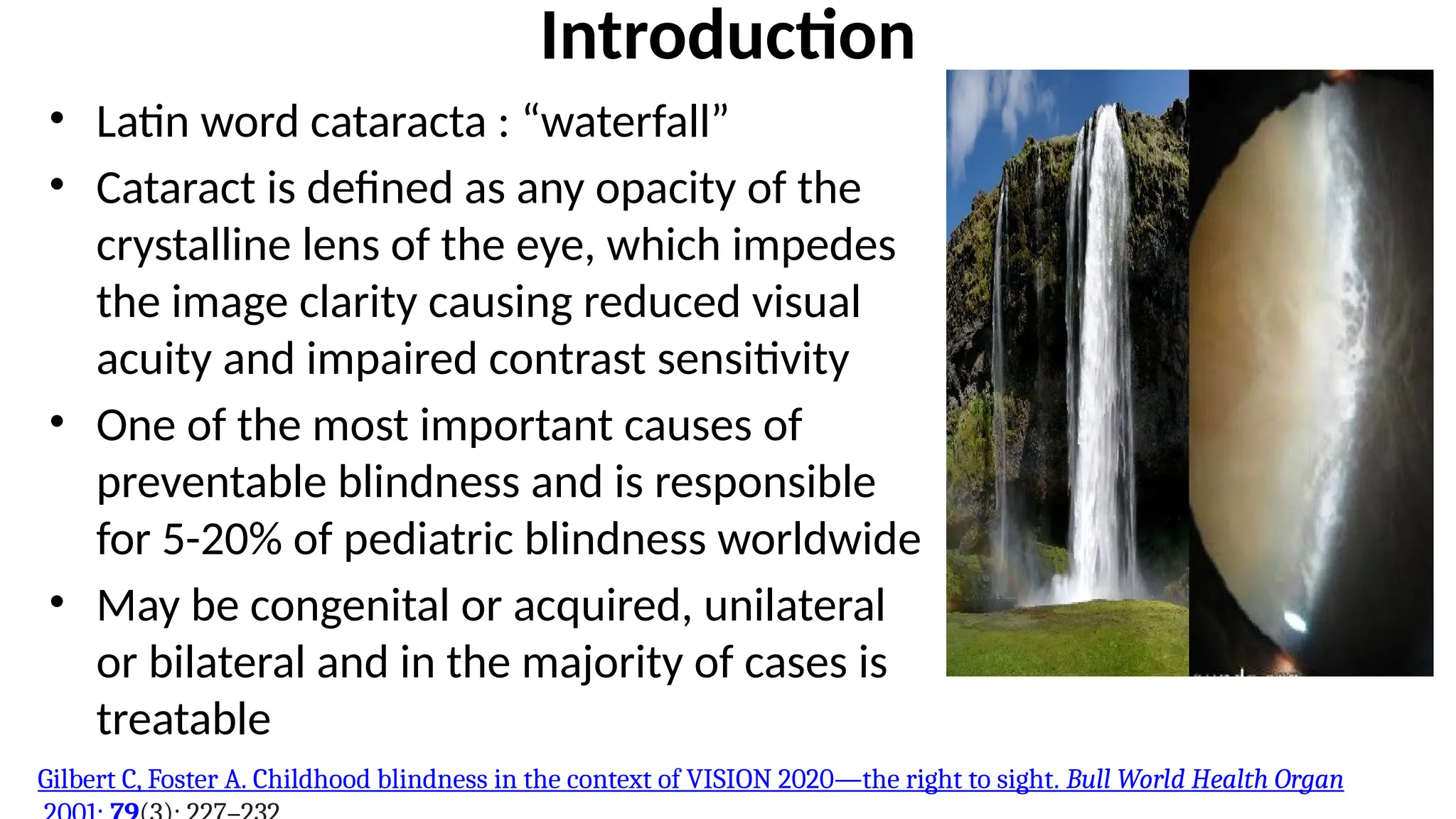


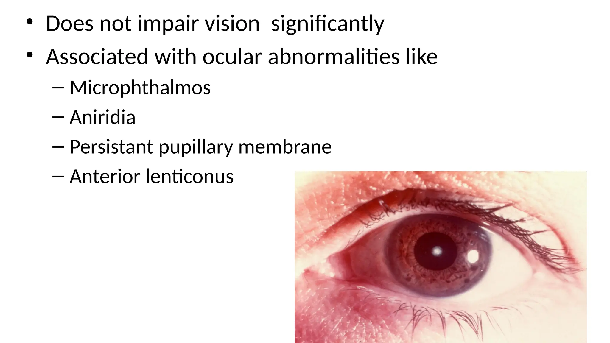


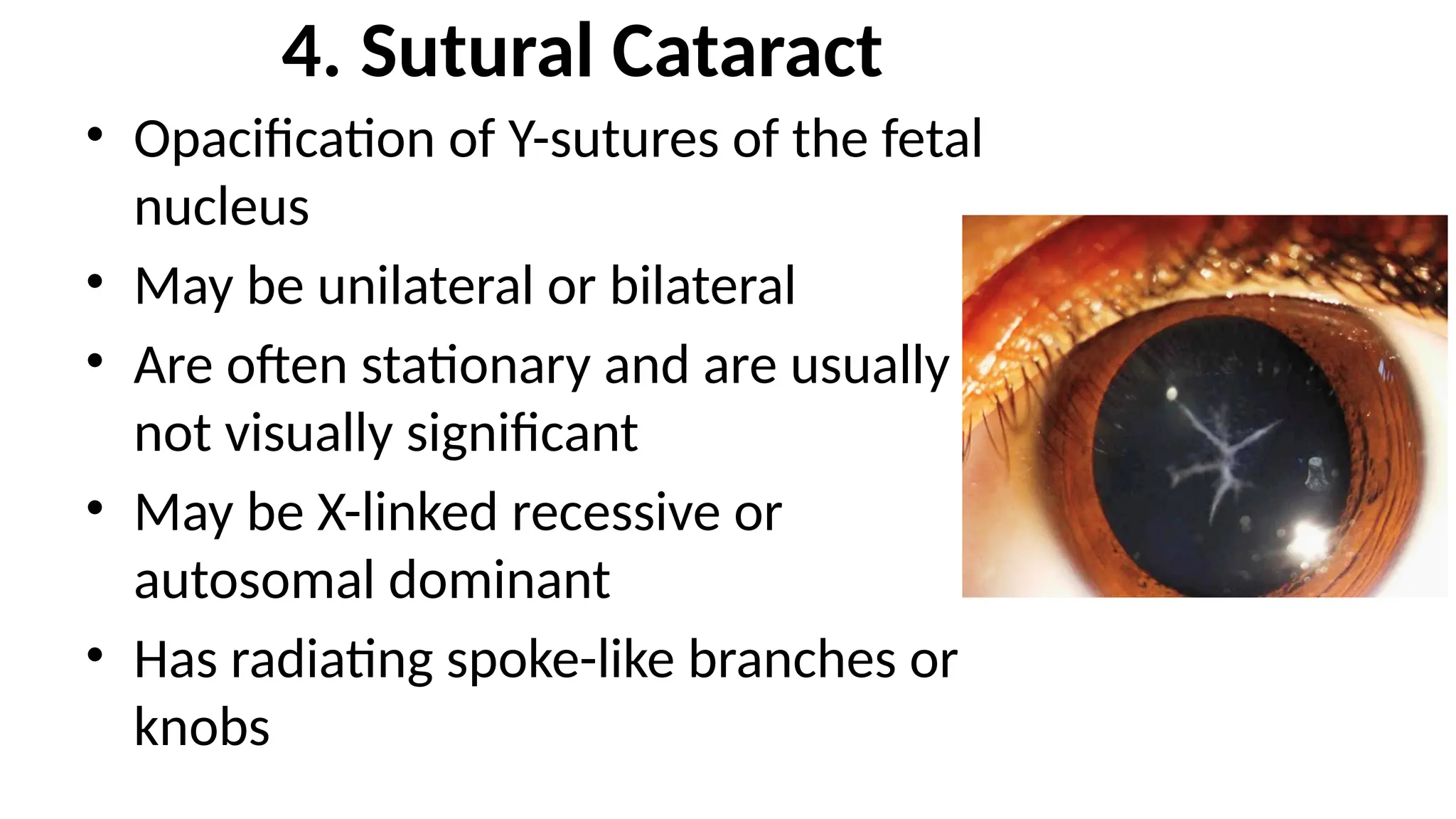


Congenital cataracts are a significant cause of preventable blindness in children, with various forms and etiologies, including genetic and acquired conditions. They can be classified based on age of onset, laterality, and morphology, leading to diverse clinical presentations. Early detection and intervention are crucial to prevent amblyopia and permanent vision loss.


