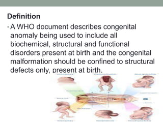
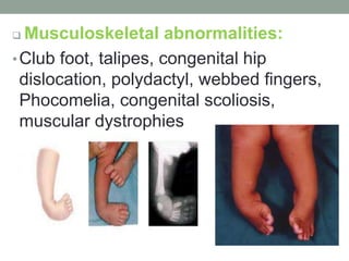
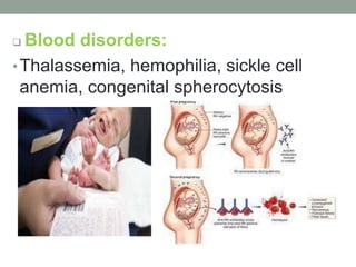
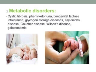
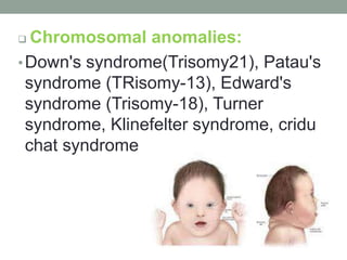
Congenital Anomalies
• Congenital anomalies are those defects
and diseases which are substantially
determined before or during birth and
recognizable in early life.
• Some disorders are detected at birth, eg;
cleft lip and cleft palate.
• Some are obvious in early life like congenital hip
dislocation which is detected until walking
commence and some may become apparent until
much later in life, e.g; patent ductus arteriosus, which
is usually diagnosed at school age or even later.
• Some defects are classified as major which may
require surgical interventions, either as emergency
or elective.
• Some are classified as minor and have no functional
implications, like skin tags in front of the ear.
Definition
• A WHO document describes congenital
anomaly being used to include all
biochemical, structural and functional
disorders present at birth and the congenital
malformation should be confined to structural
defects only, present at birth.
Incidence
• In India, the incidence of congenital defects is
about 2.5 to 4% among children most common
type of birth defects is CNS abnormalities,
approximately 22 % of all defects.
• In Northern part of India, neural tube defects are
most common, where as in the rest of India
musculoskeletal disorders are commonly found.
Risk factors
• Advanced maternal age
• Consanguinity
• Maternal malnutrition especially folic acid
deficiency can lead to CNS defects
Etiology
• Genetic factors: The anomalies may be
related to chromosomal abnormalities, single
gene disorders or polygenic inheritance.
 Single gene disorders may be either
autosomal or X-linked inheritance which may
be dominant or recessive traits.
• Chromosomal abnormalities: The most
common is Down's syndrome or Trisomy 21
• Single gene disorders: They are found as
autosomal dominant inheritance, autosomal
recessive inheritance and X-linked
inheritance.
• Autosomal dominant traits: Huntington's
chorea, polydactyly, polycystic kidney,
retinoblastoma etc;
• Autosomal recessive traits: Cystic fibrosis,
microcephaly, sickle cell anemia, thalassemia,
phenylketonuria, Hirschsprung disease etc
• Environmental factors:
 Intra uterine infections especially by
STORCH (syphilis, Toxoplasmosis, Rubella,
Cytomegalovirus and Herpes virus)
Drug intake by the mother during pregnancy
like steroid hormones, anticonvulsants,
cocaine, lithium, thalidomide etc;
 X-ray exposure during pregnancy
 Maternal diseases like DM, cardiac failure,
malnutrition, folic acid deficiency, iodine
deficiency disorders, endocrine
abnormalities.
 Abnormal intra uterine environment like bicornuate
uterus, polyhydramnios, oligohydramnios, fetal
hypoxia etc;
 Maternal addiction with alcohol, tobacco or
smoking
 Environmental pollution, especially air pollution
Diagnostic approaches
• Prenatal diagnosis
 Amniocentesis in early pregnancy
 Chorionic villus sampling
 Estimation of maternal serum alpha fetoprotein
 Ultrasonography
 Fetoscopy
 Amniography
 Fetal blood sampling
 Radiography
 Antenatal screening of maternal disease
• Postnatal diagnosis
 Maternal and family history
 Thorough physical examination.
 Biochemical assay
 Cytogenic study
 Blood test
 Hormonal assay
 Radiography
 Ultrasonography
Common congenital anomalies
 CNS defects
 Congenital include- Anencephaly, spina bifida
occulta, spina bifida cystic (meningocele,
meningomyelocele, meningoencephalocele,
encephalocele), hydrocephalus, microcephaly.
 Congenital heart diseases
• ASD, VSD, PDA, Fallot's tetralogy, pulmonic
stenosis, aortic regurgitation, tricuspid
atresia, transposition of great vessels
 Gl system Abnormalities
• Tracheo-esophageal fistula, esophageal atresia, congenital
pyloric stenosis, duodenal atresia, meconium ileus, congenital
megacolon(Hirschsprung's disease), ano-rectal
malformations, umbilical hernia, diaphragmatic hernia,
congenital intestinal obstruction.
 Respiratory system
•Trachea esophageal fistula, congenital
atelectasis, congenital cyanosis,
congenital strider
 Genitourinary system
• Congenital hydro nephrosis, congenital
polycystic kidney, horse-shoe kidney,
posterior urethral valves, hypospadias,
epispadias, congenital phimosis, congenital
hydrocele, undescended testis, congenital
inguinal hernia, ambiguous genitalia,
malformations of reproductive organs.
 Musculoskeletal abnormalities:
•Club foot, talipes, congenital hip
dislocation, polydactyl, webbed fingers,
Phocomelia, congenital scoliosis,
muscular dystrophies
 Blood disorders:
•Thalassemia, hemophilia, sickle cell
anemia, congenital spherocytosis
 Metabolic disorders:
• Cystic fibrosis, phenylketonuria, congenital lactose
intolerance, glycogen storage diseases, Tay-Sachs
disease, Gaucher disease, Wilson's disease,
galactosemia
 Endocrinal abnormalities:
•Congenital hypopituitarism (dwarfism),
congenital hypothyroidism (cretinism),
congenital adreno-genital hyperplasia,
congenital goiter, DM.
 Chromosomal anomalies:
•Down's syndrome(Trisomy21), Patau's
syndrome (TRisomy-13), Edward's
syndrome (Trisomy-18), Turner
syndrome, Klinefelter syndrome, cridu
chat syndrome
 Tracheoesophageal Fistula And Esophageal
Atresia.
 Tracheoesophageal fistula is a developmental anomaly
characterized by an abnormal connection between the
trachea and the esophagus and usually accompanies
esophageal atresia.
 Esophageal atresia is failure of the esophagus to form a
continuous passage from the pharynx to the stomach.
Signs and symptoms vary according to
location of fistula and atresia.
1) The infant appears to swallow normally but soon after
coughs, struggles, become cyanotic, and stops breathing.
2) Stomach distention may cause respiratory distress.
3) Air and gastric contents may reflux through the fistula into
the trachea resulting in chemical pneumonitis.
4) If there is esophageal atresia without a fistula, as
secretions fill the esophageal sac and overflow into the
oropharynx, the infant develops mucus in the oropharynx
and drools excessively.
5) Repeated episodes of pneumonitis, pulmonary infection
and abdominal distention may be present.
Diagnosis
1) Catheter passed through the nose meets an obstruction.
2) Chest x-ray.
3) Abdominal x-ray.
4) Cinefluorography.
Treatment
1) Tracheoesophageal fistula and esophageal atresia requires
surgical correction and are usually considered a surgical
emergency.
2) The type of surgical procedure and when it is performed
depends on the nature of the anomaly, the patient's general
condition, and the presence of coexisting congenital
defects.
Postoperative complications
1) Tracheoesophageal fistula: a) Recurrent
fistulas. b) Esophageal mobility dysfunction.
c) Esophageal stricture . d) Recurrent
bronchitis. e) Pneumothorax. f) Failure to
thrive.
2) Esophageal atresia: a) Impaired
esophageal motility. b) Hiatal hernia. c)
Reflux esophagitis.
Nursing interventions
1) Monitor respiratory status.
2) Perform pulmonary physiotherapy.
3) Suction as necessary.
4) Administer antibiotics and parenteral
fluids as ordered.
5) Observe for signs of complications
(that is pneumothorax).
6) Maintain gastrostomy tube feedings.
7) Accurate I & O.
8) Give the baby a pacifier to satisfy his sucking
needs but only when he can safely handle
secretions.
9) Offer the parents support and guidance and
encourage bonding.
10)Positioning before and after surgery varies with the
doctor's philosophy and the child's anatomy.
a) Supine with his head low to facilitate
b) Head elevated to prevent aspiration.
Prevention of congenital anomalies:
• The preventive measures should include
the following aspects:
 Genetic counseling
 Reducing and discouraging
the consanguineous marriages
 Avoiding late marriages in females and
avoidance of pregnancy beyond the age of
35 years.
 Promotion of health of girl child and pre-pregnant
health status of the females by prevention of
malnutrition, anemia, folic acid deficiency, iodine
deficiency etc;
 Encouraging the immunization of all girl child by
MMR
 Elimination of active and passive smoking of
tobacco by mothers.
 Avoidance of drug intake without consulting
physician
 Prevention of intra uterine infections and
promotion of sexual hygiene
 Efficient antenatal care especially removal of
teratogens, prevention of malnutrition by adequate
diet.
 Promotion of therapeutic abortion of abnormal
fetus, Discouraging reproduction after birth of a
baby with congenital anomalies, without genetic
counseling.
 Increasing public awareness about the risk factors
and etiological factors of congenital anomalies
 Promotion of detection of genetic carriers
 Reducing the frequency of hereditary disease
Nursing responsibilities:
• Collection of detailed history of prenatal and post natal period
along with history of family illness.
• Provide necessary information to the parents and family
members
• Motivate the family members for genetic counseling and
referring to the genetic clinic
• Provide emotional support and answer questions asked by
the counselee.
• Guide the family for rehabilitation of the child and for
available social and economical support through social
welfare agencies.
• Promote public awareness about the prevention of
congenital anomalies by individual or group health education
or by mass media informations.
congenital anomalies.pptx

congenital anomalies.pptx

  • 1.
  • 2.
    • Congenital anomaliesare those defects and diseases which are substantially determined before or during birth and recognizable in early life. • Some disorders are detected at birth, eg; cleft lip and cleft palate.
  • 3.
    • Some areobvious in early life like congenital hip dislocation which is detected until walking commence and some may become apparent until much later in life, e.g; patent ductus arteriosus, which is usually diagnosed at school age or even later. • Some defects are classified as major which may require surgical interventions, either as emergency or elective. • Some are classified as minor and have no functional implications, like skin tags in front of the ear.
  • 4.
    Definition • A WHOdocument describes congenital anomaly being used to include all biochemical, structural and functional disorders present at birth and the congenital malformation should be confined to structural defects only, present at birth.
  • 5.
    Incidence • In India,the incidence of congenital defects is about 2.5 to 4% among children most common type of birth defects is CNS abnormalities, approximately 22 % of all defects. • In Northern part of India, neural tube defects are most common, where as in the rest of India musculoskeletal disorders are commonly found.
  • 6.
    Risk factors • Advancedmaternal age • Consanguinity • Maternal malnutrition especially folic acid deficiency can lead to CNS defects
  • 7.
    Etiology • Genetic factors:The anomalies may be related to chromosomal abnormalities, single gene disorders or polygenic inheritance.  Single gene disorders may be either autosomal or X-linked inheritance which may be dominant or recessive traits. • Chromosomal abnormalities: The most common is Down's syndrome or Trisomy 21
  • 8.
    • Single genedisorders: They are found as autosomal dominant inheritance, autosomal recessive inheritance and X-linked inheritance. • Autosomal dominant traits: Huntington's chorea, polydactyly, polycystic kidney, retinoblastoma etc; • Autosomal recessive traits: Cystic fibrosis, microcephaly, sickle cell anemia, thalassemia, phenylketonuria, Hirschsprung disease etc
  • 9.
    • Environmental factors: Intra uterine infections especially by STORCH (syphilis, Toxoplasmosis, Rubella, Cytomegalovirus and Herpes virus) Drug intake by the mother during pregnancy like steroid hormones, anticonvulsants, cocaine, lithium, thalidomide etc;
  • 10.
     X-ray exposureduring pregnancy  Maternal diseases like DM, cardiac failure, malnutrition, folic acid deficiency, iodine deficiency disorders, endocrine abnormalities.
  • 11.
     Abnormal intrauterine environment like bicornuate uterus, polyhydramnios, oligohydramnios, fetal hypoxia etc;  Maternal addiction with alcohol, tobacco or smoking  Environmental pollution, especially air pollution
  • 12.
    Diagnostic approaches • Prenataldiagnosis  Amniocentesis in early pregnancy  Chorionic villus sampling  Estimation of maternal serum alpha fetoprotein  Ultrasonography  Fetoscopy  Amniography  Fetal blood sampling  Radiography  Antenatal screening of maternal disease
  • 13.
    • Postnatal diagnosis Maternal and family history  Thorough physical examination.  Biochemical assay  Cytogenic study  Blood test  Hormonal assay  Radiography  Ultrasonography
  • 14.
    Common congenital anomalies CNS defects  Congenital include- Anencephaly, spina bifida occulta, spina bifida cystic (meningocele, meningomyelocele, meningoencephalocele, encephalocele), hydrocephalus, microcephaly.
  • 15.
     Congenital heartdiseases • ASD, VSD, PDA, Fallot's tetralogy, pulmonic stenosis, aortic regurgitation, tricuspid atresia, transposition of great vessels
  • 16.
     Gl systemAbnormalities • Tracheo-esophageal fistula, esophageal atresia, congenital pyloric stenosis, duodenal atresia, meconium ileus, congenital megacolon(Hirschsprung's disease), ano-rectal malformations, umbilical hernia, diaphragmatic hernia, congenital intestinal obstruction.
  • 17.
     Respiratory system •Tracheaesophageal fistula, congenital atelectasis, congenital cyanosis, congenital strider
  • 18.
     Genitourinary system •Congenital hydro nephrosis, congenital polycystic kidney, horse-shoe kidney, posterior urethral valves, hypospadias, epispadias, congenital phimosis, congenital hydrocele, undescended testis, congenital inguinal hernia, ambiguous genitalia, malformations of reproductive organs.
  • 19.
     Musculoskeletal abnormalities: •Clubfoot, talipes, congenital hip dislocation, polydactyl, webbed fingers, Phocomelia, congenital scoliosis, muscular dystrophies
  • 20.
     Blood disorders: •Thalassemia,hemophilia, sickle cell anemia, congenital spherocytosis
  • 21.
     Metabolic disorders: •Cystic fibrosis, phenylketonuria, congenital lactose intolerance, glycogen storage diseases, Tay-Sachs disease, Gaucher disease, Wilson's disease, galactosemia
  • 22.
     Endocrinal abnormalities: •Congenitalhypopituitarism (dwarfism), congenital hypothyroidism (cretinism), congenital adreno-genital hyperplasia, congenital goiter, DM.
  • 23.
     Chromosomal anomalies: •Down'ssyndrome(Trisomy21), Patau's syndrome (TRisomy-13), Edward's syndrome (Trisomy-18), Turner syndrome, Klinefelter syndrome, cridu chat syndrome
  • 24.
     Tracheoesophageal FistulaAnd Esophageal Atresia.  Tracheoesophageal fistula is a developmental anomaly characterized by an abnormal connection between the trachea and the esophagus and usually accompanies esophageal atresia.  Esophageal atresia is failure of the esophagus to form a continuous passage from the pharynx to the stomach.
  • 26.
    Signs and symptomsvary according to location of fistula and atresia. 1) The infant appears to swallow normally but soon after coughs, struggles, become cyanotic, and stops breathing. 2) Stomach distention may cause respiratory distress. 3) Air and gastric contents may reflux through the fistula into the trachea resulting in chemical pneumonitis. 4) If there is esophageal atresia without a fistula, as secretions fill the esophageal sac and overflow into the oropharynx, the infant develops mucus in the oropharynx and drools excessively. 5) Repeated episodes of pneumonitis, pulmonary infection and abdominal distention may be present.
  • 27.
    Diagnosis 1) Catheter passedthrough the nose meets an obstruction. 2) Chest x-ray. 3) Abdominal x-ray. 4) Cinefluorography.
  • 28.
    Treatment 1) Tracheoesophageal fistulaand esophageal atresia requires surgical correction and are usually considered a surgical emergency. 2) The type of surgical procedure and when it is performed depends on the nature of the anomaly, the patient's general condition, and the presence of coexisting congenital defects.
  • 29.
    Postoperative complications 1) Tracheoesophagealfistula: a) Recurrent fistulas. b) Esophageal mobility dysfunction. c) Esophageal stricture . d) Recurrent bronchitis. e) Pneumothorax. f) Failure to thrive. 2) Esophageal atresia: a) Impaired esophageal motility. b) Hiatal hernia. c) Reflux esophagitis.
  • 30.
    Nursing interventions 1) Monitorrespiratory status. 2) Perform pulmonary physiotherapy. 3) Suction as necessary. 4) Administer antibiotics and parenteral fluids as ordered. 5) Observe for signs of complications (that is pneumothorax).
  • 31.
    6) Maintain gastrostomytube feedings. 7) Accurate I & O. 8) Give the baby a pacifier to satisfy his sucking needs but only when he can safely handle secretions. 9) Offer the parents support and guidance and encourage bonding. 10)Positioning before and after surgery varies with the doctor's philosophy and the child's anatomy. a) Supine with his head low to facilitate b) Head elevated to prevent aspiration.
  • 32.
    Prevention of congenitalanomalies: • The preventive measures should include the following aspects:  Genetic counseling  Reducing and discouraging the consanguineous marriages  Avoiding late marriages in females and avoidance of pregnancy beyond the age of 35 years.
  • 33.
     Promotion ofhealth of girl child and pre-pregnant health status of the females by prevention of malnutrition, anemia, folic acid deficiency, iodine deficiency etc;  Encouraging the immunization of all girl child by MMR  Elimination of active and passive smoking of tobacco by mothers.  Avoidance of drug intake without consulting physician  Prevention of intra uterine infections and promotion of sexual hygiene
  • 34.
     Efficient antenatalcare especially removal of teratogens, prevention of malnutrition by adequate diet.  Promotion of therapeutic abortion of abnormal fetus, Discouraging reproduction after birth of a baby with congenital anomalies, without genetic counseling.  Increasing public awareness about the risk factors and etiological factors of congenital anomalies  Promotion of detection of genetic carriers  Reducing the frequency of hereditary disease
  • 35.
    Nursing responsibilities: • Collectionof detailed history of prenatal and post natal period along with history of family illness. • Provide necessary information to the parents and family members • Motivate the family members for genetic counseling and referring to the genetic clinic
  • 36.
    • Provide emotionalsupport and answer questions asked by the counselee. • Guide the family for rehabilitation of the child and for available social and economical support through social welfare agencies. • Promote public awareness about the prevention of congenital anomalies by individual or group health education or by mass media informations.