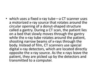
CT Scan
• The word tomography is derived from the
Greek tomos (slice) and graphien ( to write )
• Computed tomography was originally known
as the EMI scan. It was later known Computed
axial tomography CAT or CT scan
• Computed tomography is commonly referred
to as a CT scan. A CT scan is a diagnostic
imaging procedure that uses a combination of
X-rays and computer technology to produce
images of the inside of the body. It shows
detailed images of any part of the body,
including the bones, muscles, fat, organs and
blood vessels.
• The term “computed tomography,” or CT,
refers to a computerized x-ray imaging
procedure in which a narrow beam of x-rays is
aimed at a patient and quickly rotated around
the body, producing signals that are processed
by the machine’s computer to generate cross-
sectional images, or “slices.” These slices are
called tomographic images and can give a
clinician more detailed information than
conventional x-rays.
• Once a number of successive slices are
collected by the machine’s computer, they can
be digitally “stacked” together to form a
three-dimensional (3D) image of the patient
that allows for easier identification of basic
structures as well as possible tumors or
abnormalities.
• which uses a fixed x-ray tube—a CT scanner uses
a motorized x-ray source that rotates around the
circular opening of a donut-shaped structure
called a gantry. During a CT scan, the patient lies
on a bed that slowly moves through the gantry
while the x-ray tube rotates around the patient,
shooting narrow beams of x-rays through the
body. Instead of film, CT scanners use special
digital x-ray detectors, which are located directly
opposite the x-ray source. As the x-rays leave the
patient, they are picked up by the detectors and
transmitted to a computer.
• Each time the x-ray source completes one full
rotation, the CT computer uses sophisticated
mathematical techniques to construct a two-
dimensional image slice of the patient
• T scans can be used to identify disease or injury within
various regions of the body. For example, CT has
become a useful screening tool for detecting possible
tumors or lesions within the abdomen. A CT scan of the
heart may be ordered when various types of heart
disease or abnormalities are suspected. CT can also be
used to image the head in order to locate injuries,
tumors, clots leading to stroke, hemorrhage, and other
conditions. It can image the lungs in order to reveal the
presence of tumors, pulmonary embolisms (blood
clots), excess fluid, and other conditions such as
emphysema or pneumonia.
• CT scans can diagnose possibly life-threatening
conditions such as hemorrhage, blood clots, or
cancer. An early diagnosis of these conditions
could potentially be lifesaving. However, CT scans
use x-rays, and all x-rays produce
ionizing radiation. Ionizing radiation has the
potential to cause biological effects in living
tissue. This is a risk that increases with the
number of exposures added up over the life of an
individual. However, the risk of developing cancer
from x-ray radiation exposure is generally small.
Uses
• Diagnose several disorders
• Head –
• Chest
• Abdomen and pelvic

CT scan.pptx

  • 1.
  • 2.
    • The wordtomography is derived from the Greek tomos (slice) and graphien ( to write ) • Computed tomography was originally known as the EMI scan. It was later known Computed axial tomography CAT or CT scan
  • 3.
    • Computed tomographyis commonly referred to as a CT scan. A CT scan is a diagnostic imaging procedure that uses a combination of X-rays and computer technology to produce images of the inside of the body. It shows detailed images of any part of the body, including the bones, muscles, fat, organs and blood vessels.
  • 4.
    • The term“computed tomography,” or CT, refers to a computerized x-ray imaging procedure in which a narrow beam of x-rays is aimed at a patient and quickly rotated around the body, producing signals that are processed by the machine’s computer to generate cross- sectional images, or “slices.” These slices are called tomographic images and can give a clinician more detailed information than conventional x-rays.
  • 5.
    • Once anumber of successive slices are collected by the machine’s computer, they can be digitally “stacked” together to form a three-dimensional (3D) image of the patient that allows for easier identification of basic structures as well as possible tumors or abnormalities.
  • 6.
    • which usesa fixed x-ray tube—a CT scanner uses a motorized x-ray source that rotates around the circular opening of a donut-shaped structure called a gantry. During a CT scan, the patient lies on a bed that slowly moves through the gantry while the x-ray tube rotates around the patient, shooting narrow beams of x-rays through the body. Instead of film, CT scanners use special digital x-ray detectors, which are located directly opposite the x-ray source. As the x-rays leave the patient, they are picked up by the detectors and transmitted to a computer.
  • 7.
    • Each timethe x-ray source completes one full rotation, the CT computer uses sophisticated mathematical techniques to construct a two- dimensional image slice of the patient
  • 8.
    • T scanscan be used to identify disease or injury within various regions of the body. For example, CT has become a useful screening tool for detecting possible tumors or lesions within the abdomen. A CT scan of the heart may be ordered when various types of heart disease or abnormalities are suspected. CT can also be used to image the head in order to locate injuries, tumors, clots leading to stroke, hemorrhage, and other conditions. It can image the lungs in order to reveal the presence of tumors, pulmonary embolisms (blood clots), excess fluid, and other conditions such as emphysema or pneumonia.
  • 9.
    • CT scanscan diagnose possibly life-threatening conditions such as hemorrhage, blood clots, or cancer. An early diagnosis of these conditions could potentially be lifesaving. However, CT scans use x-rays, and all x-rays produce ionizing radiation. Ionizing radiation has the potential to cause biological effects in living tissue. This is a risk that increases with the number of exposures added up over the life of an individual. However, the risk of developing cancer from x-ray radiation exposure is generally small.
  • 10.
    Uses • Diagnose severaldisorders • Head – • Chest • Abdomen and pelvic