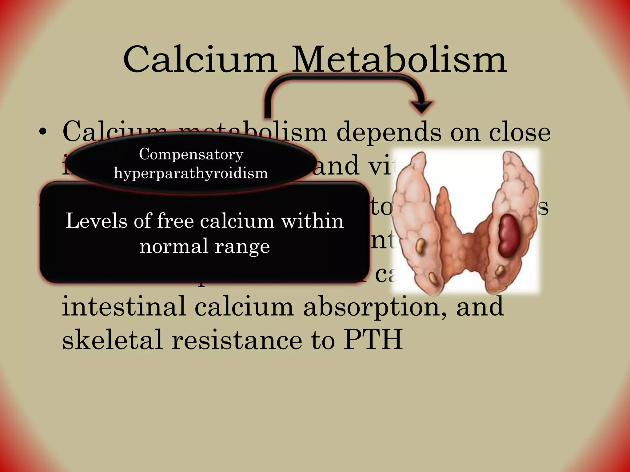
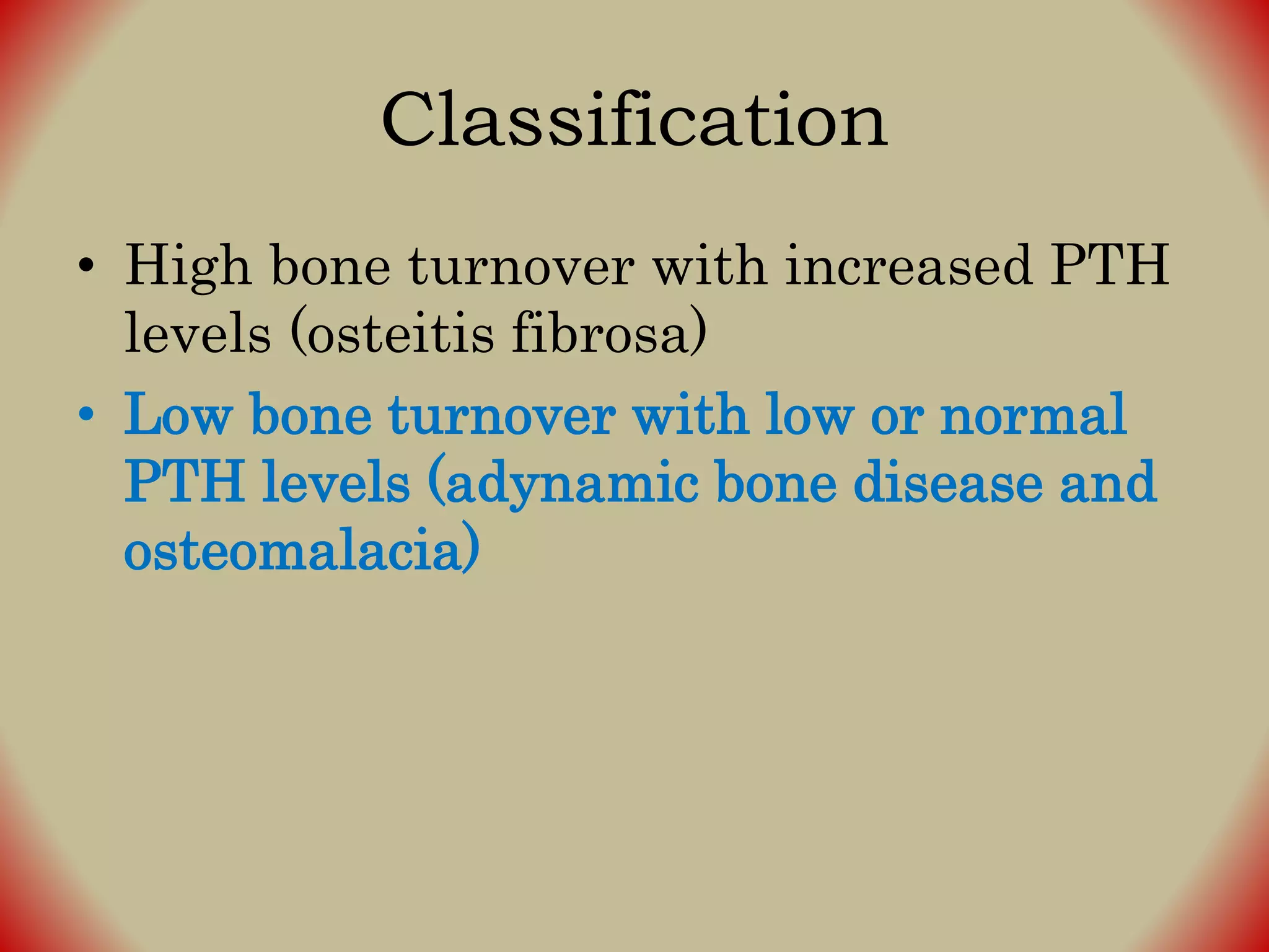
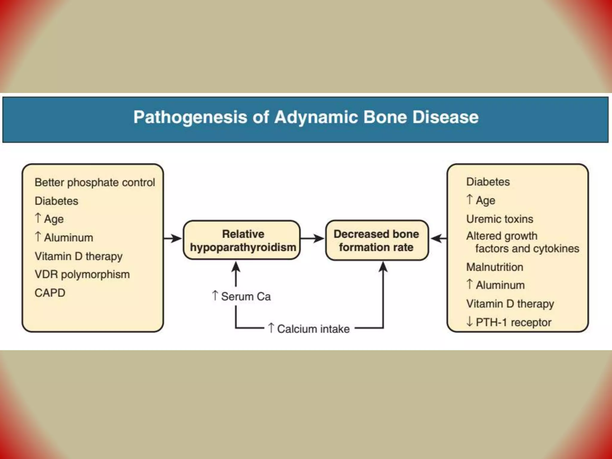
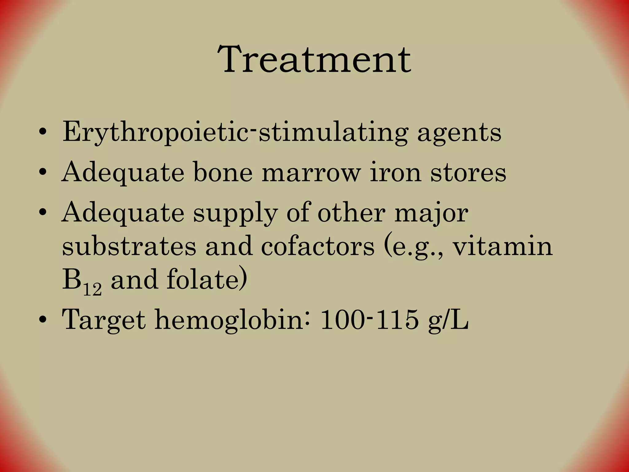
Complicationof
End StageRenalDisease
Huriah M. Putra, MD
THE CASE
General Data
• T.A.
• 24 years old
• Female
• Single
• Catholic
• Filipino
Chief Complaints
DIFFICULTY OF BREATHING
2 weeks PTC
• Complained of cough, whitish phlegm
• Associated with runny nose
• No fever
• No difficulty of breathing
• No chest pain
• No consult was done
1 week PTC
• Persistence of symptoms
• Sought consult to local clinic
• Sent home without any meds
5 days PTC
• Still with cough, whitish phlegm
• Already associated with orthopnea
(sitting position)
• Sought consult to clinic
• Given mucolytic and sent home
Day of admission
• Persistent of symptoms, patient sought
consult to local clinic
• Chest Xray was done
• Transferred to our institution
Physical Examination
• Awake, dyspnea
• BP unappreciated; HR 112; RR 42; T
36.8
• Neck vein engorgement (+)
• Crackles all over
• Abdomen soft, nontender
• No edema
In the ER
• Norepinephrine 0.1 mcg/kg/min
• Ipratropium bromide + Salbutamol
nebulization 2 doses
• 12 lead ECG ▶ sinus tachycardia
• ABG ▶ FiO2 60%
Ph 7.19
PaCO2 20
PaO2 81
HCO3 7
O2 94%
• NaHCO3 50 meq SIVP
• Intubated
• NaHCO3 150 meq in 250 cc D5W x 24o
• Piperacillin-Tazobactam 4.5 gr IV
loading then 2.25 gr IV q8
• BP 150/80
– Furosemide 80 mg IV
FiO2 100%
Ph 7.09
PaCO2 44
PaO2 50
HCO3 13
O2 71%
Leukocytes 16.27x 10⁹/L 5.0-10.0
Erythrocytes 2.87x 10¹²/L 4.2-5.4
Hemoglobin 8.7 g/dL 11.0-15.0
Hematocrit 24.4 % 37.0-47.0
Thrombocytes 559 x 109/L 150-450
Neutrophil 94.2 % 50-70
Lymphocyte 4.3 % 20-40
Monocyte 0.8 % 0-7.0
Eosinophil 0.5 % 0-5.0
Basophil 0.2 % 0-1.0
BUN 113 7-18
Creatinine 22.14 0.7-1.3
Sodium 135 136-145
Potassium 5.9 3.5-5.1
Blood Uric Acid 13.4 2.6-6.2
Calcium 6.4 8.5-10.10
Phosphorus 15.4 2.48-4.95
Magnesium 2.19 1.8-2.4
Albumin 2.6 3.4-5.0
Impression
• CKD sec to CGN in uremia
• CAP-HR
• Patient was scheduled for STAT HD
– Duration 2 hours
– UF 2L + flushing + BT
– BFR 180
– DFR 300
– Loph 18
– Flushing 50 cc Q15
CHRONIC KIDNEY DISEASE
Chronic Kidney Disease
• Kidney damage or eGFR below 60
ml/min/1.73 m2 persisting for 3 months
• Abnormalities of kidney structure
• Irrespective of the cause
Mechanism
• Chronic and sustained insults to the
kidney evolve to progressive kidney
fibrosis with destruction of the normal
microarchitecture of the kidney ▶
replaced by fibrous tissue made of
collagenous extracellular matrix ▶ loss
of function
SODIUM ANDWATER HOMEOSTASIS
• In normal renal function, tubular
reabsorption of filtered sodium and
water is adjusted; urinary excretion =
intake
• In kidney disease, dietary intake of
sodium > urinary excretion ▶ sodium
retention and ↗ extracellular fluid
volume
Sodium
retention
Expansion of
ECFV
Hypertension
Accelerate
nephron injury
Na+
Expansion of ECFV
• Presents as peripheral edema or
unresponsive hypertension
• Salt restriction
• Combination of diuretics and metolazone
can promote salt excretion
• Diuretic resistance with intractable
edema and hypertension: indication to
initiate dialysis
In face of extrarenal fluid loss..
• Prone to ECFV depletion because
inability to reclaim filtered sodium
• Compromise kidney function through
underperfusion ▶ acute-on-chronic
kidney failure
POTASSIUM HOMEOSTASIS
• Defense for decline in urinary potassium
excretion:
– Aldosterone-dependent secretion in the
distal nephron
– Augmented potassium excretion in the GI
tract
• Hyperkalemia may be precipitated:
– Increased dietary potassium intake
– Protein catabolism
– Hemolysis
– Hemorrhage
– Blood transfusion
– Metabolic acidosis
– RAS inhibitors and potassium sparing
diuretics
Hypokalemia
• Not common in CKD
• Reflects markedly reduced dietary
potassium intake, excessive diuretic,
concurrent GI losses
METABOLIC ACIDOSIS
Metabolic Acidosis in Advanced
CKD
• Caused by failure of the tubular
acidification to excrete normal daily acid
load
• As functional renal mass is reduced;
increase in adaptive ammonia
production and H+ secretion
• Overall production may be decreased
secondary to the decrease in total renal
mass
• Majority of patients can still acidify the
urine
• Less ammonia = less proton excretion
• Hyperkalemia will further depresses
ammonia production
• eGFR < 30 ml/min, patient may develop
hyperchloremic normal anion gap
metabolic acidosis associated with
normo- or mild hyperkalemia
• eGFR < 15 ml/min, acidosis change to
anion gap metabolic acidosis; inability to
excrete phosphate, sulfate
Si & Sy
• Dyspnea as a result of respiratory
compensation
• Aggravates hyperkalemia
• Inhibits protein anabolism
• Accelerates calcium loss from bone (for
buffer of hydrogen ions)
Treatment
• NaHCO3 0.5 to 1.5 mmol/kg/day
• Beginning when HCO3
- level is < 22
mmol/L
• If acidosis is refractory to medical
therapy, dialysis needs to be initiated
BONEMANIFESTATIONS
Classification
• High bone turnover with increased PTH
levels (osteitis fibrosa)
• Low bone turnover with low or normal
PTH levels (adynamic bone disease and
osteomalacia)
Classification
• High bone turnover with increased PTH
levels (osteitis fibrosa)
• Low bone turnover with low or normal
PTH levels (adynamic bone disease and
osteomalacia)
Calcium Metabolism
• Calcium metabolism depends on close
interaction of PTH and vitamin D
• Total serum Ca++ tends to decrease as
result of phosphate retention and
decreased production of calcitriol,
intestinal calcium absorption, and
skeletal resistance to PTH
Levels of free calcium within
normal range
Compensatory
hyperparathyroidism
Phosphate Metabolism
• Hyperphosphatemia does not evident
before CKD stage 4
• Compensatory hyperparathyroidism and
increases in FGF-23 result in increased
phosphaturia
Clinical Manifestations
• Aches and pains, nonspecific; lower
back, hips, legs; aggravated by weight
bearing
• May be confused with gout or
pseudogout and often respond to
NSAIDs
• Bone deformities as a consequences of
fractures; sometimes induced by brown
tumors
Treatment
• Prevention of hypocalcemia
– Calcium supplements (CaCO3) with
vitamin D
• Control of Phosphate
– Dietary phosphate restriction (0.8 g/kg/day)
– Phosphate binders (calcium-containing
antacids, magnesium salts, aluminium
hydroxide, etc.)
Classification
• High bone turnover with increased PTH
levels (osteitis fibrosa)
• Low bone turnover with low or normal
PTH levels (adynamic bone disease and
osteomalacia)
• Consequences of inadequately low PTH
levels
• Iatrogenic oversuppression of PTH
results from high dose active vitamin D,
calcium loading, or after
parathyroidectomy
Treatment
• Avoid PTH overexpression and restore
adequate PTH levels without
development of secondary
hyperparathyroidism
– Reduction or withdrawal of active
vitamin D
– Reduction of dialysate calcium
concentration
ANEMIA
• Isolated normochromic, normocytic
anemia
• Observed as early as stage 3 CKD and is
almost universal by stage 4
• Primary cause: insufficient production of
erythropoietin
Treatment
• Erythropoietic-stimulating agents
• Adequate bone marrow iron stores
• Adequate supply of other major
substrates and cofactors (e.g., vitamin
B12 and folate)
• Target hemoglobin: 100-115 g/L
Complications of ESRD

Complications of ESRD

  • 1.
  • 2.
  • 3.
    General Data • T.A. •24 years old • Female • Single • Catholic • Filipino
  • 4.
  • 5.
    2 weeks PTC •Complained of cough, whitish phlegm • Associated with runny nose • No fever • No difficulty of breathing • No chest pain • No consult was done
  • 6.
    1 week PTC •Persistence of symptoms • Sought consult to local clinic • Sent home without any meds
  • 7.
    5 days PTC •Still with cough, whitish phlegm • Already associated with orthopnea (sitting position) • Sought consult to clinic • Given mucolytic and sent home
  • 8.
    Day of admission •Persistent of symptoms, patient sought consult to local clinic • Chest Xray was done • Transferred to our institution
  • 9.
    Physical Examination • Awake,dyspnea • BP unappreciated; HR 112; RR 42; T 36.8 • Neck vein engorgement (+) • Crackles all over • Abdomen soft, nontender • No edema
  • 10.
    In the ER •Norepinephrine 0.1 mcg/kg/min • Ipratropium bromide + Salbutamol nebulization 2 doses • 12 lead ECG ▶ sinus tachycardia • ABG ▶ FiO2 60% Ph 7.19 PaCO2 20 PaO2 81 HCO3 7 O2 94%
  • 11.
    • NaHCO3 50meq SIVP • Intubated • NaHCO3 150 meq in 250 cc D5W x 24o • Piperacillin-Tazobactam 4.5 gr IV loading then 2.25 gr IV q8 • BP 150/80 – Furosemide 80 mg IV FiO2 100% Ph 7.09 PaCO2 44 PaO2 50 HCO3 13 O2 71%
  • 12.
    Leukocytes 16.27x 10⁹/L5.0-10.0 Erythrocytes 2.87x 10¹²/L 4.2-5.4 Hemoglobin 8.7 g/dL 11.0-15.0 Hematocrit 24.4 % 37.0-47.0 Thrombocytes 559 x 109/L 150-450 Neutrophil 94.2 % 50-70 Lymphocyte 4.3 % 20-40 Monocyte 0.8 % 0-7.0 Eosinophil 0.5 % 0-5.0 Basophil 0.2 % 0-1.0
  • 13.
    BUN 113 7-18 Creatinine22.14 0.7-1.3 Sodium 135 136-145 Potassium 5.9 3.5-5.1 Blood Uric Acid 13.4 2.6-6.2 Calcium 6.4 8.5-10.10 Phosphorus 15.4 2.48-4.95 Magnesium 2.19 1.8-2.4 Albumin 2.6 3.4-5.0
  • 14.
    Impression • CKD secto CGN in uremia • CAP-HR
  • 15.
    • Patient wasscheduled for STAT HD – Duration 2 hours – UF 2L + flushing + BT – BFR 180 – DFR 300 – Loph 18 – Flushing 50 cc Q15
  • 16.
  • 17.
    Chronic Kidney Disease •Kidney damage or eGFR below 60 ml/min/1.73 m2 persisting for 3 months • Abnormalities of kidney structure • Irrespective of the cause
  • 20.
    Mechanism • Chronic andsustained insults to the kidney evolve to progressive kidney fibrosis with destruction of the normal microarchitecture of the kidney ▶ replaced by fibrous tissue made of collagenous extracellular matrix ▶ loss of function
  • 21.
  • 22.
    • In normalrenal function, tubular reabsorption of filtered sodium and water is adjusted; urinary excretion = intake • In kidney disease, dietary intake of sodium > urinary excretion ▶ sodium retention and ↗ extracellular fluid volume
  • 23.
  • 24.
  • 26.
    Expansion of ECFV •Presents as peripheral edema or unresponsive hypertension • Salt restriction • Combination of diuretics and metolazone can promote salt excretion • Diuretic resistance with intractable edema and hypertension: indication to initiate dialysis
  • 27.
    In face ofextrarenal fluid loss.. • Prone to ECFV depletion because inability to reclaim filtered sodium • Compromise kidney function through underperfusion ▶ acute-on-chronic kidney failure
  • 28.
  • 29.
    • Defense fordecline in urinary potassium excretion: – Aldosterone-dependent secretion in the distal nephron – Augmented potassium excretion in the GI tract
  • 30.
    • Hyperkalemia maybe precipitated: – Increased dietary potassium intake – Protein catabolism – Hemolysis – Hemorrhage – Blood transfusion – Metabolic acidosis – RAS inhibitors and potassium sparing diuretics
  • 31.
    Hypokalemia • Not commonin CKD • Reflects markedly reduced dietary potassium intake, excessive diuretic, concurrent GI losses
  • 32.
  • 33.
    Metabolic Acidosis inAdvanced CKD • Caused by failure of the tubular acidification to excrete normal daily acid load • As functional renal mass is reduced; increase in adaptive ammonia production and H+ secretion • Overall production may be decreased secondary to the decrease in total renal mass
  • 34.
    • Majority ofpatients can still acidify the urine • Less ammonia = less proton excretion • Hyperkalemia will further depresses ammonia production
  • 35.
    • eGFR <30 ml/min, patient may develop hyperchloremic normal anion gap metabolic acidosis associated with normo- or mild hyperkalemia • eGFR < 15 ml/min, acidosis change to anion gap metabolic acidosis; inability to excrete phosphate, sulfate
  • 36.
    Si & Sy •Dyspnea as a result of respiratory compensation • Aggravates hyperkalemia • Inhibits protein anabolism • Accelerates calcium loss from bone (for buffer of hydrogen ions)
  • 37.
    Treatment • NaHCO3 0.5to 1.5 mmol/kg/day • Beginning when HCO3 - level is < 22 mmol/L • If acidosis is refractory to medical therapy, dialysis needs to be initiated
  • 38.
  • 39.
    Classification • High boneturnover with increased PTH levels (osteitis fibrosa) • Low bone turnover with low or normal PTH levels (adynamic bone disease and osteomalacia)
  • 40.
    Classification • High boneturnover with increased PTH levels (osteitis fibrosa) • Low bone turnover with low or normal PTH levels (adynamic bone disease and osteomalacia)
  • 41.
    Calcium Metabolism • Calciummetabolism depends on close interaction of PTH and vitamin D • Total serum Ca++ tends to decrease as result of phosphate retention and decreased production of calcitriol, intestinal calcium absorption, and skeletal resistance to PTH Levels of free calcium within normal range Compensatory hyperparathyroidism
  • 42.
    Phosphate Metabolism • Hyperphosphatemiadoes not evident before CKD stage 4 • Compensatory hyperparathyroidism and increases in FGF-23 result in increased phosphaturia
  • 46.
    Clinical Manifestations • Achesand pains, nonspecific; lower back, hips, legs; aggravated by weight bearing • May be confused with gout or pseudogout and often respond to NSAIDs
  • 47.
    • Bone deformitiesas a consequences of fractures; sometimes induced by brown tumors
  • 48.
    Treatment • Prevention ofhypocalcemia – Calcium supplements (CaCO3) with vitamin D • Control of Phosphate – Dietary phosphate restriction (0.8 g/kg/day) – Phosphate binders (calcium-containing antacids, magnesium salts, aluminium hydroxide, etc.)
  • 50.
    Classification • High boneturnover with increased PTH levels (osteitis fibrosa) • Low bone turnover with low or normal PTH levels (adynamic bone disease and osteomalacia)
  • 51.
    • Consequences ofinadequately low PTH levels • Iatrogenic oversuppression of PTH results from high dose active vitamin D, calcium loading, or after parathyroidectomy
  • 54.
    Treatment • Avoid PTHoverexpression and restore adequate PTH levels without development of secondary hyperparathyroidism – Reduction or withdrawal of active vitamin D – Reduction of dialysate calcium concentration
  • 55.
  • 56.
    • Isolated normochromic,normocytic anemia • Observed as early as stage 3 CKD and is almost universal by stage 4 • Primary cause: insufficient production of erythropoietin
  • 57.
    Treatment • Erythropoietic-stimulating agents •Adequate bone marrow iron stores • Adequate supply of other major substrates and cofactors (e.g., vitamin B12 and folate) • Target hemoglobin: 100-115 g/L

Editor's Notes

  • #26 Hyponatremia usually responds to water restriction
  • #49 Aluminium containing antacids are effective but is not recommended because of the risk for aluminium toxicity Calcium containing phosphate binders cant be used in hypercalcemia, extensive vascular calcification, and calciphylaxis
  • #58 Anemia resistant to ESA in the face of adequate iron stores may be due to: acute or chronic inflammation, inadequate dialysis, severe hyperparathyroidism, chronic blood loss or hemolysis, chronic infection, or malignancy