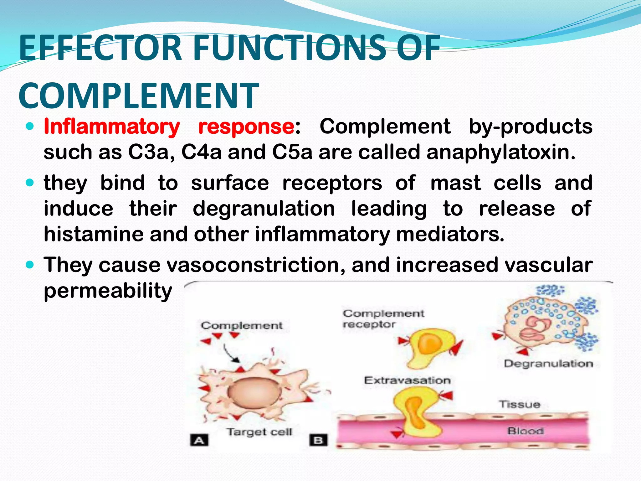
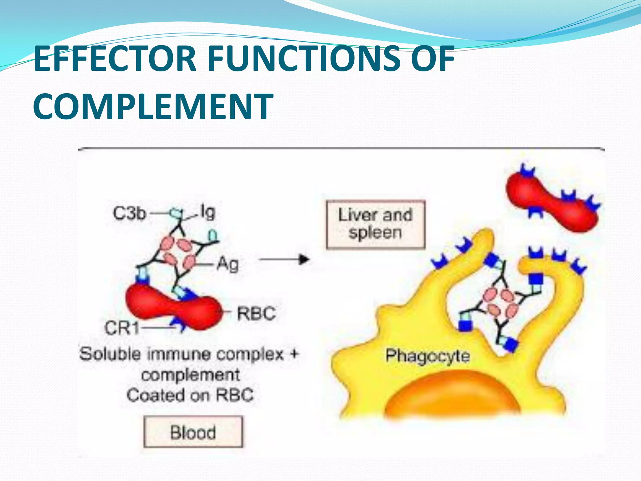
By INSHA JAN
Complement
 the term 'complement' (C) represents a group of
proteins normally found in serum in inactive form,
but when activated they augment the immune
responses.
 They constitute about 5% of normal serum proteins
and there level does not increase following either
infection or vaccination.
General properties of complement
 Complements have the following general
properties: Bind to Fc region of antibody: the
effector function of complement is mediated by
binding with Fc portion of antibody.
 the binding of complement to an antibody is
described by various terms as, fixing or
consumption (as it disappears from serum
following binding).
General properties of complement
 Role of antigen: the classical pathway complements
do not bind to free antibodies but they can only fix to
those antibodies which are bound with antigens.
However fixation of complement is not influenced by
the nature of antigens, but only by the class of
antibody.
General properties of complement
 Species nonspecific: Complements are present in the
sera of all mammals, birds, amphibians and fish.
 complements from one species can react with
antibodies from other species, though the efficiency
decreases with increase in taxonomic distance.
 Heat labile: complements get denatured by heating the
serum at 56°C for 30 minutes. Such serum with lost
complement activity is called inactivated serum.
Complement Components
 the complement system comprises of about 30 serum
proteins grouped into complement components, the
properdin system and the regulatory proteins.
 The complement components are named by numerals.
there are nine components; C1 to C9. C1 has three
subunits-C1 q, C1 r and C1 s.
 the properdin system and the regulatory proteins are
named by letter symbols, e.g. factor-B.
Synthesis
 Liver is the major site of synthesis of
complement proteins.
 Other minor sites include blood monocytes,
tissue macrophages, and epithelial cells of GIT
and genitourinary tract.
Complement Activation
 All the complement proteins are synthesized in
inactive form (e.g. zymogens) and are activated
by proteolysis.
 Complements have two unequal fragments
(large and small fragment).
 The larger fragments are usually designated as
'b' (e.g.C3b ) and the smaller fragments are
designated as 'a‘ (e.g. C3a ). An exception is C2a
which is larger fragment.
Complement Activation
 During proteolysis, the smaller fragment is
removed exposing the active site of the larger
fragments.
 the larger fragment participates in the cascade
reaction of complement pathway and the
smaller fragment diffuses away to mediate other
functions.
Complement Activation
 cascade reaction: the fragments of complements
interact in a definite sequential manner with a
cascade like effect, which leads to formation of
complex. Such complex having enzymatic activity is
designated by putting a bar over the number or
symbol (e.g. cbBb¯.
COMPLEMENT PATHWAYS
 There are three pathways of complement
activation.
 1. Classical pathway: This is an antibody dependent
pathway. Pathway is triggered by the antigen –
antibody complex formation.
 2. Alternative pathway: this is an antibody
independent pathway, triggered by the antigen
directly.
 3. Lectin pathway: This is a recently described
pathway. It resembles classical pathway, but it is
antibody independent.
Stages of complement activation
 There are four main stages in the activation of any
of the complement pathways.
 1. Initiation of the pathway
 2. Formation ofC3 convertase
 3. Formation of C5 convertase
 4. Formation of membrane attack complex (MAC)
 All the three pathways differ from each other in their
initiation till formation of C3 convertase. Then, the
remaining stages are identical in all the pathways.
CLASSICAL PATHWAY
 Classical pathway is antibody dependent.
However, not all antibodies can bind to
complements of classical pathway.
 Decreasing order of ability of antibodies to fix
complement is- lgM (most potent) > lgG3 > lgG1 >
lgG2.
 the other classes of antibodies do not fix
complements. CH2 domain on lgG, CH4 on lgM
participate in complement binding.
 The classical pathway begins with activation of C1
and binding to antigen-antibody complex.
Initiation
 the first binding portion of C1 is C1q, which
reacts with the Fc portion of lgM or lgG bound to
antigen.
 C1 q is a hexamer having six globular heads
each acting as a combining site.
 Effective activation of classical pathway begins
only when C1q is an attached to the Fc portion of
antibody by at least two of its globular binding
sites.
 lgM being pentameric, has five Fc regions, hence
one molecule of lgM can initiate the pathway.
Initiation
 Whereas lgG is monomeric, therefore two lgG
molecules are needed to initiate the process.
Hence lgM is much efficient stimulator of classical
pathway.
 C1 q binding in the presence of calcium ions, in
turn activates sequentially C1 r followed by C1 s.
Formation of C3 Convertase
 Activated C1s acts as an esterase (C1 s
esterase), which can cleave C4 to produce C4a
(an anaphylatoxin), and (C4b which binds to C1
and participates further in complement
cascade.
 C14b in the presence of magnesium ions
cleaves C2 into C2a, which remains linked to
complement complex, and C2b (has kinin like
activity), which is released outside.
 C14b2a is referred to as C3 convertase of the
classical pathway.
Formation of C5 Convertase
 C3 convertase hydrolyses many C3 molecules
into two fragments: C3a ( an anaphylatoxin) and
C3b which remains attached to C14b2a to form
C14b2a3b complex, which acts as C5
convertase of classical pathway.
Formation of Membrane Attack
Complex
 this phase begins with C5 convertase cleaving C5
into C5a (an anaphylatoxin, released into the
medium) and C5b, which continues with the
cascade.
 C5b is extremely labile, gets stabilized by binding
soon with C6 and C7 to form C5b67 followed by
addition of C8.
 the hydrophobic regions on C7 and C8 help in
penetration into the targe1 cell membrane.
 this inserted membrane complex (C5b678) has a
catalytic property to bind to C9 molecule and then it
polymerizes the C9 into a tubular channel of 10 nm
diameter.
Formation of Membrane Attack
Complex
 Penetration of C9 causes formation of channels
or pores on the targe1 cell membrane.
 Each tubular channel behaves hydrophobic
outside, but hydrophilic inside; thus allowing
free passage of ions and water into the cell
leading to cellular swelling and lysis.
Formation of Membrane Attack
Complex
 Because C5b6789 destroys the target cell by
attacking the cell membrane; hence it is called
membrane attack complex (MAC) and the
process of the cytolysis is referred to as
complement mediated cytotoxicity.
ALTERNATIVE PATHWAY
 Alternative pathway is independent of antibody;
hence is considered as a part of innate immunity.
 it also goes through the four stages; but differs from
the classical pathway in first two stages.
 Unlike the classical pathway which involves all
complement components from C1 to C9; in
alternative pathway three complement components
C1, C4 and C2 are not involved.
 Instead, it requires three other complement
proteins present in serum named factor B, factor D
and properdin.
Initiation
 the alternative complement cascade is initiated
by various cell surface constituents that are
foreign to the host, e.g. bacterial endotoxin
 The first complement component to be involved
in alternative pathway is free C3 in the
serum.(C3 hydrolyzes spontaneously, to
generate:
 C3a which diffuses out and;
 C3b fragment which attaches to foreign cell
surface antigen.
Formation of C3 Convertase
 In the next step, Factor B binds to C3b coated
foreign cells.
 Factor D, another alternative pathway
complement factor, acts on factor B, and
cleaves it into Ba (diffuses out) and Bb (remains
attached).
 C3bBb is also called C3 convertase of
alternative pathway.
Formation of C3 Convertase
 C3bBb has a very short half life of 5 minutes. If
it is stabilized by another complement protein
called properdin its half-life is increased to 30
minutes.
 The remaining two stages, i.e. formation of C5
convertase and formation of membrane attack
complex are identical to that of classical
pathway.
LECTIN PATHWAY
 Lectin pathway is another complement pathway of
innate immunity that works independent of antibody.
 It is mediated through lectin proteins of the host that
interact with mannose residues present on microbial
surface; hence the name lectin pathway.
 Among the four stages, the first stage differs from
classical pathway.
 Lectin pathway involves all complement
components used for classical pathways except
C1 (i.e. from c2 to C9);
 Instead of C1, host lectin protein called
mannose binding lectins mediate the first
'initiation' stage
Initiation
 Antigens that activate lectin pathway are the
mannose carbohydrate residues of glycoproteins
present on microbial surfaces.
 A specific host lectin protein called mannose
binding lectins (MBL) bind to mannose residues on
microbial surface.
 MBL is an acute phase reactant protein, similar to
C1q in structure.
 After binding of MBL to microbial surface,
another host protein called MBL-associated
serine protease (MASP) gets complexed with
MBL.
 MASP is similar or C1 r and C1s and mimics
their functions.
 the remaining three stages are similar to the
classical pathway.
 the MBL-MASP complex cleaves C4 which in
turn splits C2 and the MBL/MASP-C4b2a acts as
C3 convertase.
EFFECTOR FUNCTIONS OF
COMPLEMENT
 Target cell lysis by MAC: the MAC makes pores or
channels in the target cell membrane; thereby allows
the free passage of various ions and water into the
cell leading to cell swelling, lysis and death.
Bacteria, enveloped viruses, damaged cells, tumor
cells, etc. are killed by this mechanism, commonly
referred to as complement-mediated cell lysis
EFFECTOR FUNCTIONS OF
COMPLEMENT
 Inflammatory response: Complement by-products
such as C3a, C4a and C5a are called anaphylatoxin.
 they bind to surface receptors of mast cells and
induce their degranulation leading to release of
histamine and other inflammatory mediators.
 They cause vasoconstriction, and increased vascular
permeability
EFFECTOR FUNCTIONS OF
COMPLEMENT
 Opsonization: C3b and C4b act as major
opsonins that coat the immune complexes and
particulate antigens.
 Phagocytic cells express complement receptors
(CR1, CR3 and CR4) for complement
components (C3b, C4b ), and are able to bind to
complement coated antigens and enhance
phagocytosis C5a augments this process by
enhancing the CR 1 expression on phagocytes
by 10 folds.
EFFECTOR FUNCTIONS OF
COMPLEMENT
 Removing the immune complexes from blood:
 C3b plays an important role in removing immune
complexes from the blood. C3b bound immune
complexes are recognized by complement receptor
CR1 present on RBC’S Immune complexes bound to
RBCs are taken to liver and spleen where they are
phagocytosed after being separated from the RBCs
EFFECTOR FUNCTIONS OF
COMPLEMENT
EFFECTOR FUNCTIONS OF
COMPLEMENT
 Viral neutralization: Complements play a crucial role
in neutralization of the viruses; which occurs by
many ways-
 Complements coated on virus surfaces neutralize
the viral infectivity by blocking their attachment
sites.
 C3b mediated Opsonization of viral particles.
 Lysis of the enveloped viruses either by activation
of classical pathway (most viruses) or some time by
alternative or lectin pathways (by some viruses like
Epstein -Barr virus, rubella virus, etc.)
COMPLEMENT RECEPTORS
 Complement receptors (CR) play an important role in
mediating the activities of complement products as
well as in regulating their activities. They are
distributed on various cell types and bind to specific
ligands to mediate specific function
EVASION OF COMPLEMENT
SYSTEM BY MICROORGANISMS
 In order to escape from the complement mediated
effector mechanisms, micro organisms can
develop various counter mechanisms to evade the
complement system.
REGULATION OF COMPLEMENT
PATHWAYS
 Complement system are antigen non-specific;
capable of attacking micro- organisms as well as
host cells. Hence, several regulatory mechanisms
have evolved to restrict complement activity only to
the designated target cells. There are a series of
regulatory proteins, which inactivate various
complement components at different stages.
COMPLEMENT DEFICIENCIES
 Complement deficiency associated diseases fall into
two categories; diseases associated with - (1)
complement protein deficiencies and (2)
complement regulator protein deficiencies
Complenent
Complenent

Complenent

  • 1.
  • 2.
    Complement  the term'complement' (C) represents a group of proteins normally found in serum in inactive form, but when activated they augment the immune responses.  They constitute about 5% of normal serum proteins and there level does not increase following either infection or vaccination.
  • 3.
    General properties ofcomplement  Complements have the following general properties: Bind to Fc region of antibody: the effector function of complement is mediated by binding with Fc portion of antibody.  the binding of complement to an antibody is described by various terms as, fixing or consumption (as it disappears from serum following binding).
  • 4.
    General properties ofcomplement  Role of antigen: the classical pathway complements do not bind to free antibodies but they can only fix to those antibodies which are bound with antigens. However fixation of complement is not influenced by the nature of antigens, but only by the class of antibody.
  • 5.
    General properties ofcomplement  Species nonspecific: Complements are present in the sera of all mammals, birds, amphibians and fish.  complements from one species can react with antibodies from other species, though the efficiency decreases with increase in taxonomic distance.  Heat labile: complements get denatured by heating the serum at 56°C for 30 minutes. Such serum with lost complement activity is called inactivated serum.
  • 6.
    Complement Components  thecomplement system comprises of about 30 serum proteins grouped into complement components, the properdin system and the regulatory proteins.  The complement components are named by numerals. there are nine components; C1 to C9. C1 has three subunits-C1 q, C1 r and C1 s.  the properdin system and the regulatory proteins are named by letter symbols, e.g. factor-B.
  • 7.
    Synthesis  Liver isthe major site of synthesis of complement proteins.  Other minor sites include blood monocytes, tissue macrophages, and epithelial cells of GIT and genitourinary tract.
  • 8.
    Complement Activation  Allthe complement proteins are synthesized in inactive form (e.g. zymogens) and are activated by proteolysis.  Complements have two unequal fragments (large and small fragment).  The larger fragments are usually designated as 'b' (e.g.C3b ) and the smaller fragments are designated as 'a‘ (e.g. C3a ). An exception is C2a which is larger fragment.
  • 9.
    Complement Activation  Duringproteolysis, the smaller fragment is removed exposing the active site of the larger fragments.  the larger fragment participates in the cascade reaction of complement pathway and the smaller fragment diffuses away to mediate other functions.
  • 10.
    Complement Activation  cascadereaction: the fragments of complements interact in a definite sequential manner with a cascade like effect, which leads to formation of complex. Such complex having enzymatic activity is designated by putting a bar over the number or symbol (e.g. cbBb¯.
  • 11.
    COMPLEMENT PATHWAYS  Thereare three pathways of complement activation.  1. Classical pathway: This is an antibody dependent pathway. Pathway is triggered by the antigen – antibody complex formation.  2. Alternative pathway: this is an antibody independent pathway, triggered by the antigen directly.  3. Lectin pathway: This is a recently described pathway. It resembles classical pathway, but it is antibody independent.
  • 12.
    Stages of complementactivation  There are four main stages in the activation of any of the complement pathways.  1. Initiation of the pathway  2. Formation ofC3 convertase  3. Formation of C5 convertase  4. Formation of membrane attack complex (MAC)  All the three pathways differ from each other in their initiation till formation of C3 convertase. Then, the remaining stages are identical in all the pathways.
  • 13.
    CLASSICAL PATHWAY  Classicalpathway is antibody dependent. However, not all antibodies can bind to complements of classical pathway.  Decreasing order of ability of antibodies to fix complement is- lgM (most potent) > lgG3 > lgG1 > lgG2.  the other classes of antibodies do not fix complements. CH2 domain on lgG, CH4 on lgM participate in complement binding.  The classical pathway begins with activation of C1 and binding to antigen-antibody complex.
  • 14.
    Initiation  the firstbinding portion of C1 is C1q, which reacts with the Fc portion of lgM or lgG bound to antigen.  C1 q is a hexamer having six globular heads each acting as a combining site.  Effective activation of classical pathway begins only when C1q is an attached to the Fc portion of antibody by at least two of its globular binding sites.  lgM being pentameric, has five Fc regions, hence one molecule of lgM can initiate the pathway.
  • 15.
    Initiation  Whereas lgGis monomeric, therefore two lgG molecules are needed to initiate the process. Hence lgM is much efficient stimulator of classical pathway.  C1 q binding in the presence of calcium ions, in turn activates sequentially C1 r followed by C1 s.
  • 16.
    Formation of C3Convertase  Activated C1s acts as an esterase (C1 s esterase), which can cleave C4 to produce C4a (an anaphylatoxin), and (C4b which binds to C1 and participates further in complement cascade.  C14b in the presence of magnesium ions cleaves C2 into C2a, which remains linked to complement complex, and C2b (has kinin like activity), which is released outside.  C14b2a is referred to as C3 convertase of the classical pathway.
  • 17.
    Formation of C5Convertase  C3 convertase hydrolyses many C3 molecules into two fragments: C3a ( an anaphylatoxin) and C3b which remains attached to C14b2a to form C14b2a3b complex, which acts as C5 convertase of classical pathway.
  • 18.
    Formation of MembraneAttack Complex  this phase begins with C5 convertase cleaving C5 into C5a (an anaphylatoxin, released into the medium) and C5b, which continues with the cascade.  C5b is extremely labile, gets stabilized by binding soon with C6 and C7 to form C5b67 followed by addition of C8.  the hydrophobic regions on C7 and C8 help in penetration into the targe1 cell membrane.  this inserted membrane complex (C5b678) has a catalytic property to bind to C9 molecule and then it polymerizes the C9 into a tubular channel of 10 nm diameter.
  • 19.
    Formation of MembraneAttack Complex  Penetration of C9 causes formation of channels or pores on the targe1 cell membrane.  Each tubular channel behaves hydrophobic outside, but hydrophilic inside; thus allowing free passage of ions and water into the cell leading to cellular swelling and lysis.
  • 20.
    Formation of MembraneAttack Complex  Because C5b6789 destroys the target cell by attacking the cell membrane; hence it is called membrane attack complex (MAC) and the process of the cytolysis is referred to as complement mediated cytotoxicity.
  • 21.
    ALTERNATIVE PATHWAY  Alternativepathway is independent of antibody; hence is considered as a part of innate immunity.  it also goes through the four stages; but differs from the classical pathway in first two stages.  Unlike the classical pathway which involves all complement components from C1 to C9; in alternative pathway three complement components C1, C4 and C2 are not involved.  Instead, it requires three other complement proteins present in serum named factor B, factor D and properdin.
  • 22.
    Initiation  the alternativecomplement cascade is initiated by various cell surface constituents that are foreign to the host, e.g. bacterial endotoxin  The first complement component to be involved in alternative pathway is free C3 in the serum.(C3 hydrolyzes spontaneously, to generate:  C3a which diffuses out and;  C3b fragment which attaches to foreign cell surface antigen.
  • 23.
    Formation of C3Convertase  In the next step, Factor B binds to C3b coated foreign cells.  Factor D, another alternative pathway complement factor, acts on factor B, and cleaves it into Ba (diffuses out) and Bb (remains attached).  C3bBb is also called C3 convertase of alternative pathway.
  • 24.
    Formation of C3Convertase  C3bBb has a very short half life of 5 minutes. If it is stabilized by another complement protein called properdin its half-life is increased to 30 minutes.  The remaining two stages, i.e. formation of C5 convertase and formation of membrane attack complex are identical to that of classical pathway.
  • 25.
    LECTIN PATHWAY  Lectinpathway is another complement pathway of innate immunity that works independent of antibody.  It is mediated through lectin proteins of the host that interact with mannose residues present on microbial surface; hence the name lectin pathway.  Among the four stages, the first stage differs from classical pathway.
  • 26.
     Lectin pathwayinvolves all complement components used for classical pathways except C1 (i.e. from c2 to C9);  Instead of C1, host lectin protein called mannose binding lectins mediate the first 'initiation' stage
  • 27.
    Initiation  Antigens thatactivate lectin pathway are the mannose carbohydrate residues of glycoproteins present on microbial surfaces.  A specific host lectin protein called mannose binding lectins (MBL) bind to mannose residues on microbial surface.  MBL is an acute phase reactant protein, similar to C1q in structure.
  • 28.
     After bindingof MBL to microbial surface, another host protein called MBL-associated serine protease (MASP) gets complexed with MBL.  MASP is similar or C1 r and C1s and mimics their functions.  the remaining three stages are similar to the classical pathway.  the MBL-MASP complex cleaves C4 which in turn splits C2 and the MBL/MASP-C4b2a acts as C3 convertase.
  • 30.
    EFFECTOR FUNCTIONS OF COMPLEMENT Target cell lysis by MAC: the MAC makes pores or channels in the target cell membrane; thereby allows the free passage of various ions and water into the cell leading to cell swelling, lysis and death. Bacteria, enveloped viruses, damaged cells, tumor cells, etc. are killed by this mechanism, commonly referred to as complement-mediated cell lysis
  • 31.
    EFFECTOR FUNCTIONS OF COMPLEMENT Inflammatory response: Complement by-products such as C3a, C4a and C5a are called anaphylatoxin.  they bind to surface receptors of mast cells and induce their degranulation leading to release of histamine and other inflammatory mediators.  They cause vasoconstriction, and increased vascular permeability
  • 32.
    EFFECTOR FUNCTIONS OF COMPLEMENT Opsonization: C3b and C4b act as major opsonins that coat the immune complexes and particulate antigens.  Phagocytic cells express complement receptors (CR1, CR3 and CR4) for complement components (C3b, C4b ), and are able to bind to complement coated antigens and enhance phagocytosis C5a augments this process by enhancing the CR 1 expression on phagocytes by 10 folds.
  • 33.
    EFFECTOR FUNCTIONS OF COMPLEMENT Removing the immune complexes from blood:  C3b plays an important role in removing immune complexes from the blood. C3b bound immune complexes are recognized by complement receptor CR1 present on RBC’S Immune complexes bound to RBCs are taken to liver and spleen where they are phagocytosed after being separated from the RBCs
  • 34.
  • 35.
    EFFECTOR FUNCTIONS OF COMPLEMENT Viral neutralization: Complements play a crucial role in neutralization of the viruses; which occurs by many ways-  Complements coated on virus surfaces neutralize the viral infectivity by blocking their attachment sites.  C3b mediated Opsonization of viral particles.  Lysis of the enveloped viruses either by activation of classical pathway (most viruses) or some time by alternative or lectin pathways (by some viruses like Epstein -Barr virus, rubella virus, etc.)
  • 36.
    COMPLEMENT RECEPTORS  Complementreceptors (CR) play an important role in mediating the activities of complement products as well as in regulating their activities. They are distributed on various cell types and bind to specific ligands to mediate specific function
  • 38.
    EVASION OF COMPLEMENT SYSTEMBY MICROORGANISMS  In order to escape from the complement mediated effector mechanisms, micro organisms can develop various counter mechanisms to evade the complement system.
  • 40.
    REGULATION OF COMPLEMENT PATHWAYS Complement system are antigen non-specific; capable of attacking micro- organisms as well as host cells. Hence, several regulatory mechanisms have evolved to restrict complement activity only to the designated target cells. There are a series of regulatory proteins, which inactivate various complement components at different stages.
  • 42.
    COMPLEMENT DEFICIENCIES  Complementdeficiency associated diseases fall into two categories; diseases associated with - (1) complement protein deficiencies and (2) complement regulator protein deficiencies