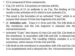
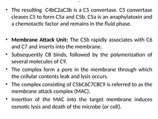
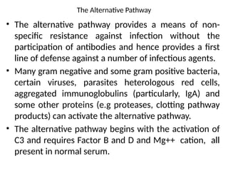
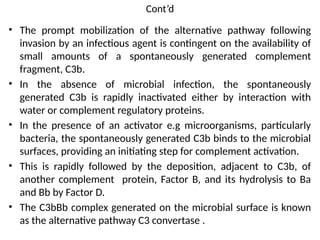
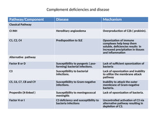
Complement System and Its
Abnormalities
Presented by
Dr. Unyime Fabian
Department of Medical Laboratory Sciences
Faculty of Allied Health Sciences
Introduction
• The complement system includes serum and
membrane bound proteins that function in both
the acquired and natural host defense system.
• These proteins are highly regulated and interact
via a series of proteolytic cascades.
• The term complement refers to the ability of
these proteins to compliment or augment the
effects of other components of the immune
system.
Cont,d
• Complements have several main effects;
• i) Lysis of cells e.g bacteria and tumour cells
• 2) Production of mediators that participate in inflammation
and attract phagocytes
• 3) Opsonization of organisms and immune complexes for
clearance by phagocytosis.
• 4) Enhancement of antibody mediated immune response.
• Complement comprises over 20 different serum proteins
that are produced by a variety of cells including,
hepatocytes, macrophages and gut epithelial cells. Some
compliment proteins bind to immunoglobulins or to
membrane components of cells.
Cont’d
• Others are pro-enzymes that when activated,
cleave one or more other complement proteins.
Upon cleavage, some of the complement protein
yield fragments that activate cells, increase
vascular permeability, or opsonize bacteria.
• Compliment is heat labile and is inactivated at
56°c for 30 minutes.
PATHWAYS OF COMPLEMENT ACTIVATION
• Complement activation can be divided into 2
pathways: the classical and alternative
pathways. Both pathways lead to the activation
of the activation of C5 convertase and result in
the production of C5b which is essential for the
activation of the membrane attack pathway.
The classical pathway
• Activation of the classical pathway of
complement occurs only after adaptive host
defenses have been recruited. Specifically,
IgM, or IgG ( subclasses 1,2 or 3) antibodies
specific for an antigen (e.g infectious agent)
must be generated and bind to that antigen. In
this antigen/antibody complex or immune
complex, antigen is bound via the Fab region
of antibody leaving the Fc regions available to
bind the complement protein, C1.
Cont’d
• The classical pathway consists of three (3) units; The
recognition, activation and membrane attack.
• The recognition unit: The pathway begins when a
specific antibody interact with its corresponding
antigen. The binding of these antibody with antigen
causes some conformational changes that will expose
the Fc portion of the antibody for the globular head of
C1q to bind.
• At least 2 molecules of IgG or 1 molecule of IgM
(pentamer) are required for binding C1q. The binding
of C1q does not occur to antibody that have not
complexed with antigen and these binding requires
calcium and magnesium ions.
• C1 is composed of three components.
• C1q, C1r and C1s. C1s possess an esterase activity.
• The binding of C1 to antibody is via C1q. The binding of C1q
results in the activation of Cir which in turn activates C1s.
• The result is the formation of an activated “C1qrs” which is an
enzyme that cleaves C4 into two fragments C4a and C4b.
• 2) Activation unit: C1qrs→→→C4a and C4b. The C4b binds to
the membrane and the C4a fragment is released into the
microenvironment.
• Activated “C1qrs” also cleaves C2 into C2a and C2b. C2a binds to
the membrane in association with C4b and C2b is released into
the microenvironment. The resulting C4bC2a complex is a C3
convertase, which cleaves C3 into C3a and C3b.
• C3b binds to the membrane in association with C4b and C2a, C3a
is an anaphylatoxin and is released into the microenvironment
Cont’d
• The resulting C4bC2aC3b is a C5 convertase. C5 convertase
cleaves C5 to form C5a and C5b. C5a is an anaphylatoxin and
a chemotactic factor and remains in the fluid phase.
• Membrane Attack Unit: The C5b rapidly associates with C6
and C7 and inserts into the membrane.
• Subsequently C8 binds, followed by the polymerization of
several molecules of C9.
• The complex form a pore in the membrane through which
the cellular contents leak and lysis occurs.
• The complex consisting of C5bC6C7C8C9 is referred to as the
membrane attack complex (MAC).
• Insertion of the MAC into the target membrane induces
osmotic lysis and death of the microbe (or cell).
The Alternative Pathway
• The alternative pathway provides a means of non-
specific resistance against infection without the
participation of antibodies and hence provides a first
line of defense against a number of infectious agents.
• Many gram negative and some gram positive bacteria,
certain viruses, parasites heterologous red cells,
aggregated immunoglobulins (particularly, IgA) and
some other proteins (e.g proteases, clotting pathway
products) can activate the alternative pathway.
• The alternative pathway begins with the activation of
C3 and requires Factor B and D and Mg++ cation, all
present in normal serum.
Cont’d
• The prompt mobilization of the alternative pathway following
invasion by an infectious agent is contingent on the availability of
small amounts of a spontaneously generated complement
fragment, C3b.
• In the absence of microbial infection, the spontaneously
generated C3b is rapidly inactivated either by interaction with
water or complement regulatory proteins.
• In the presence of an activator e.g microorganisms, particularly
bacteria, the spontaneously generated C3b binds to the microbial
surfaces, providing an initiating step for complement activation.
• This is rapidly followed by the deposition, adjacent to C3b, of
another complement protein, Factor B, and its hydrolysis to Ba
and Bb by Factor D.
• The C3bBb complex generated on the microbial surface is known
as the alternative pathway C3 convertase .
Cont’d
• This C3 convertase is stabilized by a tetrameric
protein, properdin, that extends its half life 6 to 10-
fold.
• Some of the C3b generated by the stabilized C3
convertase on the activator surface associates with
the C3bBb complex to form a C3bBbC3b complex.
• This is the C5 convertase of the alternative pathway.
• The C3 convertase generates C5b which is the
initiating step for the formation of the membrane
attack complex, which characterizes the
terminal(common) pathway of complement
activation as described in the classical pathway.
Complement Regulation
• Complement activity and deleterious complement
effects on autologous cells are minimized by a
variety of membrane-associated and soluble
proteins.
• Some are associated exclusively with one
complement activation pathway or another, while
others have a more general action.
• These proteins function in a species-specific
manner, a fact that takes on great significance in
the field of xenotransplantation ( transplantation
of organs/tissues across different species).
Cont’d
• Factor I: In the presence of any one of the cofactors,
complement receptor I (CRI), Factor H, or membrane
cofactor protein (MCP), C3b (soluble or membrane
bound) is cleaved by Factor I to yield the inactive
form, C3bi.
• Factor I also cleaves C4b to its inactive form, C4bi, in
the presence of cofactors CRI, MCP, or C4 binding
protein (C4bp).
• Factor H: As well as being a cofactor for factor I, Factor
H binds to C3b in the fluid phase thus preventing C3b
from binding to the cell surface. When the alternative
pathway C3 convertase has formed, Factor H
competitively binds with C3b, inducing the
dissociation of this convertase.
Cont’d
• Decay accelerating factor (DAF) : It binds to
membrane bound C4b and C3b, blocking the
formation of the alternative and classical pathway C3
convertase. When the C3 convertase have already
formed, DAF competitively binds with C3b or C4b
promoting their dissociation.
• C4 binding protein (C4bp): In addition to its role as a
cofactor for Factor I, C4 binding protein (C4bp) binds
to fluid phase C4b, preventing its attachment to cells.
When the classical C3 convertase has already
formed, C4bp competitively binds with C4b,
promoting its dissociation.
Cont’d
• Complement receptor I (CRI): As well as being a cofactor for
Factor I, complement receptor I (CRI) binds to membrane
bound C4b and C3b, blocking the formation of the
alternative and classical pathway C3 convertase.
• When the C3 convertases have already formed, CRI
competitively binds with C3b, or C4b, promoting their
dissociation.
• In addition, CRI binds to membrane bound C3bi and C4bi,
preventing their degradation to biological active molecules.
• Membrane cofactor protein (MCP): is a cofactor for Factor I.
• The anaphylatoxin inhibitor (AI): binds to C3a, C4a, and
C5a, inhibiting their binding to receptors on mast cells and
basophils.
Cont’d
• The CI esterase inhibitor (CI INH): It forms a complex with
CI, preventing the spontaneous activation of the classical
complement pathway.
• In addition to inhibiting CI, the CI INH inactivates the
protease, kallikrein. Kallikrein links the complement and
intrinsic coagulation pathways,
• MAC inhibitors (MAC INH): It include homologous
restriction factor (HRF), S-protein, and CD59. these inhibit
formation of MAC on autologous cells. S-protein or
vitronectin, binds to soluble (C5b, C6,C7) complexes
preventing their insertion into autologous membrane.
• CD59 and HRF bind to C8, preventing the binding and
polymerization of C9 and formation of MAC.
Complement deficiencies and disease
Pathway/Component Disease Mechanism
Classical Pathway
CI INH Hereditary angioedema Overproduction of C2b ( prokinin).
C1, C2, C4 Predisposition to SLE Opsonization of immune
complexes help keep them
soluble, deficiencies results in
increased precipitation in tissues
and inflammation
Alternative pathway
Factor B or D Susceptibility to pyogenic ( pus-
forming) bacterial infections.
Lack of sufficient opsonization of
bacteria
C3 Susceptibility to bacterial
infections.
Lack of opsonization and inability
to utilize the membrane attack
pathway
C5, C6, C7, C8 and C9 Susceptibility to Gram-negative
infections.
Inability to attack the outer
membrane of Gram-negative
bacteria.
Properdin (X-Iinked ) Susceptibility to meningococcal
meningitis
Lack of opsonization of bacteria.
Factor H or I C3 deficiency and susceptibility to
bacteria infections
Uncontrolled activation of C3 via
alternative pathway resulting in
depletion of C3.
Inherited Defects in Complement
• In humans inherited deficiencies of components of the
complement system are associated with 4 characteristic
syndromes;
• 1) Recurrent Susceptibility to bacterial infection in patients with C3
deficiency (occurring as a primary defect) or secondary deficiency
of either factor I,H or properdin.
• 2) Increased incidence of immune complex disorders or lupus like
syndromes associated with early classical pathway components
(C1, C2, C4).
• 3) Increased susceptibility to recurrent or chronic Neisseria
infections in patients with genetic defects of one of the attack
sequence proteins.
• 4) Hereditary Angioedema in patients with genetic deficiency of CI
inhibitor with the exception of CI inhibitor deficiency, complement
defects results in complete absence of hemolytic activity.
Primary C3 Deficiency
• Patients presents with recurrent infection by
pyogenic bacteria usually pneumonia,
septicaemia, otitis media, and bacterial
meningitis.
• Patients lacks C3, there is absence or depressed
complement mediated functions such as
hemolytic activity, opsonization of endotoxin,
bactericidal activity and leucocyte mobilization.
Factor I Deficiency
• Patients present with septicaemia, pneumonia,
meningitis, sinusitis, otitis media and recurrent
episodes of pyogenic infection.
• Absence of factor I leads to exhaustion of the
alternative pathway.
• Since the classical pathway is not affected, there
could be normal levels of C1 C2 and C4 but low
levels of C3 and factor B.
Cont’d
• Factor H deficiency: The patient presents with
haemolytic uremic syndromes, low levels of C3
and factor H.
• Defects of early classical pathway
components: Patients presents with lupus like
syndromes, glomerulonephritis, recurrent fever
or chronic vasculitis and sometimes recurrent
pyogenic infection. With the exception of
hereditary angioedema, C2 deficiency is the
commonest complement deficiency.
Cont’d
• Immune Complex: Deficiency of any protein needed to form the
classical pathway C3 convertase compromises the ability of the
host to eliminate antigens leading to immune complex disease.
• The classical pathway plays an important role in keeping
immune complexes soluble long enough for their safe removal
by the mononuclear phagocyte system but if not soluble, it gets
deposited in the joint causing arthritis etc.
• Deficiency of attack sequence: There is a stricking association
between deficiencies of C5, C6,C7,C8 and C9. The patient
presents with recurrent gonococcal infections particularly
septiceamia and arthritis or recurrent meningococcal meningitis.
• Deficiency of the alternative pathway: There are no cases
of inherited deficiency of factor B and D.
• CI inhibitor Deficiency: Commonest inherited deficiency
within the complement system and gives rise to a
condition of hereditary angioedema. This protein inhibits
activated C1, plasmin, kallikrein and activated factors XI
and XII of the clothing system. In its absence, these
systems are subject to low grade continous activation
allowing unrestrained activity of C1 on C2 and C4 which
are consumed to exhaustion.
• Patient presents with peripheral laryngeal or intestinal
oedema, localized oedema of the limbs, face, trunk
(neither painful nor itching). CI inhibitor,C2 and C4 are low.

Complement_System_and_its_Clinical_Correlations_lecture-2024.pptx

  • 1.
    Complement System andIts Abnormalities Presented by Dr. Unyime Fabian Department of Medical Laboratory Sciences Faculty of Allied Health Sciences
  • 2.
    Introduction • The complementsystem includes serum and membrane bound proteins that function in both the acquired and natural host defense system. • These proteins are highly regulated and interact via a series of proteolytic cascades. • The term complement refers to the ability of these proteins to compliment or augment the effects of other components of the immune system.
  • 3.
    Cont,d • Complements haveseveral main effects; • i) Lysis of cells e.g bacteria and tumour cells • 2) Production of mediators that participate in inflammation and attract phagocytes • 3) Opsonization of organisms and immune complexes for clearance by phagocytosis. • 4) Enhancement of antibody mediated immune response. • Complement comprises over 20 different serum proteins that are produced by a variety of cells including, hepatocytes, macrophages and gut epithelial cells. Some compliment proteins bind to immunoglobulins or to membrane components of cells.
  • 4.
    Cont’d • Others arepro-enzymes that when activated, cleave one or more other complement proteins. Upon cleavage, some of the complement protein yield fragments that activate cells, increase vascular permeability, or opsonize bacteria. • Compliment is heat labile and is inactivated at 56°c for 30 minutes.
  • 5.
    PATHWAYS OF COMPLEMENTACTIVATION • Complement activation can be divided into 2 pathways: the classical and alternative pathways. Both pathways lead to the activation of the activation of C5 convertase and result in the production of C5b which is essential for the activation of the membrane attack pathway.
  • 6.
    The classical pathway •Activation of the classical pathway of complement occurs only after adaptive host defenses have been recruited. Specifically, IgM, or IgG ( subclasses 1,2 or 3) antibodies specific for an antigen (e.g infectious agent) must be generated and bind to that antigen. In this antigen/antibody complex or immune complex, antigen is bound via the Fab region of antibody leaving the Fc regions available to bind the complement protein, C1.
  • 7.
    Cont’d • The classicalpathway consists of three (3) units; The recognition, activation and membrane attack. • The recognition unit: The pathway begins when a specific antibody interact with its corresponding antigen. The binding of these antibody with antigen causes some conformational changes that will expose the Fc portion of the antibody for the globular head of C1q to bind. • At least 2 molecules of IgG or 1 molecule of IgM (pentamer) are required for binding C1q. The binding of C1q does not occur to antibody that have not complexed with antigen and these binding requires calcium and magnesium ions.
  • 8.
    • C1 iscomposed of three components. • C1q, C1r and C1s. C1s possess an esterase activity. • The binding of C1 to antibody is via C1q. The binding of C1q results in the activation of Cir which in turn activates C1s. • The result is the formation of an activated “C1qrs” which is an enzyme that cleaves C4 into two fragments C4a and C4b. • 2) Activation unit: C1qrs→→→C4a and C4b. The C4b binds to the membrane and the C4a fragment is released into the microenvironment. • Activated “C1qrs” also cleaves C2 into C2a and C2b. C2a binds to the membrane in association with C4b and C2b is released into the microenvironment. The resulting C4bC2a complex is a C3 convertase, which cleaves C3 into C3a and C3b. • C3b binds to the membrane in association with C4b and C2a, C3a is an anaphylatoxin and is released into the microenvironment
  • 9.
    Cont’d • The resultingC4bC2aC3b is a C5 convertase. C5 convertase cleaves C5 to form C5a and C5b. C5a is an anaphylatoxin and a chemotactic factor and remains in the fluid phase. • Membrane Attack Unit: The C5b rapidly associates with C6 and C7 and inserts into the membrane. • Subsequently C8 binds, followed by the polymerization of several molecules of C9. • The complex form a pore in the membrane through which the cellular contents leak and lysis occurs. • The complex consisting of C5bC6C7C8C9 is referred to as the membrane attack complex (MAC). • Insertion of the MAC into the target membrane induces osmotic lysis and death of the microbe (or cell).
  • 10.
    The Alternative Pathway •The alternative pathway provides a means of non- specific resistance against infection without the participation of antibodies and hence provides a first line of defense against a number of infectious agents. • Many gram negative and some gram positive bacteria, certain viruses, parasites heterologous red cells, aggregated immunoglobulins (particularly, IgA) and some other proteins (e.g proteases, clotting pathway products) can activate the alternative pathway. • The alternative pathway begins with the activation of C3 and requires Factor B and D and Mg++ cation, all present in normal serum.
  • 11.
    Cont’d • The promptmobilization of the alternative pathway following invasion by an infectious agent is contingent on the availability of small amounts of a spontaneously generated complement fragment, C3b. • In the absence of microbial infection, the spontaneously generated C3b is rapidly inactivated either by interaction with water or complement regulatory proteins. • In the presence of an activator e.g microorganisms, particularly bacteria, the spontaneously generated C3b binds to the microbial surfaces, providing an initiating step for complement activation. • This is rapidly followed by the deposition, adjacent to C3b, of another complement protein, Factor B, and its hydrolysis to Ba and Bb by Factor D. • The C3bBb complex generated on the microbial surface is known as the alternative pathway C3 convertase .
  • 12.
    Cont’d • This C3convertase is stabilized by a tetrameric protein, properdin, that extends its half life 6 to 10- fold. • Some of the C3b generated by the stabilized C3 convertase on the activator surface associates with the C3bBb complex to form a C3bBbC3b complex. • This is the C5 convertase of the alternative pathway. • The C3 convertase generates C5b which is the initiating step for the formation of the membrane attack complex, which characterizes the terminal(common) pathway of complement activation as described in the classical pathway.
  • 13.
    Complement Regulation • Complementactivity and deleterious complement effects on autologous cells are minimized by a variety of membrane-associated and soluble proteins. • Some are associated exclusively with one complement activation pathway or another, while others have a more general action. • These proteins function in a species-specific manner, a fact that takes on great significance in the field of xenotransplantation ( transplantation of organs/tissues across different species).
  • 14.
    Cont’d • Factor I:In the presence of any one of the cofactors, complement receptor I (CRI), Factor H, or membrane cofactor protein (MCP), C3b (soluble or membrane bound) is cleaved by Factor I to yield the inactive form, C3bi. • Factor I also cleaves C4b to its inactive form, C4bi, in the presence of cofactors CRI, MCP, or C4 binding protein (C4bp). • Factor H: As well as being a cofactor for factor I, Factor H binds to C3b in the fluid phase thus preventing C3b from binding to the cell surface. When the alternative pathway C3 convertase has formed, Factor H competitively binds with C3b, inducing the dissociation of this convertase.
  • 15.
    Cont’d • Decay acceleratingfactor (DAF) : It binds to membrane bound C4b and C3b, blocking the formation of the alternative and classical pathway C3 convertase. When the C3 convertase have already formed, DAF competitively binds with C3b or C4b promoting their dissociation. • C4 binding protein (C4bp): In addition to its role as a cofactor for Factor I, C4 binding protein (C4bp) binds to fluid phase C4b, preventing its attachment to cells. When the classical C3 convertase has already formed, C4bp competitively binds with C4b, promoting its dissociation.
  • 16.
    Cont’d • Complement receptorI (CRI): As well as being a cofactor for Factor I, complement receptor I (CRI) binds to membrane bound C4b and C3b, blocking the formation of the alternative and classical pathway C3 convertase. • When the C3 convertases have already formed, CRI competitively binds with C3b, or C4b, promoting their dissociation. • In addition, CRI binds to membrane bound C3bi and C4bi, preventing their degradation to biological active molecules. • Membrane cofactor protein (MCP): is a cofactor for Factor I. • The anaphylatoxin inhibitor (AI): binds to C3a, C4a, and C5a, inhibiting their binding to receptors on mast cells and basophils.
  • 17.
    Cont’d • The CIesterase inhibitor (CI INH): It forms a complex with CI, preventing the spontaneous activation of the classical complement pathway. • In addition to inhibiting CI, the CI INH inactivates the protease, kallikrein. Kallikrein links the complement and intrinsic coagulation pathways, • MAC inhibitors (MAC INH): It include homologous restriction factor (HRF), S-protein, and CD59. these inhibit formation of MAC on autologous cells. S-protein or vitronectin, binds to soluble (C5b, C6,C7) complexes preventing their insertion into autologous membrane. • CD59 and HRF bind to C8, preventing the binding and polymerization of C9 and formation of MAC.
  • 18.
    Complement deficiencies anddisease Pathway/Component Disease Mechanism Classical Pathway CI INH Hereditary angioedema Overproduction of C2b ( prokinin). C1, C2, C4 Predisposition to SLE Opsonization of immune complexes help keep them soluble, deficiencies results in increased precipitation in tissues and inflammation Alternative pathway Factor B or D Susceptibility to pyogenic ( pus- forming) bacterial infections. Lack of sufficient opsonization of bacteria C3 Susceptibility to bacterial infections. Lack of opsonization and inability to utilize the membrane attack pathway C5, C6, C7, C8 and C9 Susceptibility to Gram-negative infections. Inability to attack the outer membrane of Gram-negative bacteria. Properdin (X-Iinked ) Susceptibility to meningococcal meningitis Lack of opsonization of bacteria. Factor H or I C3 deficiency and susceptibility to bacteria infections Uncontrolled activation of C3 via alternative pathway resulting in depletion of C3.
  • 19.
    Inherited Defects inComplement • In humans inherited deficiencies of components of the complement system are associated with 4 characteristic syndromes; • 1) Recurrent Susceptibility to bacterial infection in patients with C3 deficiency (occurring as a primary defect) or secondary deficiency of either factor I,H or properdin. • 2) Increased incidence of immune complex disorders or lupus like syndromes associated with early classical pathway components (C1, C2, C4). • 3) Increased susceptibility to recurrent or chronic Neisseria infections in patients with genetic defects of one of the attack sequence proteins. • 4) Hereditary Angioedema in patients with genetic deficiency of CI inhibitor with the exception of CI inhibitor deficiency, complement defects results in complete absence of hemolytic activity.
  • 20.
    Primary C3 Deficiency •Patients presents with recurrent infection by pyogenic bacteria usually pneumonia, septicaemia, otitis media, and bacterial meningitis. • Patients lacks C3, there is absence or depressed complement mediated functions such as hemolytic activity, opsonization of endotoxin, bactericidal activity and leucocyte mobilization.
  • 21.
    Factor I Deficiency •Patients present with septicaemia, pneumonia, meningitis, sinusitis, otitis media and recurrent episodes of pyogenic infection. • Absence of factor I leads to exhaustion of the alternative pathway. • Since the classical pathway is not affected, there could be normal levels of C1 C2 and C4 but low levels of C3 and factor B.
  • 22.
    Cont’d • Factor Hdeficiency: The patient presents with haemolytic uremic syndromes, low levels of C3 and factor H. • Defects of early classical pathway components: Patients presents with lupus like syndromes, glomerulonephritis, recurrent fever or chronic vasculitis and sometimes recurrent pyogenic infection. With the exception of hereditary angioedema, C2 deficiency is the commonest complement deficiency.
  • 23.
    Cont’d • Immune Complex:Deficiency of any protein needed to form the classical pathway C3 convertase compromises the ability of the host to eliminate antigens leading to immune complex disease. • The classical pathway plays an important role in keeping immune complexes soluble long enough for their safe removal by the mononuclear phagocyte system but if not soluble, it gets deposited in the joint causing arthritis etc. • Deficiency of attack sequence: There is a stricking association between deficiencies of C5, C6,C7,C8 and C9. The patient presents with recurrent gonococcal infections particularly septiceamia and arthritis or recurrent meningococcal meningitis.
  • 24.
    • Deficiency ofthe alternative pathway: There are no cases of inherited deficiency of factor B and D. • CI inhibitor Deficiency: Commonest inherited deficiency within the complement system and gives rise to a condition of hereditary angioedema. This protein inhibits activated C1, plasmin, kallikrein and activated factors XI and XII of the clothing system. In its absence, these systems are subject to low grade continous activation allowing unrestrained activity of C1 on C2 and C4 which are consumed to exhaustion. • Patient presents with peripheral laryngeal or intestinal oedema, localized oedema of the limbs, face, trunk (neither painful nor itching). CI inhibitor,C2 and C4 are low.