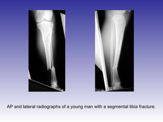
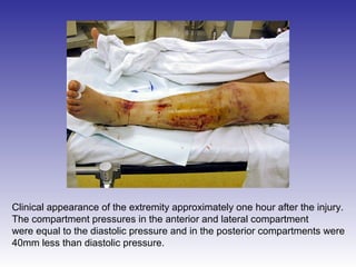
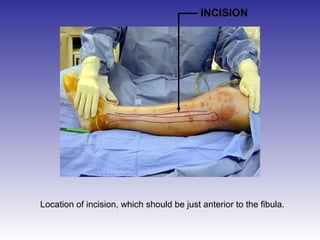
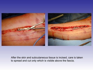
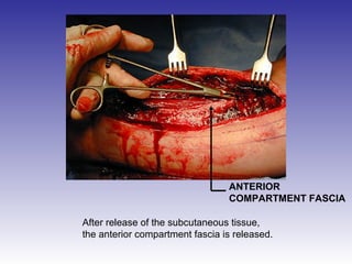
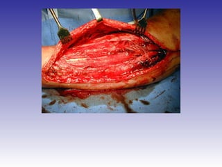
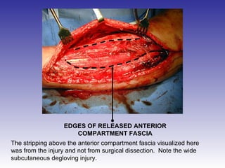
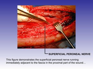
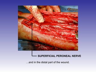
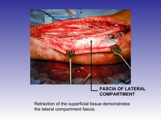
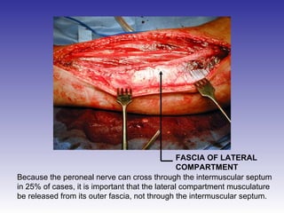
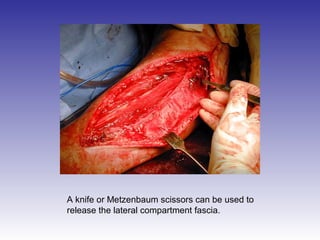
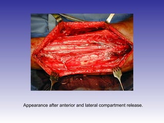
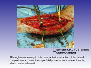
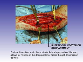
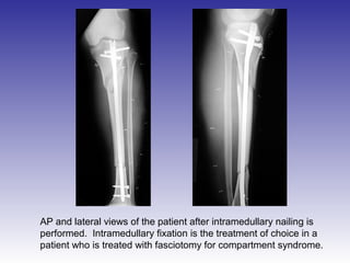
AP and lateral radiographs of a young man with a segmental tibia fracture.
Clinical appearance of the extremity approximately one hour after the injury. 
The compartment pressures in the anterior and lateral compartment 
were equal to the diastolic pressure and in the posterior compartments were 
40mm less than diastolic pressure.
INCISION 
Location of incision, which should be just anterior to the fibula.
After the skin and subcutaneous tissue is incised, care is taken 
to spread and cut only which is visible above the fascia.
ANTERIOR 
COMPARTMENT FASCIA 
After release of the subcutaneous tissue, 
the anterior compartment fascia is released.
EDGES OF RELEASED ANTERIOR 
COMPARTMENT FASCIA 
The stripping above the anterior compartment fascia visualized here 
was from the injury and not from surgical dissection. Note the wide 
subcutaneous degloving injury.
EDGES OF RELEASED ANTERIOR 
COMPARTMENT FASCIA 
Care must be taken when releasing the fascia not to damage the 
superficial peroneal nerve.
SUPERFICIAL PERONEAL NERVE 
This figure demonstrates the superficial peroneal nerve running 
immediately adjacent to the fascia in the proximal part of the wound…
SUPERFICIAL PERONEAL NERVE 
…and in the distal part of the wound.
FASCIA OF LATERAL 
COMPARTMENT 
Retraction of the superficial tissue demonstrates 
the lateral compartment fascia.
FASCIA OF LATERAL 
COMPARTMENT 
Because the peroneal nerve can cross through the intermuscular septum 
in 25% of cases, it is important that the lateral compartment musculature 
be released from its outer fascia, not through the intermuscular septum.
A knife or Metzenbaum scissors can be used to 
release the lateral compartment fascia.
Appearance after anterior and lateral compartment release.
SUPERFICIAL POSTERIOR 
COMPARTMENT 
Although unnecessary in this case, anterior retraction of the lateral 
compartment exposes the superficial posterior compartment fascia, 
which can be released.
SUPERFICIAL POSTERIOR 
COMPARTMENT 
Further dissection, as in the posterior lateral approach of Harman, 
allows for release of the deep posterior fascia through this incision 
as well.
AP and lateral views of the patient after intramedullary nailing is 
performed. Intramedullary fixation is the treatment of choice in a 
patient who is treated with fasciotomy for compartment syndrome.

Comparts

  • 2.
    AP and lateralradiographs of a young man with a segmental tibia fracture.
  • 3.
    Clinical appearance ofthe extremity approximately one hour after the injury. The compartment pressures in the anterior and lateral compartment were equal to the diastolic pressure and in the posterior compartments were 40mm less than diastolic pressure.
  • 4.
    INCISION Location ofincision, which should be just anterior to the fibula.
  • 5.
    After the skinand subcutaneous tissue is incised, care is taken to spread and cut only which is visible above the fascia.
  • 6.
    ANTERIOR COMPARTMENT FASCIA After release of the subcutaneous tissue, the anterior compartment fascia is released.
  • 8.
    EDGES OF RELEASEDANTERIOR COMPARTMENT FASCIA The stripping above the anterior compartment fascia visualized here was from the injury and not from surgical dissection. Note the wide subcutaneous degloving injury.
  • 9.
    EDGES OF RELEASEDANTERIOR COMPARTMENT FASCIA Care must be taken when releasing the fascia not to damage the superficial peroneal nerve.
  • 10.
    SUPERFICIAL PERONEAL NERVE This figure demonstrates the superficial peroneal nerve running immediately adjacent to the fascia in the proximal part of the wound…
  • 11.
    SUPERFICIAL PERONEAL NERVE …and in the distal part of the wound.
  • 12.
    FASCIA OF LATERAL COMPARTMENT Retraction of the superficial tissue demonstrates the lateral compartment fascia.
  • 13.
    FASCIA OF LATERAL COMPARTMENT Because the peroneal nerve can cross through the intermuscular septum in 25% of cases, it is important that the lateral compartment musculature be released from its outer fascia, not through the intermuscular septum.
  • 14.
    A knife orMetzenbaum scissors can be used to release the lateral compartment fascia.
  • 15.
    Appearance after anteriorand lateral compartment release.
  • 16.
    SUPERFICIAL POSTERIOR COMPARTMENT Although unnecessary in this case, anterior retraction of the lateral compartment exposes the superficial posterior compartment fascia, which can be released.
  • 17.
    SUPERFICIAL POSTERIOR COMPARTMENT Further dissection, as in the posterior lateral approach of Harman, allows for release of the deep posterior fascia through this incision as well.
  • 18.
    AP and lateralviews of the patient after intramedullary nailing is performed. Intramedullary fixation is the treatment of choice in a patient who is treated with fasciotomy for compartment syndrome.