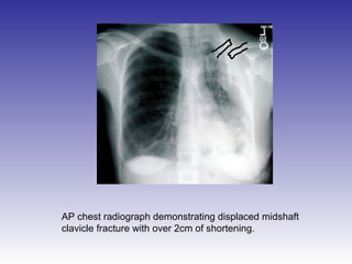
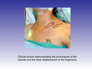
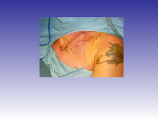
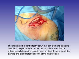
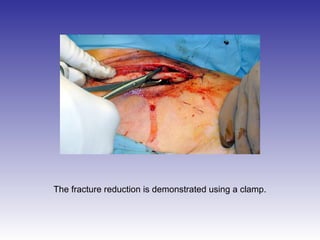
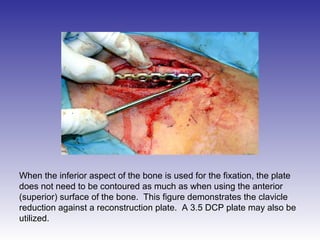
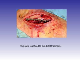
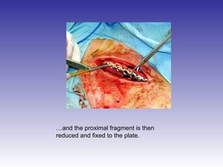
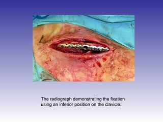
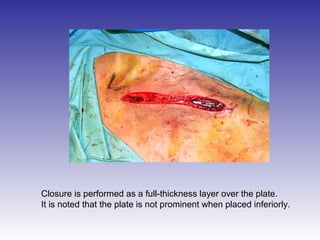
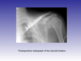
AP chest radiograph demonstrating displaced midshaft 
clavicle fracture with over 2cm of shortening.
Clinical picture demonstrating the prominence of the 
clavicle and the clear displacement of the fragments.
The incision is made over what is projected to be 
the inferior aspect of the clavicle once it is reduced.
The incision is brought directly down through skin and platysma 
muscle to the periosteum. Once the clavicle is identified, a 
subperiosteal dissection is performed on the inferior edge of the 
clavicle and circumferentially only at the fracture site.
The fracture reduction is demonstrated using a clamp.
When the inferior aspect of the bone is used for the fixation, the plate 
does not need to be contoured as much as when using the anterior 
(superior) surface of the bone. This figure demonstrates the clavicle 
reduction against a reconstruction plate. A 3.5 DCP plate may also be 
utilized.
The plate is affixed to the distal fragment…
…and the proximal fragment is then 
reduced and fixed to the plate.
The radiograph demonstrating the fixation 
using an inferior position on the clavicle.
Closure is performed as a full-thickness layer over the plate. 
It is noted that the plate is not prominent when placed inferiorly.
Postoperative radiograph of the clavicle fixation.

Clavicle

  • 3.
    AP chest radiographdemonstrating displaced midshaft clavicle fracture with over 2cm of shortening.
  • 4.
    Clinical picture demonstratingthe prominence of the clavicle and the clear displacement of the fragments.
  • 6.
    The incision ismade over what is projected to be the inferior aspect of the clavicle once it is reduced.
  • 7.
    The incision isbrought directly down through skin and platysma muscle to the periosteum. Once the clavicle is identified, a subperiosteal dissection is performed on the inferior edge of the clavicle and circumferentially only at the fracture site.
  • 8.
    The fracture reductionis demonstrated using a clamp.
  • 9.
    When the inferioraspect of the bone is used for the fixation, the plate does not need to be contoured as much as when using the anterior (superior) surface of the bone. This figure demonstrates the clavicle reduction against a reconstruction plate. A 3.5 DCP plate may also be utilized.
  • 10.
    The plate isaffixed to the distal fragment…
  • 11.
    …and the proximalfragment is then reduced and fixed to the plate.
  • 12.
    The radiograph demonstratingthe fixation using an inferior position on the clavicle.
  • 13.
    Closure is performedas a full-thickness layer over the plate. It is noted that the plate is not prominent when placed inferiorly.
  • 14.
    Postoperative radiograph ofthe clavicle fixation.