Colorimetry
Introduction
Colorimetry is oneof the most fundamental and widely used analytical techniques in biochemistry, chemistry,
and clinical laboratories for the quantitative estimation of substances based on their color intensity. The
principle behind colorimetry is straightforward yet powerful: when light passes through a colored solution, a
portion of it is absorbed, and the amount of light absorbed is directly proportional to the concentration of the
colored substance in the solution.
This technique forms the basis for many biochemical assays, including the estimation of proteins, sugars,
enzymes, nucleic acids, and metabolites in biological samples. It is especially favored for its simplicity,
accuracy, and cost-effectiveness, making it an indispensable method in teaching laboratories, hospitals, and
research institutions.
Colorimetry is grounded in the Beer–Lambert law, which quantitatively relates the absorption of light to the
properties of the material through which the light is traveling. The measurement of absorbance is performed
using an instrument known as a colorimeter, which detects light intensity before and after it passes through a
sample.
---
Principle of Colorimetry
The principle of colorimetry is based on the Beer–Lambert Law, which combines two separate but related
laws:
1. Beer’s Law (Beer, 1852):
The intensity of light absorbed by a solution is directly proportional to the concentration of the absorbing
substance.
Mathematically:
A propto c
2. Lambert’s Law (Lambert, 1760):
The amount of light absorbed is directly proportional to the path length of the light through the solution.
A propto l
Combining both gives the Beer–Lambert Law:
A = varepsilon c l
A = absorbance (unitless)
2.
ε = molarabsorptivity or molar extinction coefficient (L·mol⁻¹·cm⁻¹)
c = concentration of the solution (mol·L⁻¹)
l = path length of the cuvette (cm)
Thus, absorbance increases linearly with both concentration and path length, forming the basis for
quantitative analysis in colorimetry.
---
Basic Principle in Biochemical Applications
In biochemical colorimetry, the analyte (substance to be measured) may or may not be naturally colored.
If naturally colored, its absorbance can be measured directly.
If colorless, it must be converted into a colored compound through a chemical reaction with a suitable reagent
(called a chromogenic reagent).
For example:
Proteins are estimated using Biuret or Lowry’s reagent, forming a violet complex with copper ions.
Glucose is estimated using the glucose oxidase–peroxidase (GOD–POD) method, producing a red-colored
quinoneimine dye.
The intensity of the developed color is then measured with a colorimeter at a specific wavelength
corresponding to the maximum absorbance (λmax) of the colored complex.
---
Instrumentation of a Colorimeter
A colorimeter is an instrument designed to measure the absorbance or transmittance of light by a colored
solution. The essential components include:
1. Light Source
Provides a stable beam of light, typically from a tungsten filament lamp for visible light (400–700 nm).
3.
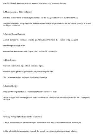
For ultraviolet (UV)measurements, a deuterium or mercury lamp may be used.
2. Monochromator (Filter or Prism)
Selects a narrow band of wavelengths suitable for the analyte’s absorbance maximum (λmax).
Simple colorimeters use glass filters, whereas advanced spectrophotometers use diffraction gratings or prisms
for higher resolution.
3. Sample Holder (Cuvette)
A small transparent container (usually quartz or glass) that holds the solution being analyzed.
Standard path length: 1 cm.
Quartz cuvettes are used for UV light; glass cuvettes for visible light.
4. Photodetector
Converts transmitted light into an electrical signal.
Common types: photocell, photodiode, or photomultiplier tube.
The current generated is proportional to light intensity.
5. Readout Device
Displays the output either as absorbance (A) or transmittance (%T).
Modern digital colorimeters provide direct readouts and often interface with computers for data storage and
analysis.
---
Working Principle (Mechanism) of a Colorimeter
1. Light from the source passes through a monochromator, which isolates the desired wavelength.
2. The selected light beam passes through the sample cuvette containing the colored solution.
4.
3. As thelight passes through, part of it is absorbed by the colored molecules, and the rest is transmitted.
4. The transmitted light reaches the photodetector, which converts it into an electrical signal proportional to
light intensity.
5. The instrument compares this signal with that from a blank solution (containing all reagents except the
analyte).
6. The resulting absorbance (A) or transmittance (%T) is displayed.
7. Using a calibration curve (standard curve) prepared from known concentrations, the concentration of the
unknown sample is determined.
---
Mechanism of Color Formation in Biochemical Reactions
In biochemical colorimetry, colored complexes are generated through specific chemical reactions. These
involve oxidation–reduction, metal chelation, or coupling reactions that yield chromophores absorbing light in
the visible spectrum.
1. Oxidation–Reduction Reactions
Many biochemical assays rely on redox reactions that produce colored products.
Example: The GOD–POD method for glucose estimation involves:
Glucose oxidized by glucose oxidase → gluconic acid + H₂O₂
H₂O₂ reacts with phenol + 4-aminophenazone in the presence of peroxidase, forming a red quinoneimine dye
measurable at 505 nm.
2. Metal–Ligand Complex Formation
Proteins and amino acids form colored complexes with metal ions.
Example: Biuret reaction – peptide bonds react with copper(II) ions in an alkaline medium to form a violet-
colored complex.
5.
3. Coupling Reactions
Phenolicor amine compounds react with diazonium salts to form azo dyes, which are intensely colored.
Example: Estimation of bilirubin or phenolic compounds using diazo coupling reactions.
The intensity of the formed color is proportional to the analyte concentration, forming the measurable
parameter in colorimetric assays.
---
Beer–Lambert Law in Practice
The Beer–Lambert Law is the foundation of quantitative colorimetry.
When monochromatic light passes through a solution of absorbing molecules:
I = I_0 e^{-varepsilon c l}
where:
I₀ = incident light intensity,
I = transmitted light intensity,
ε = molar absorptivity,
c = concentration,
l = path length.
Taking the logarithm:
A = log_{10} frac{I_0}{I} = varepsilon c l
Thus, absorbance (A) is directly proportional to concentration (c), allowing construction of a standard curve
by plotting A vs. c. The concentration of an unknown sample is then determined by interpolation.
However, the Beer–Lambert law holds true only under certain conditions:
Monochromatic light is used.
6.
The solution ishomogeneous and non-scattering.
Concentrations are low (usually < 0.01 M).
No chemical interaction or association between solute molecules.
---
Procedure of a Typical Colorimetric Experiment
1. Preparation of Standard Solutions
Prepare a series of known concentrations of the analyte.
2. Preparation of Test Solution
Treat the unknown sample under the same conditions as the standards.
3. Addition of Reagent
Add chromogenic reagent to develop color.
4. Incubation
Allow sufficient time for color development, often at specific temperature and pH.
5. Blank Preparation
Contains all reagents except the analyte to zero the instrument.
6. Measurement
Measure absorbance of standards and test at the λmax using a colorimeter.
7.
7. Calibration Curve
Plotabsorbance vs. concentration for standards.
8. Determination of Unknown
Read the test sample’s absorbance on the standard curve to find its concentration.
---
Applications of Colorimetry
Colorimetry is extensively used in clinical biochemistry, environmental science, food chemistry, and industrial
analysis.
1. Clinical Biochemistry
Estimation of Blood Glucose: Glucose oxidase–peroxidase (GOD–POD) method.
Protein Estimation: Biuret, Lowry, or Bradford methods.
Urea Determination: Diacetyl monoxime or Berthelot method.
Cholesterol and Triglyceride Analysis: Enzymatic assays producing colored end products.
Enzyme Assays: Determination of enzyme activity (e.g., alkaline phosphatase, transaminases).
2. Environmental Analysis
Determination of nitrate, phosphate, and metal ions in water samples using colorimetric reagents such as
Nessler’s reagent or molybdate complexes.
3. Food and Agricultural Chemistry
Measurement of vitamins (A, C, E) and pigments (chlorophyll, carotenoids) in food.
8.
Estimation of sugarcontent in fruit juices and honey.
4. Pharmaceutical Analysis
Quantitative estimation of drug concentration, stability, and purity using specific chromogenic reactions.
5. Industrial and Research Applications
Used in enzyme kinetics, metal ion estimation, and biochemical assays involving color changes as indicators.
---
Advantages of Colorimetry
Simple and inexpensive equipment and reagents.
Rapid analysis with minimal sample volume.
Quantitative accuracy within a suitable range.
Applicable to a wide variety of compounds (colored and colorless after derivatization).
Non-destructive – the sample can often be reused or further analyzed.
---
Limitations
Despite its widespread use, colorimetry has several limitations:
Limited sensitivity – less effective for trace amounts compared to spectrophotometry.
Interference by turbidity or suspended particles, which scatter light.
Deviations from Beer–Lambert law at high concentrations.
Color instability – some chromogenic reactions produce transient colors.
9.
Restricted wavelength selection– filter-based colorimeters cannot cover all wavelengths.
Manual handling errors may affect precision.
---
Precautions in Colorimetric Analysis
1. Always use clean, scratch-free cuvettes.
2. Handle cuvettes by the opaque sides to avoid fingerprints on optical faces.
3. Calibrate the colorimeter using a blank solution before measurements.
4. Ensure all reagents and glassware are clean and free from contamination.
5. Perform all assays under identical conditions (temperature, pH, and timing).
6. Avoid bubbles in the cuvette as they scatter light.
7. Protect light-sensitive reagents and colored solutions from direct sunlight.
---
Advancements in Colorimetry
Modern developments have improved the precision, versatility, and automation of colorimetric analysis:
1. Digital and Microprocessor-Based Colorimeters:
Provide automatic wavelength selection, temperature control, and data logging.
10.
2. Spectrophotometers:
Advanced formof colorimeter capable of measuring across UV–Vis–IR ranges.
3. Portable and Handheld Colorimeters:
Useful for on-site testing in environmental and clinical applications.
4. Microplate Colorimetry:
High-throughput measurement in 96-well plates for enzyme assays or drug screening.
5. Automation and Computer Integration:
Coupled with software for real-time data analysis and calibration curve generation.
6. Smartphone-Based Colorimetry:
Mobile applications now use smartphone cameras and apps to measure color intensity, revolutionizing field-
based testing.
7. Nanocolorimetry:
Uses nanoparticles (like gold or silver) that change color upon aggregation, enhancing sensitivity for
diagnostic biosensors.
---
Significance in Biochemistry
Colorimetry plays a central role in quantitative biochemistry and clinical diagnostics. It enables:
Monitoring of metabolic and enzymatic reactions.
Determination of blood metabolites for disease diagnosis.
11.
Standardization of biochemicalassays in research and teaching laboratories.
Evaluation of enzyme kinetics, where rate of color development reflects enzyme activity.
It bridges the gap between theoretical biochemistry and practical quantification of life’s molecular
components.
---
Conclusion
Colorimetry represents one of the earliest yet most enduring analytical techniques in biochemistry. Its
simplicity, affordability, and reliability have made it a cornerstone of quantitative analysis. The technique
translates the invisible world of molecules into measurable light, embodying the fundamental relationship
between physics and biology.
By relying on the Beer–Lambert law, colorimetry allows the estimation of diverse biological substances with
remarkable accuracy. Although advanced spectroscopic and chromatographic methods have emerged,
colorimetry continues to hold a vital place in laboratories worldwide for routine assays, teaching, and clinical
diagnostics.
Ultimately, colorimetry demonstrates how a basic physical law can unlock profound insights into biochemical
composition and function — turning color into a language through which the chemistry of life is understood.


![Polymer [ बहुलक ] Chemistry Notes PDF - Irfanullah Mehar - JJ Sir Chemistry.pdf](https://cdn.slidesharecdn.com/ss_thumbnails/polymerchemistrynotespdf-irfanullahmehar-jjsirchemistry-260210172118-3f9b37f7-thumbnail.jpg?width=640&height=640&fit=bounds)


