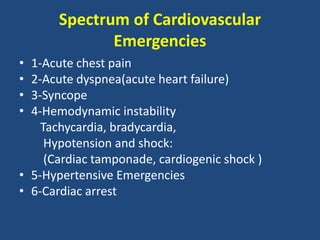
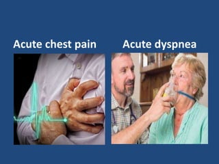
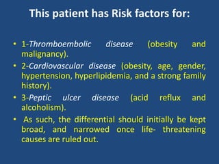
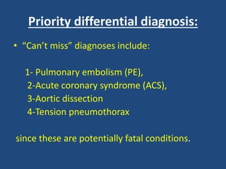
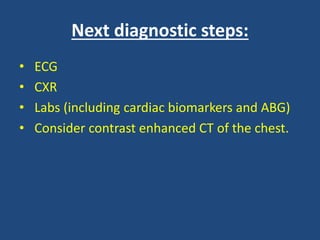
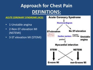
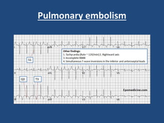
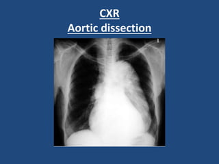
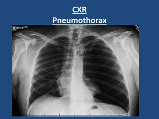
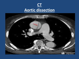
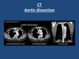
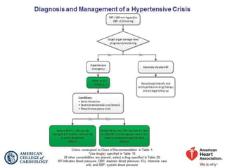
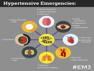
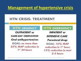
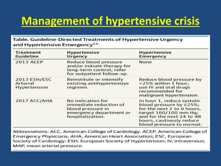
This document provides guidance on evaluating and managing common cardiovascular emergencies. It outlines the leading causes of cardiovascular emergencies, including acute chest pain, dyspnea, syncope, and hemodynamic instability. For patients presenting with chest pain, the priority is to differentiate life-threatening etiologies like pulmonary embolism, acute coronary syndrome, and aortic dissection from less serious conditions. The initial evaluation involves obtaining vital signs, performing a physical exam, 12-lead ECG, chest x-ray, and cardiac biomarker tests. Emergent conditions require urgent diagnostic testing and treatment to reduce mortality. Timely diagnosis and management is critical for improving outcomes in cardiovascular emergencies.
The document discusses uremic and dialysis-associated pericarditis. Key points include:
- Pericarditis is inflammation of the pericardium and can be caused by uremia or inadequate dialysis. It commonly causes chest pain and may develop effusions or tamponade.
- Diagnosis involves echocardiogram, EKG changes and ruling out other causes. Treatment is intensive dialysis, medications like NSAIDs or colchicine, and pericardiocentesis for large effusions or tamponade.
- Prognosis is generally good with early management but pericarditis was once common in renal failure and can still cause morbidity or mortality if
ECG- Atrial Fibrillation, CXR-P/A view-Cardiomegaly,
Echocardiogram-severe mitral stenosis with severe MR with
moderate pulmonary hypertension. Patient underwent MVR and
she is doing well.
This document discusses acute coronary syndromes and ischemic heart disease. It begins with an overview of heart anatomy and physiology. It then defines acute coronary syndrome and myocardial infarction, describing signs and symptoms. Risk factors for ischemic heart disease are outlined. The document concludes with descriptions of nursing assessments, diagnoses, and interventions for patients with acute coronary syndromes or ischemic heart disease, focusing on pain management, improving perfusion, and reducing anxiety through education.
Pulmonary embolism - Diagnosis and managementDr Vivek Baliga
Pulmonary embolism is a common problem seen in medical practice. This presentation by Dr Vivek Baliga discusses the basic aspects and evidence behind current management.
Resistant hypertension is defined as blood pressure that remains above goal despite treatment with three antihypertensive agents of different classes, one being a diuretic. Refractory hypertension is when blood pressure cannot be controlled despite four or more drugs at maximal doses. Pseudoresistant hypertension occurs when poor control is due to non-adherence or suboptimal treatment rather than true treatment resistance. Evaluation of patients with resistant hypertension includes screening for secondary causes like primary aldosteronism or renal artery stenosis through tests of electrolytes, renal function, and imaging studies. Treatment involves optimizing the current three-drug regimen before adding supplementary drugs like beta blockers or aldosterone antagonists.
Acute coronary syndrome is a term used to describe a range of conditions associated with sudden, reduced blood flow to the heart.
One such condition is a heart attack (myocardial infarction) — when cell death results in damaged or destroyed heart tissue. Even when acute coronary syndrome causes no cell death, the reduced blood flow changes how your heart works and is a sign of a high risk of heart attack.
Acute coronary syndrome often causes severe chest pain or discomfort. It is a medical emergency that requires prompt diagnosis and care. The goals of treatment include improving blood flow, treating complications and preventing future problems.
Acute Coronary Syndrome (ACS) refers to a range of conditions caused by reduced blood flow in the coronary arteries. It includes Unstable Angina (UA), Non-ST-segment elevation myocardial infarction (NSTEMI), and ST-segment elevation myocardial infarction (STEMI). ACS is diagnosed based on electrocardiogram (ECG) findings and cardiac enzyme levels. STEMI shows ST elevations and enzyme elevations, while NSTEMI shows ST depressions/inversions and enzyme elevations without ST elevations. UA shows non-specific ECG changes and normal enzymes. Risk stratification systems like the TIMI score are used for NSTEMI/UA patients to guide management, which may
Ventricular tachycardia is a fast heart rhythm originating from the ventricles with a rate over 100 bpm. It is classified based on duration (sustained vs non-sustained), morphology (monomorphic, polymorphic, sinusoidal), and symptoms. Causes include structural heart disease, electrolyte abnormalities, drugs, and prolonged QT interval. Diagnosis involves ECG criteria showing ventricular origin. Treatment depends on hemodynamic stability and may include antiarrhythmic drugs, implantable cardioverter-defibrillator, catheter ablation, or surgery. Recurrent ventricular tachycardia is managed long term with devices, drugs, and treatment of underlying causes.
Syncope is a transient loss of consciousness due to transient global cerebral hypoperfusion. It is characterized by rapid onset, short duration, and spontaneous recovery. The most common causes are reflex-mediated syncope and orthostatic hypotension, which account for one-third of syncopal episodes. Evaluation involves detailed history taking and physical examination, including orthostatic vital signs and carotid sinus massage. Tilt table testing can be used to confirm neurogenic causes when initial evaluation is insufficient.
The document outlines guidelines for the management of arterial hypertension from the 2018 ESC/ESH conference. It discusses definitions of hypertension, recommendations for blood pressure measurement, classifications of hypertension, screening and diagnosis, assessment of hypertension-mediated organ damage, and initiation of blood pressure-lowering treatment. Key points include defining hypertension as a blood pressure over 140/90 mmHg, outlining options for office and out-of-office blood pressure measurement, stratifying cardiovascular risk, and recommending prompt initiation of treatment for grade 2 or 3 hypertension or grade 1 hypertension with high risk or organ damage.
Non st elevation myocardial infarction and unstable anginaGrerk Sutamtewagul
This document outlines the definition, pathophysiology, clinical presentation, risk stratification, and management of unstable angina and non-ST elevation myocardial infarction (NSTEMI). It defines unstable angina and NSTEMI and describes the pathophysiologic process involving plaque rupture, thrombosis, and vasoconstriction. It discusses the clinical examination, electrocardiogram, cardiac markers, and high-risk subgroups. Scoring systems like the TIMI risk score and GRACE risk score are presented to aid in risk stratification and prognostication.
ARNI as new standard of care in Heart Failure SYEDRAZA56411
Angiotensin Receptor Blocker -Neprilysin Inhibitor combination has an important role to play in patients with Heart Failure with reduced ejection fraction. ARNI is now first line medication in HRrEF
Chest pain has many potential causes, both cardiac and non-cardiac. A thorough history and physical exam are important to establish a pre-test probability of different diseases. Clinical decision rules can help quantify the likelihood of conditions like myocardial infarction, pneumonia, and pulmonary embolism. No single diagnostic test is perfect, so likelihood ratios provide a framework for interpreting test results in the context of the pre-test probability. An evidence-based approach uses all available information to guide appropriate testing and diagnosis.
The document defines and classifies different types of arrhythmias. It discusses the etiology, symptoms, electrocardiogram characteristics and treatment options for various arrhythmias including sinus arrhythmia, atrial fibrillation, ventricular tachycardia, premature contractions, and more. Anti-arrhythmia medications are also categorized based on their mechanisms of action.
This document discusses supraventricular tachycardias (SVT). It defines different types of SVT including paroxysmal SVT, which is common in emergency rooms. Quality of life is often poor for those with paroxysmal SVT. The document discusses mechanisms of SVT including reentry circuits, enhanced automaticity, and triggered activity. It provides details on differentiating AV nodal reentrant tachycardia from AV reentrant tachycardia using electrocardiogram findings. Treatment options discussed include carotid sinus massage, adenosine, and catheter ablation.
This document discusses acute pulmonary embolism (PE), which results from blood clots (deep vein thromboses or DVTs) breaking off and traveling to the lungs. PE is a leading cause of preventable hospital death. The document covers risk factors for PE like immobility, surgery, cancer, and inherited conditions. It also discusses methods for diagnosing PE like the Wells criteria, D-dimer testing, chest imaging like CT scans, and treatment including anticoagulation and thrombolysis for hemodynamically unstable patients. Poor prognostic signs of PE include hypotension, cardiac biomarkers indicating injury, and imaging findings of right ventricular dysfunction. Prevention through appropriate DVT prophylaxis is emphasized.
The document discusses various clinical trials related to cardiovascular diseases. It summarizes the ACCORD BP study which found that targeting a SBP of <120 mm Hg compared to <140 mm Hg in patients with type 2 diabetes did not reduce cardiovascular events. It also summarizes the HOPE trial which found that ramipril reduced cardiovascular deaths, myocardial infarction, and stroke in high-risk patients without low ejection fraction or heart failure. Finally, it summarizes the EUROPA trial which found that perindopril reduced the primary endpoint of cardiovascular mortality, non-fatal MI, and cardiac arrest in patients with stable coronary artery disease.
This document describes different types of supraventricular tachycardias (SVTs), which are rapid heart rhythms originating above the ventricles. It defines SVTs and paroxysmal supraventricular tachycardia (PSVT), and lists common symptoms. The types of SVTs are categorized based on their origin in the sinoatrial node, atria, or atrioventricular node/junction. Each type has a distinct electrocardiogram appearance and cause, such as reentry circuits, ectopic foci, or increased node automaticity. Common examples include AV nodal reentrant tachycardia, atrial fibrillation, atrial flutter, and Wolff-Parkinson-
The document summarizes guidelines from the Canadian Cardiovascular Society (CCS) for the diagnosis and management of stable ischemic heart disease. It provides recommendations on establishing diagnosis and prognosis through history, physical exam, testing. It recommends non-invasive testing such as exercise ECG or imaging to diagnose patients with chest pain symptoms. The guidelines also discuss assessing prognosis based on factors like anatomical disease burden and left ventricular function. It provides guidance on selecting initial diagnostic tests and interpreting high risk features of test results.
This document discusses pulmonary embolism (PE), which occurs when blood clots travel to the lungs. It describes the pathophysiology and risk factors of PE, including deep vein thrombosis. Symptoms can range from faintness to chest pain and difficulty breathing. Investigations include chest x-rays, ECGs, blood tests and imaging like CT scans. Treatment involves oxygen, anticoagulation with heparin or warfarin, and potentially thrombolysis for massive PE. Anticoagulation aims to prevent further clots and emboli and lower mortality.
New class of therapeutic agents called soluble guanylate cyclase (sGC) stimulators.
Impairment of NO synthesis and signaling through the NO-sGC–cGMP pathway is involved in the pathogenesis of pulmonary hypertension.
Dual mode of action,
Directly stimulating sGC independently of NO, and
Increasing the sensitivity of sGC to NO.
vasorelaxation , antiproliferative and antifibrotic effects
Complications of Myocardial Infarction (MI)Eneutron
Ischemic complications are common after acute myocardial infarction and include infarct extension, recurrent infarction, and recurrent angina. Left ventricular failure is a major predictor of mortality, ranging from mild congestive heart failure to cardiogenic shock. Other mechanical complications include ventricular septal rupture, mitral regurgitation, and cardiac wall rupture. Prompt treatment with medications, percutaneous coronary intervention, or surgery can help manage complications and reduce mortality risks.
1) Acute coronary syndrome includes unstable angina and myocardial infarction, characterized by chest pain and elevated cardiac biomarkers.
2) Clinical features include severe, prolonged chest pain that may radiate to other areas, as well as syncope, vomiting, and arrhythmias.
3) Complications include heart failure, arrhythmias like ventricular fibrillation, cardiac rupture, and remodelling. Diagnosis involves ECG, cardiac enzymes, and angiography. Management focuses on reperfusion therapy, anticoagulation, and risk factor reduction.
Hypertension is common in diabetics, affecting 20-60% of those with diabetes. It is the leading cause of morbidity and mortality worldwide. While only 25% of hypertensive patients have adequate blood pressure control, tight control of both blood pressure and glucose levels can significantly reduce cardiovascular and mortality risks for diabetics. Hypertension and diabetes frequently occur together due to their shared risk factors and pathophysiology, with each condition increasing the risks associated with the other. Proper management of both is important for preventing diabetes and hypertension-related complications.
The document discusses acute coronary syndrome (ACS), including the clinical presentation, risk factors, diagnostic testing such as electrocardiograms and cardiac enzymes, and treatment approaches for ACS depending on whether it presents with ST-elevation myocardial infarction (STEMI) or non-ST-elevation ACS such as unstable angina or non-STEMI. For STEMI patients, reperfusion therapy through either fibrinolysis or primary percutaneous coronary intervention is recommended to open the blocked vessel within specific time goals in order to reduce mortality.
Pulmonary hypertension is an abnormal elevation in pulmonary artery pressure. It is classified into 5 groups based on underlying causes. Group 1 includes pulmonary arterial hypertension which is characterized by pre-capillary pulmonary hypertension in the absence of other causes. Molecular abnormalities in pulmonary arterial hypertension include decreased prostacyclin and nitric oxide, and increased endothelin-1. Genetic mutations like in the BMPR2 gene are also associated. Idiopathic pulmonary arterial hypertension has no known cause. Symptoms include fatigue, chest pain and syncope with exertion. Signs include increased pulmonary component of heart sound and murmurs.
This document discusses the management of persistent asthma using a single inhaler for both maintenance and rescue treatment (SMART). It provides background on asthma as a global health problem, describes current treatment approaches, and outlines the SMART method. With SMART, patients use a single inhaler containing budesonide and formoterol for both regular maintenance doses and additional as-needed doses to control symptoms. This approach aims to improve asthma control with one easy-to-use inhaler instead of multiple devices.
- Chest pain is a common complaint accounting for 5% of emergency department visits. It can be caused by conditions affecting the heart, lungs, esophagus or other structures in the chest.
- The document outlines the epidemiology, characteristics, diagnostic workup and differential diagnosis for various potential causes of chest pain. These include acute coronary syndromes, pulmonary embolism, aortic dissection, pneumothorax and esophageal rupture.
- Key aspects of the history, physical exam and initial testing are discussed to help guide diagnosis and emergency management of life-threatening conditions causing chest pain.
Acute myocardial infarction (MI) results from occlusion of a coronary artery causing death of cardiac myocytes in the supplied region. It is usually caused by atherosclerotic plaque rupture and superimposed thrombus. Risk factors include those associated with coronary artery disease like smoking, hypertension, diabetes, and high cholesterol. MI is classified based on location of damaged tissue as STEMI or NSTEMI/unstable angina and diagnosed through symptoms, electrocardiogram (ECG) changes, and cardiac enzyme levels. Treatment involves pain management, oxygen, nitroglycerin, aspirin, fibrinolytic therapy if indicated, and long-term management of underlying risk factors.
Chest pain is a common presenting complaint that can be caused by many cardiac and non-cardiac conditions. A thorough history, physical exam, EKG, biomarkers, imaging and stress testing are often needed to determine the underlying cause. The differential diagnosis includes acute coronary syndrome, pulmonary embolism, aortic dissection, pneumonia, gastroesophageal reflux disease, musculoskeletal disorders and anxiety. Location, radiation, relieving/worsening factors, associated symptoms and risk factors can help distinguish between potential life-threatening versus more benign causes of chest pain.
A 60-year-old man presented with chest pain and was found to have risk factors for coronary artery disease including a previous myocardial infarction and diabetes. Diagnostic testing showed elevated biomarkers and ECG changes consistent with an acute myocardial infarction. The goals of treatment were to restore blood flow to the heart with percutaneous coronary intervention, reduce ischemia and complications with medications, and modify risk factors to prevent future events.
This document provides an overview of the approach to evaluating and managing chest pain. It discusses the anatomy and pathophysiology of chest pain, including the differences between somatic and visceral pain. Common causes of chest pain are reviewed, including acute coronary syndrome (ACS), pulmonary embolism, thoracic aortic dissection, and others. Risk stratification tools for ACS are described. The document then outlines the stepwise approach to a patient with chest pain, including history, physical exam, ECG, imaging, labs, and potential treatments.
Cardiovascular Emergencies discusses coronary artery disease, which is the leading cause of death in US adults. The document covers risk factors for CAD like smoking, diabetes, and family history. It also discusses signs and symptoms of a heart attack, including chest pain and assessments like an ECG. Treatment for a suspected heart attack includes oxygen, aspirin, nitroglycerin, and morphine. Fibrinolysis and percutaneous coronary intervention are discussed as alternatives to treat a heart attack. Congestive heart failure and associated pathophysiology, signs, and prehospital treatment are also summarized.
The document provides information on cardiovascular epidemiology and atherosclerotic plaque development, coronary artery disease, myocardial infarction, hypertension, and congestive heart failure. It defines key terms and describes the pathological progression and clinical manifestations of various cardiovascular conditions. It also outlines risk factors, investigations, management approaches, and complications for these conditions.
This case presentation discusses a 52-year-old male smoker who presented to the emergency department with chest pain and syncope. Initial workup found ST elevation on ECG consistent with an ST-segment elevation myocardial infarction (STEMI). The patient underwent coronary angiography which found significant coronary artery disease. He was started on guideline directed medical therapy and underwent coronary artery bypass grafting during his hospital stay. The case highlights the evaluation, management, and educational review of STEMI with a focus on reperfusion strategies, antiplatelet and anticoagulation therapy, and secondary prevention goals.
Chest pain can be caused by many serious cardiac and pulmonary conditions. It is considered a medical emergency and requires prompt evaluation and treatment. Common causes of chest pain include acute coronary syndrome, aortic dissection, pulmonary embolism, pericarditis, gastroesophageal reflux disease, and psychological factors like panic attacks and anxiety. A careful history, physical exam, electrocardiogram, cardiac enzyme tests, imaging studies, and other diagnostics are used to determine the underlying cause and guide management.
[Int. med] chest pain 3rd year class from SIMS LahoreMuhammad Ahmad
Chest pain can be caused by many serious cardiac and pulmonary conditions. It is considered a medical emergency and requires prompt evaluation and treatment. Common causes of chest pain include acute coronary syndrome, aortic dissection, pulmonary embolism, pericarditis, gastroesophageal reflux disease, and psychological factors like panic attacks and anxiety. A careful history, physical exam, electrocardiogram, cardiac enzyme tests, imaging studies, and other diagnostics are used to determine the underlying cause and guide management.
This document discusses acute coronary syndrome (ACS), including its definition, symptoms, risk factors, gender differences, pathophysiology, and treatment protocols. ACS is an umbrella term for conditions caused by reduced blood flow in the coronary arteries, including unstable angina and myocardial infarction. It provides statistics on worldwide ACS cases and reviews the medical management of ACS, including use of medications like nitroglycerin and oxygen as well as protocols for patients experiencing chest pain.
Cardiology 1.1. Chest pain - by Dr. Farjad IkramFarjad Ikram
Introduction to one of the most common symptoms that can represent a wide range of diseases, from benign to life-threatening, covering number of systems including gastrointestinal, cardiovascular, pulmonary, musculoskeletal and psychiatric. Includes a brief explanation of anti-anginal therapy.
Template design credits - http://www.slidescarnival.com
This document discusses the assessment, investigation, and treatment of chronic stable angina. It defines chronic stable angina as chest pain or discomfort that is reproducibly associated with exertion or stress and relieved by rest. The document outlines how to evaluate patients presenting with chest pain through history, physical exam, risk factor assessment, and probability estimation models. It recommends initial tests like ECG, cardiac biomarkers, and stress testing. Treatment focuses on lifestyle changes, medications like aspirin, beta-blockers, calcium channel blockers, and revascularization if needed. Regular patient follow up and education are also emphasized.
Approach to a patient with cardiovascular diseasedrfarhatbashir
This document provides guidance on evaluating patients presenting with cardiovascular complaints such as chest pain, shortness of breath, palpitations, syncope, and edema. It emphasizes taking a thorough history as the key to diagnosis, as initial investigations may be normal. Common life-threatening causes of these symptoms include myocardial infarction, aortic dissection, pulmonary embolism, and tension pneumothorax. The document outlines approaches to categorizing different types of chest pain, dyspnea, palpitations, syncope, and edema. It provides diagnostic criteria and recommends focused physical exams and initial tests such as ECG, CXR, and cardiac enzymes.
Ischemic heart disease (IHD) is caused by atherosclerosis that leads to inadequate blood flow to the heart muscle. The main symptoms are angina (chest pain), myocardial infarction (heart attack), and heart failure. Risk factors include age, male sex, family history, smoking, hypertension, diabetes, and high cholesterol. Diagnosis involves assessing symptoms, risk factors, electrocardiograms at rest and during exercise, and imaging tests. Treatment focuses on controlling symptoms, managing risk factors, and identifying high-risk patients for procedures to improve blood flow.
This document provides guidance on evaluating and managing different causes of chest pain in the emergency department. It begins by outlining the wide range of potential diagnoses for chest pain, from life-threatening conditions like heart attacks, pulmonary embolisms, and aortic dissections to more benign causes. Key parts of the history and physical exam are described for accurately diagnosing acute coronary syndrome, pulmonary embolism, aortic dissection, pneumothorax, and esophageal rupture. Diagnostic testing and treatment approaches are also summarized for each of these critical differential diagnoses. The goal is to help emergency physicians quickly recognize life-threatening conditions and provide appropriate care.
The document provides guidance on evaluating and diagnosing the causes of chest pain, including cardiac conditions like acute coronary syndrome (ACS) and pulmonary embolism. It describes approaches to obtaining a history, performing a physical exam, and ordering diagnostic tests like electrocardiograms (ECGs) and cardiac biomarkers. The goal is to identify high-risk conditions and accurately diagnose the underlying cause of the patient's chest discomfort.
- Pregnancy can place significant strain on a woman's heart and is a leading cause of maternal mortality. The most common cardiac issues in pregnancy are rheumatic heart disease in developing countries and corrected congenital heart diseases in developed countries.
- Precise monitoring and management of cardiac patients during pregnancy is required due to significant hemodynamic changes that occur. Risk is highest during labor/delivery and postpartum. Functional classification, echocardiogram, and risk score can help determine risk level.
- Management involves multidisciplinary care with focus on prevention of heart failure through activity restriction, salt/fluid control, medications if needed. Delivery plan depends on cardiac status and risk level, with caesarean
Ischemic heart disease, also known as coronary artery disease, is caused by a reduced blood supply to the heart muscle due to atherosclerosis or plaque buildup in the coronary arteries. The main symptoms include chest pain or angina on exertion. Diagnosis involves electrocardiograms, blood tests, and cardiac stress tests. Treatment focuses on controlling risk factors like high cholesterol, high blood pressure, diabetes and smoking through medications, lifestyle changes and procedures like angioplasty or bypass surgery to restore blood flow. Complications can include heart damage, heart attack and arrhythmias if not properly managed.
Similar to Clinical tips in cardiovascular emergencies (20)
TEST BANK For Community Health Nursing A Canadian Perspective, 5th Edition by...Donc Test
TEST BANK For Community Health Nursing A Canadian Perspective, 5th Edition by Stamler, Verified Chapters 1 - 33, Complete Newest Version Community Health Nursing A Canadian Perspective, 5th Edition by Stamler, Verified Chapters 1 - 33, Complete Newest Version Community Health Nursing A Canadian Perspective, 5th Edition by Stamler Community Health Nursing A Canadian Perspective, 5th Edition TEST BANK by Stamler Test Bank For Community Health Nursing A Canadian Perspective, 5th Edition Pdf Chapters Download Test Bank For Community Health Nursing A Canadian Perspective, 5th Edition Pdf Download Stuvia Test Bank For Community Health Nursing A Canadian Perspective, 5th Edition Study Guide Test Bank For Community Health Nursing A Canadian Perspective, 5th Edition Ebook Download Stuvia Test Bank For Community Health Nursing A Canadian Perspective, 5th Edition Questions and Answers Quizlet Test Bank For Community Health Nursing A Canadian Perspective, 5th Edition Studocu Test Bank For Community Health Nursing A Canadian Perspective, 5th Edition Quizlet Test Bank For Community Health Nursing A Canadian Perspective, 5th Edition Stuvia Community Health Nursing A Canadian Perspective, 5th Edition Pdf Chapters Download Community Health Nursing A Canadian Perspective, 5th Edition Pdf Download Course Hero Community Health Nursing A Canadian Perspective, 5th Edition Answers Quizlet Community Health Nursing A Canadian Perspective, 5th Edition Ebook Download Course hero Community Health Nursing A Canadian Perspective, 5th Edition Questions and Answers Community Health Nursing A Canadian Perspective, 5th Edition Studocu Community Health Nursing A Canadian Perspective, 5th Edition Quizlet Community Health Nursing A Canadian Perspective, 5th Edition Stuvia Community Health Nursing A Canadian Perspective, 5th Edition Test Bank Pdf Chapters Download Community Health Nursing A Canadian Perspective, 5th Edition Test Bank Pdf Download Stuvia Community Health Nursing A Canadian Perspective, 5th Edition Test Bank Study Guide Questions and Answers Community Health Nursing A Canadian Perspective, 5th Edition Test Bank Ebook Download Stuvia Community Health Nursing A Canadian Perspective, 5th Edition Test Bank Questions Quizlet Community Health Nursing A Canadian Perspective, 5th Edition Test Bank Studocu Community Health Nursing A Canadian Perspective, 5th Edition Test Bank Quizlet Community Health Nursing A Canadian Perspective, 5th Edition Test Bank Stuvia
share - Lions, tigers, AI and health misinformation, oh my!.pptxTina Purnat
• Pitfalls and pivots needed to use AI effectively in public health
• Evidence-based strategies to address health misinformation effectively
• Building trust with communities online and offline
• Equipping health professionals to address questions, concerns and health misinformation
• Assessing risk and mitigating harm from adverse health narratives in communities, health workforce and health system
Cell Therapy Expansion and Challenges in Autoimmune DiseaseHealth Advances
There is increasing confidence that cell therapies will soon play a role in the treatment of autoimmune disorders, but the extent of this impact remains to be seen. Early readouts on autologous CAR-Ts in lupus are encouraging, but manufacturing and cost limitations are likely to restrict access to highly refractory patients. Allogeneic CAR-Ts have the potential to broaden access to earlier lines of treatment due to their inherent cost benefits, however they will need to demonstrate comparable or improved efficacy to established modalities.
In addition to infrastructure and capacity constraints, CAR-Ts face a very different risk-benefit dynamic in autoimmune compared to oncology, highlighting the need for tolerable therapies with low adverse event risk. CAR-NK and Treg-based therapies are also being developed in certain autoimmune disorders and may demonstrate favorable safety profiles. Several novel non-cell therapies such as bispecific antibodies, nanobodies, and RNAi drugs, may also offer future alternative competitive solutions with variable value propositions.
Widespread adoption of cell therapies will not only require strong efficacy and safety data, but also adapted pricing and access strategies. At oncology-based price points, CAR-Ts are unlikely to achieve broad market access in autoimmune disorders, with eligible patient populations that are potentially orders of magnitude greater than the number of currently addressable cancer patients. Developers have made strides towards reducing cell therapy COGS while improving manufacturing efficiency, but payors will inevitably restrict access until more sustainable pricing is achieved.
Despite these headwinds, industry leaders and investors remain confident that cell therapies are poised to address significant unmet need in patients suffering from autoimmune disorders. However, the extent of this impact on the treatment landscape remains to be seen, as the industry rapidly approaches an inflection point.
Adhd Medication Shortage Uk - trinexpharmacy.comreignlana06
The UK is currently facing a Adhd Medication Shortage Uk, which has left many patients and their families grappling with uncertainty and frustration. ADHD, or Attention Deficit Hyperactivity Disorder, is a chronic condition that requires consistent medication to manage effectively. This shortage has highlighted the critical role these medications play in the daily lives of those affected by ADHD. Contact : +1 (747) 209 – 3649 E-mail : sales@trinexpharmacy.com
Promoting Wellbeing - Applied Social Psychology - Psychology SuperNotesPsychoTech Services
A proprietary approach developed by bringing together the best of learning theories from Psychology, design principles from the world of visualization, and pedagogical methods from over a decade of training experience, that enables you to: Learn better, faster!
Basavarajeeyam is a Sreshta Sangraha grantha (Compiled book ), written by Neelkanta kotturu Basavaraja Virachita. It contains 25 Prakaranas, First 24 Chapters related to Rogas& 25th to Rasadravyas.
2. Introduction
• Cardiovascular disease is the most prevalent
disease worldwide.
• It is the leading global cause of death, accounting
for 15 million deaths in 2015 .
• Cardiovascular disease often presents in
emergency situations; prompt treatment is
essential to reduce mortality.
3. • Out-of-hospital cardiac arrest is one of the most
dreadful conditions leading to over 90% mortality
rate
• “Time is gold” has always been The cornerstone
of cardiovascular emergency management ;For
example in patients with STEMI, every 30-minute
delay in door to balloon time translates into 7.5%
relative increase in mortality
14. Case scenario
• A 68- year -old obese man is brought in by ambulance
to the emergency room complaining of abrupt onset
of chest discomfort for the past hour .
• He describes “severe aching ” under the distal aspect
of his sternum with radiation into the inferior left side
of his chest.
• His symptoms started at rest, have been constant, an
worsen when he takes a deep breath.
• He has a history of acid reflux disease, alcoholism,
hyperlipidemia, hypertension, prostate cancer, and
strong family history of myocardial infarction
15. • On examination, the patient appears restless and in
modest distress.
• Vital signs are temperature 37°c, heart ate 112 bpm,
blood pressure 80/ 60 mmHg in the left arm and 85/
65mmHg in the right arm, respirations 26 breaths/min,
and oxygen saturation 90% on room air .
• The patient’s breathing is labored , with normal breath
sounds.
• He has a tachycardic regular rhythm without
murmurs, rubs, o gallops.
• The epigastrium is mildly tender to palpation, and his
stool is negative for occult blood.
16. • What are the priority diagnosis to evaluate?
• What are your next diagnostic steps?
18. Summary:
• This 68-year-old man presents with vague
substernal and left-sided chest pain for 1 hour.
• His pain is associated with dyspnea, tachypnea,
and unstable vital signs, including hypotension,
tachycardia, and relative hypoxia.
• He is currently in respiratory distress, and triage
should focus on differentiating between possible
life threatening etiologies of his symptoms that
require urgent attention.
19. This patient has Risk factors for:
• 1-Thromboembolic disease (obesity and
malignancy).
• 2-Cardiovascular disease (obesity, age, gender,
hypertension, hyperlipidemia, and a strong family
history).
• 3-Peptic ulcer disease (acid reflux and
alcoholism).
• As such, the differential should initially be kept
broad, and narrowed once life- threatening
causes are ruled out.
20. Priority differential diagnosis:
• “Can’t miss” diagnoses include:
1- Pulmonary embolism (PE),
2-Acute coronary syndrome (ACS),
3-Aortic dissection
4-Tension pneumothorax
since these are potentially fatal conditions.
21. Next diagnostic steps:
• ECG
• CXR
• Labs (including cardiac biomarkers and ABG)
• Consider contrast enhanced CT of the chest.
22. Approach for Chest Pain
DEFINITIONS:
ACUTE CORONARY SYNDROME (ACS):
• 1-Unstable angina
• 2-Non-ST elevation MI
(NSTEMI)
• 3-ST elevation MI (STEMI)
23. Approach for Chest Pain
DEFINITIONS:
Percutaneous coronary intervention
Catheter-based therapy by
which blood flow is restored
to an occluded coronary artery
by balloon angioplasty or
stenting
24. Differential Diagnosis
• The differential diagnosis of chest pain is
extensive, and although it is usually due to
benign causes, some causes of chest pain may
be life-threatening.
• As such, for each patient presenting with
chest pain, serious causes should be ruled out
before less dangerous conditions are
considered
25.
26. • Non-emergent chest pain evaluated in the primary
care office is most often due to musculoskeletal pain
followed by gastrointestinal issues and is less
commonly due to cardiac causes (most of which are
stable angina).
• Acute chest pain in patients with risk factors for
coronary artery disease will be more likely to be
cardiac in origin (in patients older than 40, up to 50%
of cases will be due to a cardiac cause) .
27. History
Chest pain analysis
• Onset, course duration
• Site and radiation
• Character and duration
• Precipitating and relieving
factors
• Associated symptoms
• Severity
Risk factors and type of patient
• DM
• Hypertension
• Smoking
• Obesity
• Family history
• Age
• Prior similar attacks
• Prolonged Immobilization
• Recent surgery
• Oral contraceptive pills
28. History suggestive of MI
Chest pain analysis
• Site: Retrosternal
• Radiation :left or right shoulder or both ,back,
lower jaw, epigastrium
• Character: Compressing, heaviness, Burning
• Duration: More than 20 minutes
• Precipitating factors: Physical or emotional stress
• Relieving factors: Rest or SL nitrates
• Associated symptoms: Vomiting, sweating
dyspnea and syncope
29. History suggestive of MI
Risk factors and type of patient
• DM
• Hypertension
• Smoking
• Obesity
• Family history
• Age
• Prior similar attacks
30. Important tips
• Normal ECG does not rule out ACS
• Do not allow patient presenting to ER at night with
acute chest pain to go home
• Presentations may be atypical in the elderly, women,
and diabetics.
• Up to one-third of these patients may not experience
classic ischemic chest pain with myocardial infarction
(MI).
• They can present with dyspnea(angina equivalent) or
fatigue, syncope, arrhythmia, acute HF or even silent
infarction.
• Epigastric pain may be sign of inferior infarction
31. History suggestive of aortic dissection
Chest pain analysis
• Sudden tearing chest
pain refer to the
back in interscapular
region
• Severe pain from
the start
Risk factors and type of patient
• Usually male patient
,smoker with
uncontrolled
hypertension
• Marfan syndrome
32. Important tip
• The possibility of aortic dissection should be
excluded in every patient with ACS as
antiplatelet and anticoagulant as well as
thrombolytic therapy are contraindicated and
will be catastrophic in patients with aortic
dissection
33. History suggestive of pneumothorax
Chest pain analysis
• Pleuritic chest pain:
stitching localized chest
pain that increase with
cough or deep
inspiration or positional
pain
• Associated with severe
dyspnea
Risk factors and type of patient
• Spontaneous
pneumothorax classically
occurs in tall patients,
those with cystic fibrosis,
α1-antitrypsin deficiency,
following trauma to the
chest, or iatrogenically
• Patient with long history
of chest problems(COPD,
BA)
34. Important tip
• Any patient with chest pain and normal ECG
should have a CXR to look for pneumothorax
or wide mediastinum
35. History suggestive of pulmonary
embolism
Chest pain analysis
• Typical chest pain
or
• Pleuritic chest pain:
stitching localized chest
pain that increase with
cough or deep inspiration
or positional pain
• Associated with severe
unexplained dyspnea
Risk factors and type of patient
• Prolonged
Immobilization
• Recent surgery
• Oral contraceptive
pills
• Malignancy
• Pregnancy
36. Important tip
• Any patient with unexplained dyspnea with
normal CXR should be considered pulmonary
embolism until proved otherwise
37. Less urgent causes of chest pain that
may mimic MI include the following
• Pericarditis (pain is typically better when leaning forward, and may
be pleuritic)
• Myocarditis (may be preceded by a recent flulike illness)
• Pneumonia (may be associated with fevers, chills, cough, and
leukocytosis)
• Peptic ulcer (pain is more epigastric, is reproducible, and may be
associated with peritoneal signs if perforated)
• Pancreatitis
• Cholecystitis
• Musculoskeletal pain (always a diagnosis of exclusion).
38. Physical Exam
1-Vital signs
• Assessment of the vital signs is essential in the early evaluation of
chest pain.
• Pulmonary embolism:
Tachycardia and tachypnea may be early signs of a pulmonary
embolism, even if the patient is not hypoxic.
• Aortic dissection:
• Blood pressure differential of >20 mmHg between the arms is
suggestive of an aortic dissection.
40. Chest examination
1-Acute coronary syndrome:
• Final bilateral basal crepitation if complicated
with heart failure
2-Pneumothorax:
• Unilateral bulge or limited chest expansion
• Hyper-resonance by percussion
• Diminished breath sounds
41. Important tips
• The physical examination may be completely
normal in a patient with life-threatening chest
pain.
• As such, a normal exam may be falsely
assuring, and diagnostic testing should be
done.
42. Diagnostic Testing
1-ECG:
• An ECG should be obtained within 10 minutes of arrival to
the ED to rule out acute MI
2-CXR:
(Wide mediastinum, pneumothorax)
3-Echocardiography:
(RWMAs or dissection flap)
4-CT:
(Triple rule out, CT aortography, CT pulmonary
angiography, CT coronary angiography)
5-Labs:
D-dimer and cardiac enzymes ,ABG
55. Important tips
• Do not wait for cardiac enzymes in patients
with STEMI
• D-dimer is a good negative test but it should
be only used in patients with low or
intermediate probability of pulmonary
embolism
• First set of cardiac enzymes may be normal
and you ask for serial cardiac enzymes
56. Management of ACS
• 1-Loading dose of dual antiplatelet therapy :
4 tablets Acetyl salicylic acid(4 tablet Aspocid 75mg)
and
4 tablets Clopidgrel ( 4 tablet Plavix 75 mg)
or
2 tablet Ticagrelor( 2 tablets of Birlique 90 mg)
• 2-Pain relief by morphia or SL nitrates(Dinitra 5 mg SL
tab)
57. Management of ACS
• 3-Reperfusion or revascularization
STEMI
Patients with STEMI should immediately proceed to PCI, and patients
with STEMI who cannot receive PCI within 120 minutes should be
considered for thrombolysis (with an agent such as streptokinase or
alteplase), whereas lytic agents are contraindicated in NSTEMI.
NSTEMI
For NSTEMI, if not high-risk, PCI can be delayed for up to 72 hours,
and patients with high-risk NSTEMI (persistent chest pain, heart
failure, or electrical instability) should proceed immediately to PCI.
4-Anticoagulation,ACEI, Betablocker ,statins and PPI
58. Management of aortic dissection
• Patients with aortic dissection are typically emergently
treated with IV beta-blockers (which decrease heart rate,
blood pressure, and shear force of blood along the arterial
wall) and afterload reduction with nitroprusside.
• Type A dissections (involving the ascending aorta to the
left subclavian artery) are typically managed with
immediate surgery
• Type B dissections (involving the descending aorta distal to
the left subclavian artery) may be initially managed
medically with surgery reserved for patients with refractory
pain or evidence of end-organ hypoperfusion.
59. Management of pneumothorax
In the case of simple, uncomplicated pneumothorax:
• patients are typically monitored closely with serial
CXR, and 100% oxygen may be empirically
administered to increase the rate of absorption.
Patients with tension pneumothorax
• Usually unstable on presentation, and require a needle
thoracotomy to the 2nd intercostal space,
midclavicular line.
• This immediately relieves the pressure, and a chest
tube may be placed surgically immediately thereafter.
60. Management of pulmonary embolism
• Parenteral and oral anticoagulation
• Thrombolytic therapy(If there is hemodynamic
instability or shock)
• Ogygen
61. Tachyarrhythmia
• Any patient presenting with tachyarrhythmia
and hemodynamically unstable you must go
synchronized DC cardioversion
62. Bradyarrhythmia
• You can give up to 3 mg atropine(0.5mg every 5 minutes)
• Always suspect hyperkalemia and if so you should give
anti-hyperkalemic measures
Slow IV calcium gluconate over 15 minutes
100 cc glucose 25% with 10 units of rapid acting insulin)
Nebulizer with beta agonist (farcoline)
Lasix
Sodium bicarbonate if there is acidosis)
• Refer for possible temporary or permanent pacemaker
70. Important tips
• SL nifidipine (Epilat) is absolutely
contraindicated and no longer used as it can
lead to acute severe lowering of BP with
subsequent cerebral hypoperfusion and
stroke
• Lasix is not used in hypertensive urgency, it is
used only in hypertensive emergency in form
of acute pulmonary edema
72. Cardiac arrest
You should follow BLS and ALS algorithm
putting in mind the difference between:
• 1-Shockable rhythms(VF or pulseless VT):
you should give non-synchronized DC
cardioversion
• 2-Non-Shockable rhythms(Bardy-Asystole)
74. COMPREHENSION QUESTIONS
• 1-A 68-year-old man with no medical history presents
to a rural emergency department with chest pain for
the past 30 minutes.
• The ECG shows ST elevation in V3–V6 and I, and aVL.
• The hospital is not equipped for PCI, and the closest
hospital that performs PCI is 3 hours away.
• Vital signs are HR 110 bpm, BP 150/84 mmHg, RR 18
per minute, and O2 saturation 98% on room air (RA).
• In addition to aspirin and IV heparin, what is the most
appropriate next step?
75. • A. Administration of full-dose thrombolysis, and transfer to
the nearest PCI capable hospital for angiography
• B. Administration of full-dose thrombolysis, and
subsequent transfer only if patient is unstable
• C. Administration of half-dose thrombolysis, and transfer to
the nearest PCI capable hospital for immediate PCI
• D. Medical management with the addition of clopidogrel
76. • 1 A.
• Patients who present to a hospital not equipped
for PCI who are more than 120 minutes from the
nearest PCI-capable hospital should be given
thrombolysis unless contraindicated.
• Angiography can then be performed, and PCI
carried out if reperfusion is not complete.
• Trials of half-dose lytic and immediate PCI (called
“facilitated PCI”) have not shown favorable
results, and this strategy is not advocated.
77. • 2 -A 70-year-old woman with a history of hypertension,
coronary artery disease, and smoking presents with
tearing chest pain across the chest that radiates to the
back for the past 1 hour.
• Vitals are HR 100 bpm , BP 190/110 mmHg, RR 18 per
minute, and O2 saturation 97% on RA.
• A chest CT with contrast shows an aortic dissection
extending 1 cm distal to the left subclavian artery to 2
cm superior to the renal arteries.
• What is the most appropriate management strategy?
78. • A. Immediate surgery
• B. Administration of IV labetalol, nitroglycerine,
and surgery when stable
• C. Administration of IV heparin, IV metoprolol,
and continued monitoring
• D. Administration of IV heparin, IV nitroprusside,
IV metoprolol, and continued monitoring
• E. Administration of IV metoprolol, IV
nitroprusside, and continued monitoring
79. • 2 E.
• This patient has a type B aortic dissection, which may
be managed medically with IV metoprolol and IV
nitroprusside.
• Intravenous labetalol does not reduce shear force of
blood along the arterial wall as well as metoprolol, and
• nitroglycerine is generally considered inferior to
nitroprusside for afterload reduction.
• Surgery is not required unless the aneurysm continues
to extend or there are complications, and IV heparin is
contraindicated.
80. • 3 An 18-year-old man presents with chest pain
and dyspnea with deep breathing for the past
1 hour. Vitals are stable. CXR shows a small
pneumothorax involving 10% of area of the
left lung.
• What is the most appropriate management
strategy?
81. • A. Needle thoracotomy of the 2nd left
intercostal space, midclavicular line
• B. Placement of a chest tube
• C. 100% oxygen and serial CXR over the next
24 hours
• D. Albuterol inhaler, 100% oxygen, and chest
physical therapy
82. • 3 C.
• This young man has a simple, uncomplicated
pneumothorax, which may be monitored with
serial CXR for stability.
• No urgent intervention is required, and 100%
oxygen may help it resorb.
• Needle thoracotomy and chest tube are
therapies reserved for tension pneumothorax.
83. • 4 A 45-year-old man with a history of
hypertension and lung cancer presents with
pleuritic chest pain, and left calf swelling after
a 4-hour plane flight.
• He is tachycardic, hypoxic, but otherwise
stable.
• What is the most appropriate next step in
management?
84. • A. Obtain a left lower extremity venous
ultrasound
• B. Obtain a chest CT scan with contrast
• C. Obtain a bedside transthoracic
echocardiogram
• D. Check a d -dimer
• E. Empiric administration of IV unfractionated
heparin
85. • 21.4 B.
• This patient likely has a pulmonary embolism,
caused by a left lower extremity deep-vein
thrombosis (DVT).
• The next best step is to obtain a chest CT with
contrast to confirm the diagnosis.
• In patients with renal insufficiency, a venous
ultrasound to confirm a DVT may be sufficient
to infer a diagnosis, but is less ideal.
86. • A bedside echocardiogram is typically unnecessary
unless the right heart needs to be assessed in a patient
with signs of hemodynamic instability.
• A d-dimer is reasonable to rule out a PE in a patient
with low to intermediate probability for PE; however,
this may be falsely elevated in this patient with lung
cancer.
• IV heparin should not be administered without a
diagnosis if this can be avoided.