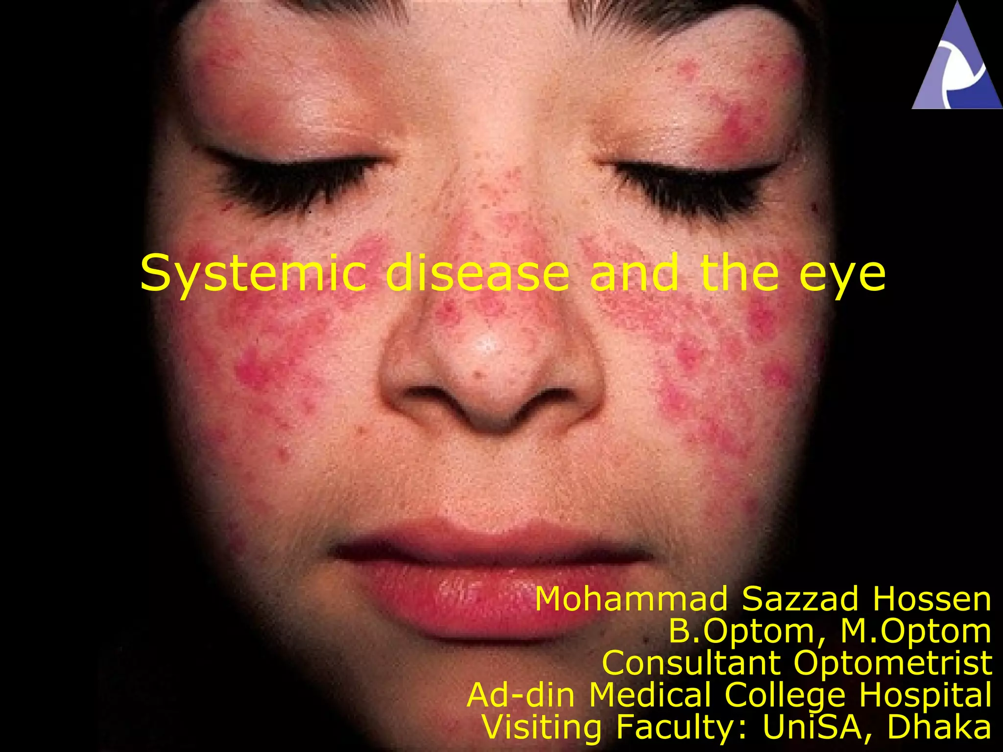
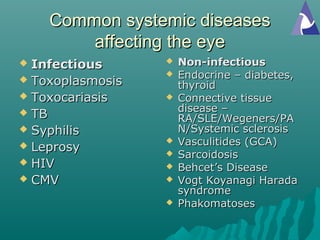
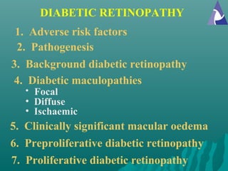
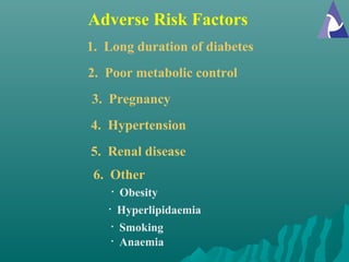
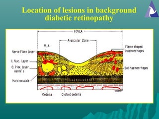
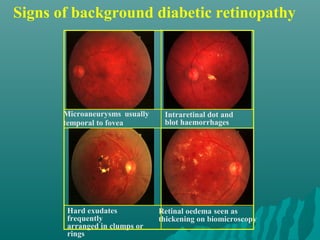
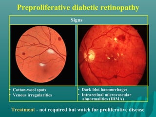
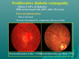
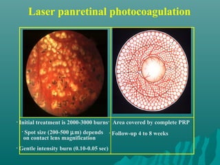
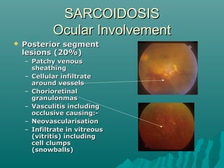
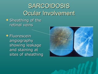
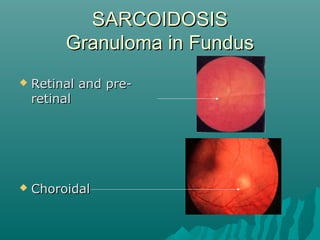
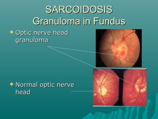
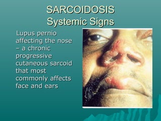
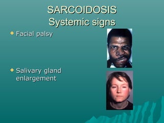
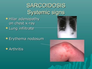
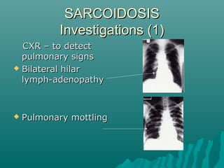
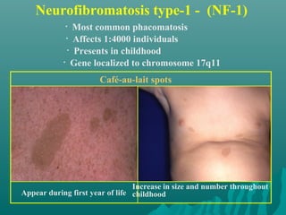
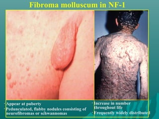
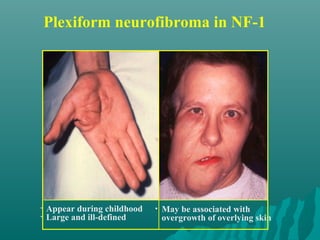
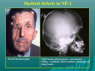
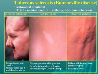
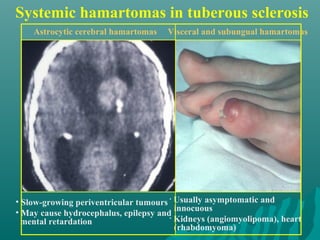
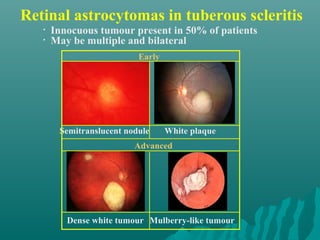
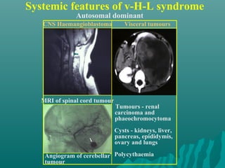
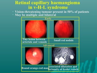
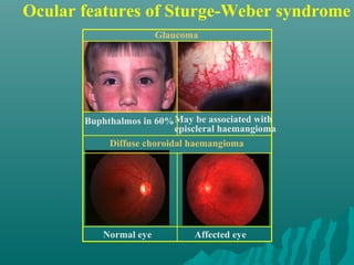
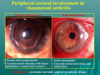
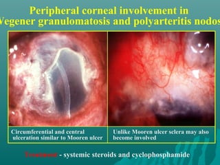
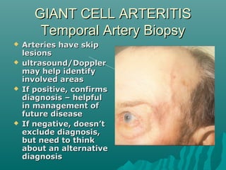
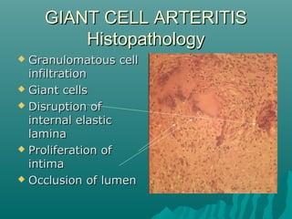
Mohammad Sazzad Hossen
B.Optom, M.Optom
Consultant Optometrist
Ad-din Medical College Hospital
Visiting Faculty: UniSA, Dhaka
Systemic disease and the eye
Common systemic diseasesCommon systemic diseases
affecting the eyeaffecting the eye
 InfectiousInfectious
 ToxoplasmosisToxoplasmosis
 ToxocariasisToxocariasis
 TBTB
 SyphilisSyphilis
 LeprosyLeprosy
 HIVHIV
 CMVCMV
 Non-infectiousNon-infectious
 Endocrine – diabetes,Endocrine – diabetes,
thyroidthyroid
 Connective tissueConnective tissue
disease –disease –
RA/SLE/Wegeners/PARA/SLE/Wegeners/PA
N/Systemic sclerosisN/Systemic sclerosis
 Vasculitides (GCA)Vasculitides (GCA)
 SarcoidosisSarcoidosis
 Behcet’s DiseaseBehcet’s Disease
 Vogt Koyanagi HaradaVogt Koyanagi Harada
syndromesyndrome
 PhakomatosesPhakomatoses
DIABETIC RETINOPATHY
1. Adverse risk factors
2. Pathogenesis
5. Clinically significant macular oedema
6. Preproliferative diabetic retinopathy
3. Background diabetic retinopathy
4. Diabetic maculopathies
• Focal
• Diffuse
• Ischaemic
7. Proliferative diabetic retinopathy
Adverse Risk Factors
1. Long duration of diabetes
• Obesity
• Hyperlipidaemia
2. Poor metabolic control
3. Pregnancy
4. Hypertension
5. Renal disease
6. Other
• Smoking
• Anaemia
Location of lesions in background
diabetic retinopathy
Signs of background diabetic retinopathy
Microaneurysms usually
temporal to fovea
Intraretinal dot and
blot haemorrhages
Hard exudates
frequently
arranged in clumps or
rings
Retinal oedema seen as
thickening on biomicroscopy
Preproliferative diabetic retinopathy
Treatment - not required but watch for proliferative disease
• Cotton-wool spots
• Venous irregularities
• Dark blot haemorrhages
• Intraretinal microvascular
abnormalities (IRMA)
Signs
Proliferative diabetic retinopathy
• Flat or elevated
• Severity determined by comparing with area of disc
Neovascularization
Neovascularization of disc = NVD
• Affects 5-10% of diabetics
• IDD at increased risk (60% after 30 years)
Neovascularization elsewhere = NVE
• Spot size (200-500 µm) depends
on contact lens magnification
• Gentle intensity burn (0.10-0.05 sec)
• Follow-up 4 to 8 weeks
• Area covered by complete PRP• Initial treatment is 2000-3000 burns
Laser panretinal photocoagulation
Retinal Vein OcclusionRetinal Vein Occlusion
Second most common cause of vascular-related visual loss.
Risk factors: hypertension, age, blood dyscrasias (OCP,HRT) and
vasculitis (Behcets,sarcoidosis,AIDS,SLE)
Retinal Artery OcclusionRetinal Artery Occlusion
Risk factors: Carotid artery atherosclerosis (CRAO), carotid emboli
(BRAO), vasculitis (GCA,SLE,PAN), coagulopathy.
OCULAR EMERGENCY - Immediate referral to ophthalmologist
1. Soft tissue involvement
• Periorbital and lid swelling
• Conjunctival hyperaemia
• Chemosis
• Superior limbic keratoconjunctivitis
2. Eyelid retraction
3. Proptosis
4. Optic neuropathy
5. Restrictive myopathy
THYROID EYE DISEASE
Soft tissue involvement
Periorbital and lid swelling
Chemosis
Conjunctival hyperaemia
Superior limbic
keratoconjunctivitis
Signs of eyelid retraction
Occurs in about 50%
• Bilateral lid retraction
• No associated proptosis
• Bilateral lid retraction
• Bilateral proptosis
• Lid lag in downgaze• Unilateral lid retraction
• Unilateral proptosis
Proptosis
Treatment options
• Systemic steroids
• Radiotherapy
• Surgical decompression
• Occurs in about 50%
• Uninfluenced by treatment of hyperthyroidism
Axial and permanent in about 70% May be associated with choroidal folds
Optic neuropathy
• Occurs in about 5%
• Early defective colour vision
• Usually normal disc appearance
Caused by optic nerve
compression at
orbital apex by enlarged recti
Often occurs in absence of significant
proptosis
• Occurs in about 40%
• Due to fibrotic contracture
Restrictive myopathy
Elevation defect - most common Abduction defect - less common
Depression defect - uncommon Adduction defect - rare
SARCOIDOSISSARCOIDOSIS
 Idiopathic multisystem disorderIdiopathic multisystem disorder
 Characterised by non-caseatingCharacterised by non-caseating
granulomatagranulomata
 More common in women 20-50 yrsMore common in women 20-50 yrs
 More common in blacks and AsiansMore common in blacks and Asians
 ? Related to mycobacteria? Related to mycobacteria
SARCOIDOSISSARCOIDOSIS
Systemic InvolvementSystemic Involvement
 Lung lesions –Lung lesions –
95%95%
 Thoracic lymphThoracic lymph
nodes – 50%nodes – 50%
 Skin lesions – 30%Skin lesions – 30%
→→
 Eyes – 30%Eyes – 30%
SARCOIDOSISSARCOIDOSIS
Ocular InvolvementOcular Involvement
 Anterior segmentAnterior segment
lesions (30%)lesions (30%)
– ConjunctivalConjunctival
granulomagranuloma
– Lacrimal glandLacrimal gland
involvement/dry eyeinvolvement/dry eye
– Acute or chronicAcute or chronic
uveitisuveitis →→
– KPs described asKPs described as
‘mutton fat’ because‘mutton fat’ because
they are large andthey are large and
greasygreasy
SARCOIDOSISSARCOIDOSIS
Ocular InvolvementOcular Involvement
 Posterior segmentPosterior segment
lesions (20%)lesions (20%)
– Patchy venousPatchy venous
sheathingsheathing
– Cellular infiltrateCellular infiltrate
around vesselsaround vessels
– ChorioretinalChorioretinal
granulonmasgranulonmas
– Vasculitis includingVasculitis including
occlusive causing:-occlusive causing:-
– NeovascularisationNeovascularisation
– Infiltrate in vitreousInfiltrate in vitreous
(vitritis) including(vitritis) including
cell clumpscell clumps
(snowballs)(snowballs)
SARCOIDOSISSARCOIDOSIS
Ocular InvolvementOcular Involvement
 Sheathing of theSheathing of the
retinal veinsretinal veins
 FluoresceinFluorescein
angiographyangiography
showing leakageshowing leakage
and staining atand staining at
sites of sheathingsites of sheathing
SARCOIDOSISSARCOIDOSIS
Granuloma in FundusGranuloma in Fundus
 Retinal and pre-Retinal and pre-
retinalretinal
 ChoroidalChoroidal
SARCOIDOSISSARCOIDOSIS
Granuloma in FundusGranuloma in Fundus
 Optic nerve headOptic nerve head
granulomagranuloma
 Normal optic nerveNormal optic nerve
headhead
SARCOIDOSISSARCOIDOSIS
Systemic SignsSystemic Signs
Lupus pernioLupus pernio
affecting the noseaffecting the nose
– a chronic– a chronic
progressiveprogressive
cutaneous sarcoidcutaneous sarcoid
that mostthat most
commonly affectscommonly affects
face and earsface and ears
SARCOIDOSISSARCOIDOSIS
Systemic signsSystemic signs
 Facial palsyFacial palsy
 Salivary glandSalivary gland
enlargementenlargement
SARCOIDOSISSARCOIDOSIS
Systemic signsSystemic signs
 Hilar adenopathyHilar adenopathy
on chest x-rayon chest x-ray
 Lung infiltrateLung infiltrate
 Erythema nodosumErythema nodosum
 ArthritisArthritis
SARCOIDOSISSARCOIDOSIS
Investigations (1)Investigations (1)
CXR – to detectCXR – to detect
pulmonary signspulmonary signs
 Bilateral hilarBilateral hilar
lymph-adenopathylymph-adenopathy
 Pulmonary mottlingPulmonary mottling
SARCOIDOSISSARCOIDOSIS
Investigations (2)Investigations (2)
 Serum angiotensin-convertingSerum angiotensin-converting
enzyme (ACE) – elevated in activeenzyme (ACE) – elevated in active
sarcoidosissarcoidosis
 Mantoux test – caution in patientsMantoux test – caution in patients
who have had BCG vaccination. Testwho have had BCG vaccination. Test
may be negativemay be negative
 Lung function testsLung function tests
SARCOIDOSISSARCOIDOSIS
Investigations (3)Investigations (3)
Gallium scanGallium scan
showing increasedshowing increased
uptake in theuptake in the
lacrimal andlacrimal and
parotid glands andparotid glands and
pulmonary regionspulmonary regions
in a patient within a patient with
active sarcoidosisactive sarcoidosis
SARCOIDOSISSARCOIDOSIS
TreatmentTreatment
Systemic steroids may be necessarySystemic steroids may be necessary
in patients with posterior segmentin patients with posterior segment
disease where vision is threatened,disease where vision is threatened,
especially if optic nerve is involvedespecially if optic nerve is involved
PHACOMATOSES
1. Neurofibromatosis
2. Tuberous sclerosis (Bourneville disease)
3. von-Hippel-Lindau syndrome
4. Sturge-Weber syndrome
• Type I (NF-1) - von Recklinghausen disease
• Type II (NF-2) - bilateral acoustic neuromas
Neurofibromatosis type-1 - (NF-1)
Appear during first year of life
Café-au-lait spots
• Most common phacomatosis
Increase in size and number throughout
childhood
• Affects 1:4000 individuals
• Presents in childhood
• Gene localized to chromosome 17q11
Fibroma molluscum in NF-1
• Appear at puberty
• Pedunculated, flabby nodules consisting of
neurofibromas or schwannomas
• Increase in number
throughout life
• Frequently widely distributed
Plexiform neurofibroma in NF-1
• May be associated with
overgrowth of overlying skin
• Appear during childhood
• Large and ill-defined
Skeletal defects in NF-1
• Mild head enlargement - uncommon
• Other - scoliosis, short stature, thinning of
long bones
• Facial hemiatrophy
Orbital lesions in NF-1
Spheno-orbital encephaloceleOptic nerve glioma in about 15%
• Sagittal MRI scan of optic nerve glioma
invading hypothalamus
• Glioma may be unilateral or bilateral
• Axial CT scan of congenital absence of
left greater wing of sphenoid bone
• Causes pulsating proptosis without bruit
Eyelid neurofibromas in NF-1
Nodular Plexiform
May cause mechanical ptosis May be associated with glaucoma
Intraocular lesions in NF-1
Lisch nodules
Very common - eventually present
in 95% of cases
Congenital ectropion uveae
Uncommon - may be associated
with glaucoma
Retinal astrocytomas
Rare - identical to those seen in
tuberous sclerosis
Choroidal naevi
Common - may be multifocal
and bilateral
Ocular features of NF-2
Common - combined hamartomas of RPE
and retina
Very common -presenile cataract
Tuberous sclerosis (Bourneville disease)
• Diffuse thickening over
lumbar region
• Present in 40%
Shagreen patches
• Autosomal dominant
• Triad - mental handicap, epilepsy, adenoma sebaceum
Adenoma sebaceum
• Around nose and
cheeks
• Appear after age 1
and slowly enlarge
Ash leaf spots
• Hypopigmented skin patches
• In infants best detected using
ultraviolet light (Wood’s lamp)
Systemic hamartomas in tuberous sclerosis
Astrocytic cerebral hamartomas
• Slow-growing periventricular tumours
• May cause hydrocephalus, epilepsy and
mental retardation
• Usually asymptomatic and
innocuous
• Kidneys (angiomyolipoma), heart
(rhabdomyoma)
Visceral and subungual hamartomas
Retinal astrocytomas in tuberous scleritis
Dense white tumour Mulberry-like tumour
Early
• Innocuous tumour present in 50% of patients
• May be multiple and bilateral
Semitranslucent nodule White plaque
Advanced
Systemic features of v-H-L syndrome
Autosomal dominant
• Tumours - renal
carcinoma and
phaeochromocytoma
• Cysts - kidneys, liver,
pancreas, epididymis,
ovary and lungs
• Polycythaemia
CNS Haemangioblastoma
MRI of spinal cord tumour
Angiogram of cerebellar
tumour
Visceral tumours
Retinal capillary haemangioma
in v-H-L syndrome
Round orange-red mass
Early
• Vision-threatening tumour present in 50% of patients
• May be multiple and bilateral
Tiny lesion between
arteriole and venuole
Small red nodule
Associated dilatation and
tortuosity of feeder vessels
Advanced
Systemic features of Sturge-Weber syndrome
• Congenital, does not blanche
with pressure
• Associated with ipsilateral
glaucoma in 30% of cases
Naevus flammeus
• CT scan showing left
parietal haemangioma
• Complications - mental handicap,
epilepsy and hemiparesis
Meningeal haemangioma
Ocular features of Sturge-Weber syndrome
Normal eye
Buphthalmos in 60%May be associated with
episcleral haemangioma
Affected eye
Diffuse choroidal haemangioma
Glaucoma
Peripheral corneal involvement in
rheumatoid arthritis
• Chronic and asymptomatic
• Circumferential thinning with intact
epithelium (‘contact lens cornea’)
• Acute and painful
• Circumferential ulceration and
infiltration
Treatment - systemic steroids and/or cytotoxic drugs
Without inflammation With inflammation
Peripheral corneal involvement in
Wegener granulomatosis and polyarteritis nodos
Circumferential and central
ulceration similar to Mooren ulcer
Unlike Mooren ulcer sclera may also
become involved
Treatment - systemic steroids and cyclophosphamide
GIANT CELL ARTERITISGIANT CELL ARTERITIS
(Temporal or Cranial Arteritis)(Temporal or Cranial Arteritis)
 Idiopathic vasculitisIdiopathic vasculitis
 Same disease spectrum asSame disease spectrum as
polymyalgia rheumaticapolymyalgia rheumatica
 Mainly women 65-80 years oldMainly women 65-80 years old
 Medium and large arteries in head &Medium and large arteries in head &
neck involvedneck involved
GIANT CELL ARTERITISGIANT CELL ARTERITIS
PresentationPresentation
 HeadacheHeadache
 Scalp tendernessScalp tenderness
 ThickenedThickened
temporal arteriestemporal arteries
 Jaw claudicationJaw claudication
 Acute visual lossAcute visual loss
 Weight loss,Weight loss,
anorexia, fever,anorexia, fever,
night sweats,night sweats,
malaise &malaise &
depressiondepression
GIANT CELL ARTERITISGIANT CELL ARTERITIS
Ocular ComplicationsOcular Complications
 TransientTransient
monocular visualmonocular visual
loss (amaurosisloss (amaurosis
fugax)fugax)
 Visual loss due toVisual loss due to
– Central retinalCentral retinal
artery occlusionartery occlusion
(CRAO) or(CRAO) or
– AnteriorAnterior
ischaemic opticischaemic optic
neuropathyneuropathy
(AION)(AION)
 Visual field defectsVisual field defects
GIANT CELL ARTERITISGIANT CELL ARTERITIS
ManagementManagement
 ESR if suspectedESR if suspected
 Start high dose steroids immediatelyStart high dose steroids immediately
to prevent stroke or second eyeto prevent stroke or second eye
involvementinvolvement
 Temporal artery biopsy within aTemporal artery biopsy within a
week of starting steroidsweek of starting steroids
GIANT CELL ARTERITISGIANT CELL ARTERITIS
Temporal Artery BiopsyTemporal Artery Biopsy
 Arteries have skipArteries have skip
lesionslesions
 ultrasound/Dopplerultrasound/Doppler
may help identifymay help identify
involved areasinvolved areas
 If positive, confirmsIf positive, confirms
diagnosis – helpfuldiagnosis – helpful
in management ofin management of
future diseasefuture disease
 If negative, doesn’tIf negative, doesn’t
exclude diagnosis,exclude diagnosis,
but need to thinkbut need to think
about an alternativeabout an alternative
diagnosisdiagnosis
GIANT CELL ARTERITISGIANT CELL ARTERITIS
HistopathologyHistopathology
 Granulomatous cellGranulomatous cell
infiltrationinfiltration
 Giant cellsGiant cells
 Disruption ofDisruption of
internal elasticinternal elastic
laminalamina
 Proliferation ofProliferation of
intimaintima
 Occlusion of lumenOcclusion of lumen
GIANT CELL ARTERITISGIANT CELL ARTERITIS
TreatmentTreatment
 Intravenous and oral steroids –Intravenous and oral steroids –
prolonged course of steroids oftenprolonged course of steroids often
necessarynecessary
Ocular manifestationsOcular manifestations
of HIV infectionof HIV infection
IntroductionIntroduction
 AIDS is an infectious disease caused by theAIDS is an infectious disease caused by the
gradual decrease ingradual decrease in CD4+ T lymphocytesCD4+ T lymphocytes
causing subsequent opportunistic infectionscausing subsequent opportunistic infections
and neoplasia. It is a blood borne and sexuallyand neoplasia. It is a blood borne and sexually
transmitted infection caused by the HIVtransmitted infection caused by the HIV
(Human Immunodeficiency Virus)(Human Immunodeficiency Virus)
 Approximately 36 million persons around theApproximately 36 million persons around the
world are infected. Up to 70% of patientsworld are infected. Up to 70% of patients
infected with HIV will develop some form ofinfected with HIV will develop some form of
ocular involvement, ie: direct infection byocular involvement, ie: direct infection by
HIV,opportunistic infections and neoplasia.HIV,opportunistic infections and neoplasia.
 HIV infection progresses thoughHIV infection progresses though
different phasesdifferent phases
Ophthalmic Manifestations of HIV InfectionOphthalmic Manifestations of HIV Infection
 AROUND THE EYEAROUND THE EYE
– Molluscum ContagiosumMolluscum Contagiosum
– Herpes Zoster OphthalmicusHerpes Zoster Ophthalmicus
– Kaposi’s SarcomaKaposi’s Sarcoma
– Conjunctival Squamous Cell CarcinomaConjunctival Squamous Cell Carcinoma
– TrichomegalyTrichomegaly
 FRONT OF THE EYEFRONT OF THE EYE
– Dry EyeDry Eye
– Anterior UveitisAnterior Uveitis
 BACK OF THE EYEBACK OF THE EYE
– Retinal MicrovasculopathyRetinal Microvasculopathy
– CMV RetinitisCMV Retinitis
– Acute Retinal NecrosisAcute Retinal Necrosis
– Progressive Outer Retinal NeProgressive Outer Retinal Ne
– Toxoplasmosis RetinochoroidToxoplasmosis Retinochoroid
– Syphilis RetinitisSyphilis Retinitis
– Candida albicans endophthalCandida albicans endophthal
 NEURO-OPHTHALMICNEURO-OPHTHALMIC
Molluscum ContagiosumMolluscum Contagiosum
 Molluscum contagiosum isMolluscum contagiosum is
a viral infection of the skin.a viral infection of the skin.
 Affects up to 20% ofAffects up to 20% of
symptomatic HIV infectedsymptomatic HIV infected
patients.patients.
 Clinically appears likeClinically appears like
painless, small, umbilicatedpainless, small, umbilicated
nodules, which produce anodules, which produce a
waxy discharge whenwaxy discharge when
pressured.pressured.
 Treatment consists onTreatment consists on
excision of the lesion,excision of the lesion,
curettage or cryotherapycurettage or cryotherapy
Herpes Zoster OphthalmicusHerpes Zoster Ophthalmicus
 Due to the reactivation of a latent infection byDue to the reactivation of a latent infection by
Varicella Zoster Virus in the dorsal root ofVaricella Zoster Virus in the dorsal root of
trigeminal nerve ganglion.trigeminal nerve ganglion.
 It manifests with a maculo-papulo-vesicular rashIt manifests with a maculo-papulo-vesicular rash
which often is preceded by pain. Usually involveswhich often is preceded by pain. Usually involves
the upper lid and does not cross the midlinethe upper lid and does not cross the midline
 Treatment consists on oral Aciclovir 800mg 5Treatment consists on oral Aciclovir 800mg 5
times /day. In immunocompromised patientstimes /day. In immunocompromised patients
Aciclovir is given intravenously for two weeks.Aciclovir is given intravenously for two weeks.
Ocular manifestations such as anterior uveitis,Ocular manifestations such as anterior uveitis,
are treated with topical steroids and mydriatics.are treated with topical steroids and mydriatics.
Kaposi’s SarcomaKaposi’s Sarcoma
 Kaposi’s sarcoma is a vascular neoplasm which isKaposi’s sarcoma is a vascular neoplasm which is
almost exclusively seen in patients with AIDS.almost exclusively seen in patients with AIDS.
 KS is the commonest anterior segment lesion seen inKS is the commonest anterior segment lesion seen in
AIDS; appears as a violaceous non-tender nodule onAIDS; appears as a violaceous non-tender nodule on
the eyelid or conjunctiva.the eyelid or conjunctiva.
 Typically KS involves only the skin but when there isTypically KS involves only the skin but when there is
a reduced CD4 count it can progress rapidly to othera reduced CD4 count it can progress rapidly to other
sites such as the gastrointestinal tract and CNSsites such as the gastrointestinal tract and CNS
 Treatment of ocular adnexal KS may be necessaryTreatment of ocular adnexal KS may be necessary
for cosmesis and to relieve functional difficulties. Thefor cosmesis and to relieve functional difficulties. The
mainstay of treatment is radiotherapy. Other optionsmainstay of treatment is radiotherapy. Other options
include cryotherapy or chemotherapy.include cryotherapy or chemotherapy.
Conjunctival Squamous Cell CarcinomaConjunctival Squamous Cell Carcinoma
 Squamous cell carcinoma (SCC) is the third mostSquamous cell carcinoma (SCC) is the third most
common neoplasm associated to HIV infection.common neoplasm associated to HIV infection.
This may be due to an interaction between HIV,This may be due to an interaction between HIV,
sunlight and Human Papilloma Virus infection.sunlight and Human Papilloma Virus infection.
 SCC appears as a pink, gelatinous growth, usuallySCC appears as a pink, gelatinous growth, usually
in the interpalpebral area. Often an engorgedin the interpalpebral area. Often an engorged
blood vessel feeding the tumour is seen. It mayblood vessel feeding the tumour is seen. It may
extend onto the cornea, but deep invasion andextend onto the cornea, but deep invasion and
metastasis are rare.metastasis are rare.
 The treatment of choice is local excision andThe treatment of choice is local excision and
cryotherapy but the presence of orbital invasioncryotherapy but the presence of orbital invasion
is an indication of exenterationis an indication of exenteration
TrichomegalyTrichomegaly
 Trichomegaly orTrichomegaly or
hypertrichosis is anhypertrichosis is an
exaggerated growth ofexaggerated growth of
the eye lashes foundthe eye lashes found
in the later stages ofin the later stages of
the diseasethe disease
 The cause is notThe cause is not
knownknown
 When symptomatic orWhen symptomatic or
for cosmetic reasonsfor cosmetic reasons
the eyelashes can bethe eyelashes can be
trimmed or pluckedtrimmed or plucked
Dry EyeDry Eye
 Sicca syndrome isSicca syndrome is
frequent amongfrequent among
patients with HIVpatients with HIV
infectioninfection
 Patients complain ofPatients complain of
burningburning
uncomfortable reduncomfortable red
eyes.eyes.
 There are severalThere are several
causes of dry eye incauses of dry eye in
HIV infection fromHIV infection from
blepharitis toblepharitis to
destruction of thedestruction of the
lacrimal glands.lacrimal glands.
 Treatment is withTreatment is with
Anterior UveitisAnterior Uveitis
 HIV related anteriorHIV related anterior uveitisuveitis
can be:can be:
– Direct manifestation of theDirect manifestation of the
human immunodeficiencyhuman immunodeficiency
virus infectionvirus infection
– autoimmnune in originautoimmnune in origin
– drug induced ie: rifabutin,drug induced ie: rifabutin,
secondary to direct toxicsecondary to direct toxic
effect upon the non-effect upon the non-
pigmented epithelium ofpigmented epithelium of
the ciliary bodythe ciliary body
– Any of the differentAny of the different
infections associated withinfections associated with
AIDS, ie: Herpes ZosterAIDS, ie: Herpes Zoster
Virus, Herpes SimplexVirus, Herpes Simplex
Rifabutin induced anterior uveitisRifabutin induced anterior uveitis
Retinal microvasculitisRetinal microvasculitis
 Retinal microvasculopathy occurs in more than half ofRetinal microvasculopathy occurs in more than half of
the patients with HIVthe patients with HIV
 It is seen as transient cotton wool spots (CWS), intra-It is seen as transient cotton wool spots (CWS), intra-
retinal haemorrhages and microaneurysm, which occursretinal haemorrhages and microaneurysm, which occurs
in 50-70% of patients. It is usually asymptomatic.in 50-70% of patients. It is usually asymptomatic.
 It has an unclear pathogenesis, but it is thought to beIt has an unclear pathogenesis, but it is thought to be
HIV infection of retinal vascular cells.HIV infection of retinal vascular cells.
 In an otherwise healthy individual the presence ofIn an otherwise healthy individual the presence of
CWS, should be differentiated from other forms ofCWS, should be differentiated from other forms of
retinopathy, such as diabetic or hypertensiveretinopathy, such as diabetic or hypertensive
retinopathy. Serological test for HIV will confirm theretinopathy. Serological test for HIV will confirm the
diagnosisdiagnosis
 Treatment is based in delaying the progression of theTreatment is based in delaying the progression of the
Cotton Wool SpotsCotton Wool Spots
CMV RetinitisCMV Retinitis
 IntroductionIntroduction
– CMV Retinitis is the commonest intraocular ocularCMV Retinitis is the commonest intraocular ocular
opportunistic infection seen in patients with AIDSopportunistic infection seen in patients with AIDS
– Antibodies are found in almost 95% of adults,Antibodies are found in almost 95% of adults,
causing a trivial illness in immunocompetent adults,causing a trivial illness in immunocompetent adults,
however severe immunosuppression causes viralhowever severe immunosuppression causes viral
reactivation and tissue invasive diseasereactivation and tissue invasive disease
 PathogenesisPathogenesis
– Reactivation from extraocular sites leads to seedingReactivation from extraocular sites leads to seeding
in other sites such as the retinain other sites such as the retina
 EpidemiologyEpidemiology
– The number of newly diagnosed cases of CMVR hasThe number of newly diagnosed cases of CMVR has
decreased since the introduction of the HAARTdecreased since the introduction of the HAART
Highly Active Antiretroviral TherapyHighly Active Antiretroviral Therapy
CMV RetinitisCMV Retinitis
 Clinical manifestationsClinical manifestations
– Patients may complain of minor visual symptoms such asPatients may complain of minor visual symptoms such as
floaters, flashing lights or mild blurred vision, or be totallyfloaters, flashing lights or mild blurred vision, or be totally
asymptomatic.asymptomatic.
– It presents with a wide range of clinical appearances. FromIt presents with a wide range of clinical appearances. From
cotton wool spots which may look like HIV Retinopathy tocotton wool spots which may look like HIV Retinopathy to
confluent areas of full thickness retinal necrosis and vasculitis.confluent areas of full thickness retinal necrosis and vasculitis.
CMVR can progress in a “brushfire” pattern from the activeCMVR can progress in a “brushfire” pattern from the active
edge of an active lesion. The retinal vessels in an affected areaedge of an active lesion. The retinal vessels in an affected area
show attenuation, becoming ghost vessels eventually.show attenuation, becoming ghost vessels eventually.
 TreatmentTreatment
– The treatment of CMVR in patients with AIDS requires the useThe treatment of CMVR in patients with AIDS requires the use
of specific antiviral agents, ganciclovir, foscarnet or cidovir inof specific antiviral agents, ganciclovir, foscarnet or cidovir in
conjunction with HAART.conjunction with HAART.
– These treatments can be administered orally, intravenously orThese treatments can be administered orally, intravenously or
intravitreally. Systemic treatment has the advantage of treatingintravitreally. Systemic treatment has the advantage of treating
infection elsewhere in the body as well as the other eye but hasinfection elsewhere in the body as well as the other eye but has
CMV RetinitisCMV Retinitis
Acute Retinal NecrosisAcute Retinal Necrosis
 ARN is a confluent peripheral whitening of theARN is a confluent peripheral whitening of the
retina with marked vitritis and blood vesselretina with marked vitritis and blood vessel
closure. Optic neuritis and retinal detachment areclosure. Optic neuritis and retinal detachment are
frequent complications.frequent complications.
 ARN is usually due to Varicella-Zoster infection,ARN is usually due to Varicella-Zoster infection,
but it can also be caused by Herpes Simplex virusbut it can also be caused by Herpes Simplex virus
or Cytomegalovirus.or Cytomegalovirus.
 Initially described in the immunocompetent, it hasInitially described in the immunocompetent, it has
also been described in the immunosuppressed.also been described in the immunosuppressed.
 The diagnosis is mainly clinical and is confirmedThe diagnosis is mainly clinical and is confirmed
by PCR assays on vitreous samples.by PCR assays on vitreous samples.
 Patients are treated with high doses ofPatients are treated with high doses of
intravenous aciclovir or famciclovir, combined withintravenous aciclovir or famciclovir, combined with
laser treatment to prevent retinal detachment.laser treatment to prevent retinal detachment.
Acute Retinal NecrosisAcute Retinal Necrosis
Progressive Outer RetinalProgressive Outer Retinal
NecrosisNecrosis
(Varicella-Zoster Retinitis)(Varicella-Zoster Retinitis)
 PORN is a devastating viral retinitis caused by Varicella-PORN is a devastating viral retinitis caused by Varicella-
Zoster virus, without vitritis or retinal vasculitis.Zoster virus, without vitritis or retinal vasculitis.
 The retinitis can be located anywhere but it is commonThe retinitis can be located anywhere but it is common
for the lesions to coalesce and spread posteriorly in afor the lesions to coalesce and spread posteriorly in a
rapid fashion.rapid fashion.
 The main symptom is rapid loss of vision.The retinaThe main symptom is rapid loss of vision.The retina
shows typically a white lesion with no haemorrhages orshows typically a white lesion with no haemorrhages or
exudates.exudates.
 Treatment is often unsatisfactory and usually requiresTreatment is often unsatisfactory and usually requires
combination of Ganciclovir and Aciclovir. The prognosiscombination of Ganciclovir and Aciclovir. The prognosis
is very poor and retinal detachment is common.is very poor and retinal detachment is common.
Resolution may leave a white plaque with theResolution may leave a white plaque with the
appearance of “cracked mud”.appearance of “cracked mud”.
Toxoplasma RetinochoroiditisToxoplasma Retinochoroiditis
 Toxoplasmosis retinochoroiditis is an uncommonToxoplasmosis retinochoroiditis is an uncommon
infection of the eye in AIDS. Ocular toxoplasmosisinfection of the eye in AIDS. Ocular toxoplasmosis
in HIV positive patients is different in appearancein HIV positive patients is different in appearance
from immunocompetent patients. Unlike infrom immunocompetent patients. Unlike in
immunocompetent patients, HIV infected patientsimmunocompetent patients, HIV infected patients
often have bilateral and multifocal diseaseoften have bilateral and multifocal disease
associated with anterior uveitis and vitritis butassociated with anterior uveitis and vitritis but
unlike immunocompetent patients, in HIVunlike immunocompetent patients, in HIV
infected patients often have with no pigmentedinfected patients often have with no pigmented
scars adjacent to the areas of retinal necrosis.scars adjacent to the areas of retinal necrosis.
Toxoplasmosis in immunocompromised patientsToxoplasmosis in immunocompromised patients
is not self-limiting as it is in imunocompetentis not self-limiting as it is in imunocompetent
patients.patients.
Toxoplasma RetinochoroiditisToxoplasma Retinochoroiditis
 When testing patients for antibodies toWhen testing patients for antibodies to
toxoplasmosis both IgG and IgM levels may betoxoplasmosis both IgG and IgM levels may be
raised, but in immunocompromised patientsraised, but in immunocompromised patients
these tests may be negative.these tests may be negative.
 Treatment in immunocompromised patientsTreatment in immunocompromised patients
consists in the association of sulphadiazine orconsists in the association of sulphadiazine or
clindamycin, pyrimethamine and folinic acidclindamycin, pyrimethamine and folinic acid
(triple therapy).(triple therapy).
 Long term maintenance treatment may beLong term maintenance treatment may be
needed in order to prevent relapses.needed in order to prevent relapses.
 Often associated with toxoplasma lesions in theOften associated with toxoplasma lesions in the
Central Nervous System.Central Nervous System.
MRI T1 showing an uniformly
enhancing lesion in the
midbrain
One week later, the lesion
showing ring enhancement
ImmunocompetentImmunocompetent ImmunocompromisedImmunocompromised
Syphilis RetinitisSyphilis Retinitis
 There is a strong association betweenThere is a strong association between
syphilis and HIV infection.syphilis and HIV infection.
 It can manifest as a retinitis with denseIt can manifest as a retinitis with dense
vitritis, retinal vasculitis, serous retinalvitritis, retinal vasculitis, serous retinal
detachment or neuroretinitis, as well as otherdetachment or neuroretinitis, as well as other
types of ocular involvement such as,types of ocular involvement such as,
conjunctivitis, anterior uveitis, cranial nerveconjunctivitis, anterior uveitis, cranial nerve
palsies and optic neuritis.palsies and optic neuritis.
 Treatment consists in high dose ofTreatment consists in high dose of
intravenous Penicillin for 2 weeks.intravenous Penicillin for 2 weeks.
Candida albicansCandida albicans
endophthalmitisendophthalmitis
 Infection with candida albicans is rare. CandidaInfection with candida albicans is rare. Candida
albicans is the commonest cause of fungalalbicans is the commonest cause of fungal
endophthalmitisendophthalmitis
 Affected patients usually have a history of drugAffected patients usually have a history of drug
abuse or indwelling central linesabuse or indwelling central lines
 In the initial stages, floaters are the mainIn the initial stages, floaters are the main
symptom. As the condition progresses, whitishsymptom. As the condition progresses, whitish
“puff-balls” and vitreous strands develop. Later,“puff-balls” and vitreous strands develop. Later,
similar infiltrates appear in the choroid and retinasimilar infiltrates appear in the choroid and retina
 The treatment depends on the severity of theThe treatment depends on the severity of the
ocular involvement and systemic disease. Theocular involvement and systemic disease. The
original foci should be removed. The drugs oforiginal foci should be removed. The drugs of
choice are Amphotericine B and Fluconazolchoice are Amphotericine B and Fluconazol
Candida albicansCandida albicans
endophthalmitisendophthalmitis
GlossaryGlossary
 CD4CD4: Director of the immune response. When activated: Director of the immune response. When activated
it releasesit releases cytokines which in turn will activate thecytokines which in turn will activate the
immune systemimmune system
 Cotton Wool SpotsCotton Wool Spots: Light-coloured deposits in the: Light-coloured deposits in the
retina secondaryretina secondary to infarcts of the nerve fibre layerto infarcts of the nerve fibre layer
 HAARTHAART: Highly Active Antiretroviral Therapy: Highly Active Antiretroviral Therapy
 ImmunoblogulinImmunoblogulin: Protein in charge of fighting foreign: Protein in charge of fighting foreign
substances insubstances in our body.our body. IgGIgG is the commonest type ofis the commonest type of
immunoglobulin andimmunoglobulin and IgMIgM is theis the
earliest class of immunoglobulin.earliest class of immunoglobulin.
 PCRPCR: Polymerase Chain Reaction is a technique used to: Polymerase Chain Reaction is a technique used to
makemake numerous copies of an specific portionnumerous copies of an specific portion
of DNAof DNA
 VDRLVDRL: Venereal Disease Research Laboratory. The test: Venereal Disease Research Laboratory. The test
Questions?

Systemic disease SR

  • 1.
    Mohammad Sazzad Hossen B.Optom,M.Optom Consultant Optometrist Ad-din Medical College Hospital Visiting Faculty: UniSA, Dhaka Systemic disease and the eye
  • 2.
    Common systemic diseasesCommonsystemic diseases affecting the eyeaffecting the eye  InfectiousInfectious  ToxoplasmosisToxoplasmosis  ToxocariasisToxocariasis  TBTB  SyphilisSyphilis  LeprosyLeprosy  HIVHIV  CMVCMV  Non-infectiousNon-infectious  Endocrine – diabetes,Endocrine – diabetes, thyroidthyroid  Connective tissueConnective tissue disease –disease – RA/SLE/Wegeners/PARA/SLE/Wegeners/PA N/Systemic sclerosisN/Systemic sclerosis  Vasculitides (GCA)Vasculitides (GCA)  SarcoidosisSarcoidosis  Behcet’s DiseaseBehcet’s Disease  Vogt Koyanagi HaradaVogt Koyanagi Harada syndromesyndrome  PhakomatosesPhakomatoses
  • 3.
    DIABETIC RETINOPATHY 1. Adverserisk factors 2. Pathogenesis 5. Clinically significant macular oedema 6. Preproliferative diabetic retinopathy 3. Background diabetic retinopathy 4. Diabetic maculopathies • Focal • Diffuse • Ischaemic 7. Proliferative diabetic retinopathy
  • 4.
    Adverse Risk Factors 1.Long duration of diabetes • Obesity • Hyperlipidaemia 2. Poor metabolic control 3. Pregnancy 4. Hypertension 5. Renal disease 6. Other • Smoking • Anaemia
  • 5.
    Location of lesionsin background diabetic retinopathy
  • 6.
    Signs of backgrounddiabetic retinopathy Microaneurysms usually temporal to fovea Intraretinal dot and blot haemorrhages Hard exudates frequently arranged in clumps or rings Retinal oedema seen as thickening on biomicroscopy
  • 7.
    Preproliferative diabetic retinopathy Treatment- not required but watch for proliferative disease • Cotton-wool spots • Venous irregularities • Dark blot haemorrhages • Intraretinal microvascular abnormalities (IRMA) Signs
  • 8.
    Proliferative diabetic retinopathy •Flat or elevated • Severity determined by comparing with area of disc Neovascularization Neovascularization of disc = NVD • Affects 5-10% of diabetics • IDD at increased risk (60% after 30 years) Neovascularization elsewhere = NVE
  • 9.
    • Spot size(200-500 µm) depends on contact lens magnification • Gentle intensity burn (0.10-0.05 sec) • Follow-up 4 to 8 weeks • Area covered by complete PRP• Initial treatment is 2000-3000 burns Laser panretinal photocoagulation
  • 10.
    Retinal Vein OcclusionRetinalVein Occlusion Second most common cause of vascular-related visual loss. Risk factors: hypertension, age, blood dyscrasias (OCP,HRT) and vasculitis (Behcets,sarcoidosis,AIDS,SLE)
  • 11.
    Retinal Artery OcclusionRetinalArtery Occlusion Risk factors: Carotid artery atherosclerosis (CRAO), carotid emboli (BRAO), vasculitis (GCA,SLE,PAN), coagulopathy. OCULAR EMERGENCY - Immediate referral to ophthalmologist
  • 12.
    1. Soft tissueinvolvement • Periorbital and lid swelling • Conjunctival hyperaemia • Chemosis • Superior limbic keratoconjunctivitis 2. Eyelid retraction 3. Proptosis 4. Optic neuropathy 5. Restrictive myopathy THYROID EYE DISEASE
  • 13.
    Soft tissue involvement Periorbitaland lid swelling Chemosis Conjunctival hyperaemia Superior limbic keratoconjunctivitis
  • 14.
    Signs of eyelidretraction Occurs in about 50% • Bilateral lid retraction • No associated proptosis • Bilateral lid retraction • Bilateral proptosis • Lid lag in downgaze• Unilateral lid retraction • Unilateral proptosis
  • 15.
    Proptosis Treatment options • Systemicsteroids • Radiotherapy • Surgical decompression • Occurs in about 50% • Uninfluenced by treatment of hyperthyroidism Axial and permanent in about 70% May be associated with choroidal folds
  • 16.
    Optic neuropathy • Occursin about 5% • Early defective colour vision • Usually normal disc appearance Caused by optic nerve compression at orbital apex by enlarged recti Often occurs in absence of significant proptosis
  • 17.
    • Occurs inabout 40% • Due to fibrotic contracture Restrictive myopathy Elevation defect - most common Abduction defect - less common Depression defect - uncommon Adduction defect - rare
  • 18.
    SARCOIDOSISSARCOIDOSIS  Idiopathic multisystemdisorderIdiopathic multisystem disorder  Characterised by non-caseatingCharacterised by non-caseating granulomatagranulomata  More common in women 20-50 yrsMore common in women 20-50 yrs  More common in blacks and AsiansMore common in blacks and Asians  ? Related to mycobacteria? Related to mycobacteria
  • 19.
    SARCOIDOSISSARCOIDOSIS Systemic InvolvementSystemic Involvement Lung lesions –Lung lesions – 95%95%  Thoracic lymphThoracic lymph nodes – 50%nodes – 50%  Skin lesions – 30%Skin lesions – 30% →→  Eyes – 30%Eyes – 30%
  • 20.
    SARCOIDOSISSARCOIDOSIS Ocular InvolvementOcular Involvement Anterior segmentAnterior segment lesions (30%)lesions (30%) – ConjunctivalConjunctival granulomagranuloma – Lacrimal glandLacrimal gland involvement/dry eyeinvolvement/dry eye – Acute or chronicAcute or chronic uveitisuveitis →→ – KPs described asKPs described as ‘mutton fat’ because‘mutton fat’ because they are large andthey are large and greasygreasy
  • 21.
    SARCOIDOSISSARCOIDOSIS Ocular InvolvementOcular Involvement Posterior segmentPosterior segment lesions (20%)lesions (20%) – Patchy venousPatchy venous sheathingsheathing – Cellular infiltrateCellular infiltrate around vesselsaround vessels – ChorioretinalChorioretinal granulonmasgranulonmas – Vasculitis includingVasculitis including occlusive causing:-occlusive causing:- – NeovascularisationNeovascularisation – Infiltrate in vitreousInfiltrate in vitreous (vitritis) including(vitritis) including cell clumpscell clumps (snowballs)(snowballs)
  • 22.
    SARCOIDOSISSARCOIDOSIS Ocular InvolvementOcular Involvement Sheathing of theSheathing of the retinal veinsretinal veins  FluoresceinFluorescein angiographyangiography showing leakageshowing leakage and staining atand staining at sites of sheathingsites of sheathing
  • 23.
    SARCOIDOSISSARCOIDOSIS Granuloma in FundusGranulomain Fundus  Retinal and pre-Retinal and pre- retinalretinal  ChoroidalChoroidal
  • 24.
    SARCOIDOSISSARCOIDOSIS Granuloma in FundusGranulomain Fundus  Optic nerve headOptic nerve head granulomagranuloma  Normal optic nerveNormal optic nerve headhead
  • 25.
    SARCOIDOSISSARCOIDOSIS Systemic SignsSystemic Signs LupuspernioLupus pernio affecting the noseaffecting the nose – a chronic– a chronic progressiveprogressive cutaneous sarcoidcutaneous sarcoid that mostthat most commonly affectscommonly affects face and earsface and ears
  • 26.
    SARCOIDOSISSARCOIDOSIS Systemic signsSystemic signs Facial palsyFacial palsy  Salivary glandSalivary gland enlargementenlargement
  • 27.
    SARCOIDOSISSARCOIDOSIS Systemic signsSystemic signs Hilar adenopathyHilar adenopathy on chest x-rayon chest x-ray  Lung infiltrateLung infiltrate  Erythema nodosumErythema nodosum  ArthritisArthritis
  • 28.
    SARCOIDOSISSARCOIDOSIS Investigations (1)Investigations (1) CXR– to detectCXR – to detect pulmonary signspulmonary signs  Bilateral hilarBilateral hilar lymph-adenopathylymph-adenopathy  Pulmonary mottlingPulmonary mottling
  • 29.
    SARCOIDOSISSARCOIDOSIS Investigations (2)Investigations (2) Serum angiotensin-convertingSerum angiotensin-converting enzyme (ACE) – elevated in activeenzyme (ACE) – elevated in active sarcoidosissarcoidosis  Mantoux test – caution in patientsMantoux test – caution in patients who have had BCG vaccination. Testwho have had BCG vaccination. Test may be negativemay be negative  Lung function testsLung function tests
  • 30.
    SARCOIDOSISSARCOIDOSIS Investigations (3)Investigations (3) GalliumscanGallium scan showing increasedshowing increased uptake in theuptake in the lacrimal andlacrimal and parotid glands andparotid glands and pulmonary regionspulmonary regions in a patient within a patient with active sarcoidosisactive sarcoidosis
  • 31.
    SARCOIDOSISSARCOIDOSIS TreatmentTreatment Systemic steroids maybe necessarySystemic steroids may be necessary in patients with posterior segmentin patients with posterior segment disease where vision is threatened,disease where vision is threatened, especially if optic nerve is involvedespecially if optic nerve is involved
  • 32.
    PHACOMATOSES 1. Neurofibromatosis 2. Tuberoussclerosis (Bourneville disease) 3. von-Hippel-Lindau syndrome 4. Sturge-Weber syndrome • Type I (NF-1) - von Recklinghausen disease • Type II (NF-2) - bilateral acoustic neuromas
  • 33.
    Neurofibromatosis type-1 -(NF-1) Appear during first year of life Café-au-lait spots • Most common phacomatosis Increase in size and number throughout childhood • Affects 1:4000 individuals • Presents in childhood • Gene localized to chromosome 17q11
  • 34.
    Fibroma molluscum inNF-1 • Appear at puberty • Pedunculated, flabby nodules consisting of neurofibromas or schwannomas • Increase in number throughout life • Frequently widely distributed
  • 35.
    Plexiform neurofibroma inNF-1 • May be associated with overgrowth of overlying skin • Appear during childhood • Large and ill-defined
  • 36.
    Skeletal defects inNF-1 • Mild head enlargement - uncommon • Other - scoliosis, short stature, thinning of long bones • Facial hemiatrophy
  • 37.
    Orbital lesions inNF-1 Spheno-orbital encephaloceleOptic nerve glioma in about 15% • Sagittal MRI scan of optic nerve glioma invading hypothalamus • Glioma may be unilateral or bilateral • Axial CT scan of congenital absence of left greater wing of sphenoid bone • Causes pulsating proptosis without bruit
  • 38.
    Eyelid neurofibromas inNF-1 Nodular Plexiform May cause mechanical ptosis May be associated with glaucoma
  • 39.
    Intraocular lesions inNF-1 Lisch nodules Very common - eventually present in 95% of cases Congenital ectropion uveae Uncommon - may be associated with glaucoma Retinal astrocytomas Rare - identical to those seen in tuberous sclerosis Choroidal naevi Common - may be multifocal and bilateral
  • 40.
    Ocular features ofNF-2 Common - combined hamartomas of RPE and retina Very common -presenile cataract
  • 41.
    Tuberous sclerosis (Bournevilledisease) • Diffuse thickening over lumbar region • Present in 40% Shagreen patches • Autosomal dominant • Triad - mental handicap, epilepsy, adenoma sebaceum Adenoma sebaceum • Around nose and cheeks • Appear after age 1 and slowly enlarge Ash leaf spots • Hypopigmented skin patches • In infants best detected using ultraviolet light (Wood’s lamp)
  • 42.
    Systemic hamartomas intuberous sclerosis Astrocytic cerebral hamartomas • Slow-growing periventricular tumours • May cause hydrocephalus, epilepsy and mental retardation • Usually asymptomatic and innocuous • Kidneys (angiomyolipoma), heart (rhabdomyoma) Visceral and subungual hamartomas
  • 43.
    Retinal astrocytomas intuberous scleritis Dense white tumour Mulberry-like tumour Early • Innocuous tumour present in 50% of patients • May be multiple and bilateral Semitranslucent nodule White plaque Advanced
  • 44.
    Systemic features ofv-H-L syndrome Autosomal dominant • Tumours - renal carcinoma and phaeochromocytoma • Cysts - kidneys, liver, pancreas, epididymis, ovary and lungs • Polycythaemia CNS Haemangioblastoma MRI of spinal cord tumour Angiogram of cerebellar tumour Visceral tumours
  • 45.
    Retinal capillary haemangioma inv-H-L syndrome Round orange-red mass Early • Vision-threatening tumour present in 50% of patients • May be multiple and bilateral Tiny lesion between arteriole and venuole Small red nodule Associated dilatation and tortuosity of feeder vessels Advanced
  • 46.
    Systemic features ofSturge-Weber syndrome • Congenital, does not blanche with pressure • Associated with ipsilateral glaucoma in 30% of cases Naevus flammeus • CT scan showing left parietal haemangioma • Complications - mental handicap, epilepsy and hemiparesis Meningeal haemangioma
  • 47.
    Ocular features ofSturge-Weber syndrome Normal eye Buphthalmos in 60%May be associated with episcleral haemangioma Affected eye Diffuse choroidal haemangioma Glaucoma
  • 48.
    Peripheral corneal involvementin rheumatoid arthritis • Chronic and asymptomatic • Circumferential thinning with intact epithelium (‘contact lens cornea’) • Acute and painful • Circumferential ulceration and infiltration Treatment - systemic steroids and/or cytotoxic drugs Without inflammation With inflammation
  • 49.
    Peripheral corneal involvementin Wegener granulomatosis and polyarteritis nodos Circumferential and central ulceration similar to Mooren ulcer Unlike Mooren ulcer sclera may also become involved Treatment - systemic steroids and cyclophosphamide
  • 50.
    GIANT CELL ARTERITISGIANTCELL ARTERITIS (Temporal or Cranial Arteritis)(Temporal or Cranial Arteritis)  Idiopathic vasculitisIdiopathic vasculitis  Same disease spectrum asSame disease spectrum as polymyalgia rheumaticapolymyalgia rheumatica  Mainly women 65-80 years oldMainly women 65-80 years old  Medium and large arteries in head &Medium and large arteries in head & neck involvedneck involved
  • 51.
    GIANT CELL ARTERITISGIANTCELL ARTERITIS PresentationPresentation  HeadacheHeadache  Scalp tendernessScalp tenderness  ThickenedThickened temporal arteriestemporal arteries  Jaw claudicationJaw claudication  Acute visual lossAcute visual loss  Weight loss,Weight loss, anorexia, fever,anorexia, fever, night sweats,night sweats, malaise &malaise & depressiondepression
  • 52.
    GIANT CELL ARTERITISGIANTCELL ARTERITIS Ocular ComplicationsOcular Complications  TransientTransient monocular visualmonocular visual loss (amaurosisloss (amaurosis fugax)fugax)  Visual loss due toVisual loss due to – Central retinalCentral retinal artery occlusionartery occlusion (CRAO) or(CRAO) or – AnteriorAnterior ischaemic opticischaemic optic neuropathyneuropathy (AION)(AION)  Visual field defectsVisual field defects
  • 53.
    GIANT CELL ARTERITISGIANTCELL ARTERITIS ManagementManagement  ESR if suspectedESR if suspected  Start high dose steroids immediatelyStart high dose steroids immediately to prevent stroke or second eyeto prevent stroke or second eye involvementinvolvement  Temporal artery biopsy within aTemporal artery biopsy within a week of starting steroidsweek of starting steroids
  • 54.
    GIANT CELL ARTERITISGIANTCELL ARTERITIS Temporal Artery BiopsyTemporal Artery Biopsy  Arteries have skipArteries have skip lesionslesions  ultrasound/Dopplerultrasound/Doppler may help identifymay help identify involved areasinvolved areas  If positive, confirmsIf positive, confirms diagnosis – helpfuldiagnosis – helpful in management ofin management of future diseasefuture disease  If negative, doesn’tIf negative, doesn’t exclude diagnosis,exclude diagnosis, but need to thinkbut need to think about an alternativeabout an alternative diagnosisdiagnosis
  • 55.
    GIANT CELL ARTERITISGIANTCELL ARTERITIS HistopathologyHistopathology  Granulomatous cellGranulomatous cell infiltrationinfiltration  Giant cellsGiant cells  Disruption ofDisruption of internal elasticinternal elastic laminalamina  Proliferation ofProliferation of intimaintima  Occlusion of lumenOcclusion of lumen
  • 56.
    GIANT CELL ARTERITISGIANTCELL ARTERITIS TreatmentTreatment  Intravenous and oral steroids –Intravenous and oral steroids – prolonged course of steroids oftenprolonged course of steroids often necessarynecessary
  • 57.
    Ocular manifestationsOcular manifestations ofHIV infectionof HIV infection
  • 58.
    IntroductionIntroduction  AIDS isan infectious disease caused by theAIDS is an infectious disease caused by the gradual decrease ingradual decrease in CD4+ T lymphocytesCD4+ T lymphocytes causing subsequent opportunistic infectionscausing subsequent opportunistic infections and neoplasia. It is a blood borne and sexuallyand neoplasia. It is a blood borne and sexually transmitted infection caused by the HIVtransmitted infection caused by the HIV (Human Immunodeficiency Virus)(Human Immunodeficiency Virus)  Approximately 36 million persons around theApproximately 36 million persons around the world are infected. Up to 70% of patientsworld are infected. Up to 70% of patients infected with HIV will develop some form ofinfected with HIV will develop some form of ocular involvement, ie: direct infection byocular involvement, ie: direct infection by HIV,opportunistic infections and neoplasia.HIV,opportunistic infections and neoplasia.  HIV infection progresses thoughHIV infection progresses though different phasesdifferent phases
  • 60.
    Ophthalmic Manifestations ofHIV InfectionOphthalmic Manifestations of HIV Infection  AROUND THE EYEAROUND THE EYE – Molluscum ContagiosumMolluscum Contagiosum – Herpes Zoster OphthalmicusHerpes Zoster Ophthalmicus – Kaposi’s SarcomaKaposi’s Sarcoma – Conjunctival Squamous Cell CarcinomaConjunctival Squamous Cell Carcinoma – TrichomegalyTrichomegaly  FRONT OF THE EYEFRONT OF THE EYE – Dry EyeDry Eye – Anterior UveitisAnterior Uveitis  BACK OF THE EYEBACK OF THE EYE – Retinal MicrovasculopathyRetinal Microvasculopathy – CMV RetinitisCMV Retinitis – Acute Retinal NecrosisAcute Retinal Necrosis – Progressive Outer Retinal NeProgressive Outer Retinal Ne – Toxoplasmosis RetinochoroidToxoplasmosis Retinochoroid – Syphilis RetinitisSyphilis Retinitis – Candida albicans endophthalCandida albicans endophthal  NEURO-OPHTHALMICNEURO-OPHTHALMIC
  • 61.
    Molluscum ContagiosumMolluscum Contagiosum Molluscum contagiosum isMolluscum contagiosum is a viral infection of the skin.a viral infection of the skin.  Affects up to 20% ofAffects up to 20% of symptomatic HIV infectedsymptomatic HIV infected patients.patients.  Clinically appears likeClinically appears like painless, small, umbilicatedpainless, small, umbilicated nodules, which produce anodules, which produce a waxy discharge whenwaxy discharge when pressured.pressured.  Treatment consists onTreatment consists on excision of the lesion,excision of the lesion, curettage or cryotherapycurettage or cryotherapy
  • 62.
    Herpes Zoster OphthalmicusHerpesZoster Ophthalmicus  Due to the reactivation of a latent infection byDue to the reactivation of a latent infection by Varicella Zoster Virus in the dorsal root ofVaricella Zoster Virus in the dorsal root of trigeminal nerve ganglion.trigeminal nerve ganglion.  It manifests with a maculo-papulo-vesicular rashIt manifests with a maculo-papulo-vesicular rash which often is preceded by pain. Usually involveswhich often is preceded by pain. Usually involves the upper lid and does not cross the midlinethe upper lid and does not cross the midline  Treatment consists on oral Aciclovir 800mg 5Treatment consists on oral Aciclovir 800mg 5 times /day. In immunocompromised patientstimes /day. In immunocompromised patients Aciclovir is given intravenously for two weeks.Aciclovir is given intravenously for two weeks. Ocular manifestations such as anterior uveitis,Ocular manifestations such as anterior uveitis, are treated with topical steroids and mydriatics.are treated with topical steroids and mydriatics.
  • 64.
    Kaposi’s SarcomaKaposi’s Sarcoma Kaposi’s sarcoma is a vascular neoplasm which isKaposi’s sarcoma is a vascular neoplasm which is almost exclusively seen in patients with AIDS.almost exclusively seen in patients with AIDS.  KS is the commonest anterior segment lesion seen inKS is the commonest anterior segment lesion seen in AIDS; appears as a violaceous non-tender nodule onAIDS; appears as a violaceous non-tender nodule on the eyelid or conjunctiva.the eyelid or conjunctiva.  Typically KS involves only the skin but when there isTypically KS involves only the skin but when there is a reduced CD4 count it can progress rapidly to othera reduced CD4 count it can progress rapidly to other sites such as the gastrointestinal tract and CNSsites such as the gastrointestinal tract and CNS  Treatment of ocular adnexal KS may be necessaryTreatment of ocular adnexal KS may be necessary for cosmesis and to relieve functional difficulties. Thefor cosmesis and to relieve functional difficulties. The mainstay of treatment is radiotherapy. Other optionsmainstay of treatment is radiotherapy. Other options include cryotherapy or chemotherapy.include cryotherapy or chemotherapy.
  • 66.
    Conjunctival Squamous CellCarcinomaConjunctival Squamous Cell Carcinoma  Squamous cell carcinoma (SCC) is the third mostSquamous cell carcinoma (SCC) is the third most common neoplasm associated to HIV infection.common neoplasm associated to HIV infection. This may be due to an interaction between HIV,This may be due to an interaction between HIV, sunlight and Human Papilloma Virus infection.sunlight and Human Papilloma Virus infection.  SCC appears as a pink, gelatinous growth, usuallySCC appears as a pink, gelatinous growth, usually in the interpalpebral area. Often an engorgedin the interpalpebral area. Often an engorged blood vessel feeding the tumour is seen. It mayblood vessel feeding the tumour is seen. It may extend onto the cornea, but deep invasion andextend onto the cornea, but deep invasion and metastasis are rare.metastasis are rare.  The treatment of choice is local excision andThe treatment of choice is local excision and cryotherapy but the presence of orbital invasioncryotherapy but the presence of orbital invasion is an indication of exenterationis an indication of exenteration
  • 68.
    TrichomegalyTrichomegaly  Trichomegaly orTrichomegalyor hypertrichosis is anhypertrichosis is an exaggerated growth ofexaggerated growth of the eye lashes foundthe eye lashes found in the later stages ofin the later stages of the diseasethe disease  The cause is notThe cause is not knownknown  When symptomatic orWhen symptomatic or for cosmetic reasonsfor cosmetic reasons the eyelashes can bethe eyelashes can be trimmed or pluckedtrimmed or plucked
  • 69.
    Dry EyeDry Eye Sicca syndrome isSicca syndrome is frequent amongfrequent among patients with HIVpatients with HIV infectioninfection  Patients complain ofPatients complain of burningburning uncomfortable reduncomfortable red eyes.eyes.  There are severalThere are several causes of dry eye incauses of dry eye in HIV infection fromHIV infection from blepharitis toblepharitis to destruction of thedestruction of the lacrimal glands.lacrimal glands.  Treatment is withTreatment is with
  • 70.
    Anterior UveitisAnterior Uveitis HIV related anteriorHIV related anterior uveitisuveitis can be:can be: – Direct manifestation of theDirect manifestation of the human immunodeficiencyhuman immunodeficiency virus infectionvirus infection – autoimmnune in originautoimmnune in origin – drug induced ie: rifabutin,drug induced ie: rifabutin, secondary to direct toxicsecondary to direct toxic effect upon the non-effect upon the non- pigmented epithelium ofpigmented epithelium of the ciliary bodythe ciliary body – Any of the differentAny of the different infections associated withinfections associated with AIDS, ie: Herpes ZosterAIDS, ie: Herpes Zoster Virus, Herpes SimplexVirus, Herpes Simplex
  • 71.
    Rifabutin induced anterioruveitisRifabutin induced anterior uveitis
  • 72.
    Retinal microvasculitisRetinal microvasculitis Retinal microvasculopathy occurs in more than half ofRetinal microvasculopathy occurs in more than half of the patients with HIVthe patients with HIV  It is seen as transient cotton wool spots (CWS), intra-It is seen as transient cotton wool spots (CWS), intra- retinal haemorrhages and microaneurysm, which occursretinal haemorrhages and microaneurysm, which occurs in 50-70% of patients. It is usually asymptomatic.in 50-70% of patients. It is usually asymptomatic.  It has an unclear pathogenesis, but it is thought to beIt has an unclear pathogenesis, but it is thought to be HIV infection of retinal vascular cells.HIV infection of retinal vascular cells.  In an otherwise healthy individual the presence ofIn an otherwise healthy individual the presence of CWS, should be differentiated from other forms ofCWS, should be differentiated from other forms of retinopathy, such as diabetic or hypertensiveretinopathy, such as diabetic or hypertensive retinopathy. Serological test for HIV will confirm theretinopathy. Serological test for HIV will confirm the diagnosisdiagnosis  Treatment is based in delaying the progression of theTreatment is based in delaying the progression of the
  • 73.
  • 74.
    CMV RetinitisCMV Retinitis IntroductionIntroduction – CMV Retinitis is the commonest intraocular ocularCMV Retinitis is the commonest intraocular ocular opportunistic infection seen in patients with AIDSopportunistic infection seen in patients with AIDS – Antibodies are found in almost 95% of adults,Antibodies are found in almost 95% of adults, causing a trivial illness in immunocompetent adults,causing a trivial illness in immunocompetent adults, however severe immunosuppression causes viralhowever severe immunosuppression causes viral reactivation and tissue invasive diseasereactivation and tissue invasive disease  PathogenesisPathogenesis – Reactivation from extraocular sites leads to seedingReactivation from extraocular sites leads to seeding in other sites such as the retinain other sites such as the retina  EpidemiologyEpidemiology – The number of newly diagnosed cases of CMVR hasThe number of newly diagnosed cases of CMVR has decreased since the introduction of the HAARTdecreased since the introduction of the HAART Highly Active Antiretroviral TherapyHighly Active Antiretroviral Therapy
  • 75.
    CMV RetinitisCMV Retinitis Clinical manifestationsClinical manifestations – Patients may complain of minor visual symptoms such asPatients may complain of minor visual symptoms such as floaters, flashing lights or mild blurred vision, or be totallyfloaters, flashing lights or mild blurred vision, or be totally asymptomatic.asymptomatic. – It presents with a wide range of clinical appearances. FromIt presents with a wide range of clinical appearances. From cotton wool spots which may look like HIV Retinopathy tocotton wool spots which may look like HIV Retinopathy to confluent areas of full thickness retinal necrosis and vasculitis.confluent areas of full thickness retinal necrosis and vasculitis. CMVR can progress in a “brushfire” pattern from the activeCMVR can progress in a “brushfire” pattern from the active edge of an active lesion. The retinal vessels in an affected areaedge of an active lesion. The retinal vessels in an affected area show attenuation, becoming ghost vessels eventually.show attenuation, becoming ghost vessels eventually.  TreatmentTreatment – The treatment of CMVR in patients with AIDS requires the useThe treatment of CMVR in patients with AIDS requires the use of specific antiviral agents, ganciclovir, foscarnet or cidovir inof specific antiviral agents, ganciclovir, foscarnet or cidovir in conjunction with HAART.conjunction with HAART. – These treatments can be administered orally, intravenously orThese treatments can be administered orally, intravenously or intravitreally. Systemic treatment has the advantage of treatingintravitreally. Systemic treatment has the advantage of treating infection elsewhere in the body as well as the other eye but hasinfection elsewhere in the body as well as the other eye but has
  • 76.
  • 77.
    Acute Retinal NecrosisAcuteRetinal Necrosis  ARN is a confluent peripheral whitening of theARN is a confluent peripheral whitening of the retina with marked vitritis and blood vesselretina with marked vitritis and blood vessel closure. Optic neuritis and retinal detachment areclosure. Optic neuritis and retinal detachment are frequent complications.frequent complications.  ARN is usually due to Varicella-Zoster infection,ARN is usually due to Varicella-Zoster infection, but it can also be caused by Herpes Simplex virusbut it can also be caused by Herpes Simplex virus or Cytomegalovirus.or Cytomegalovirus.  Initially described in the immunocompetent, it hasInitially described in the immunocompetent, it has also been described in the immunosuppressed.also been described in the immunosuppressed.  The diagnosis is mainly clinical and is confirmedThe diagnosis is mainly clinical and is confirmed by PCR assays on vitreous samples.by PCR assays on vitreous samples.  Patients are treated with high doses ofPatients are treated with high doses of intravenous aciclovir or famciclovir, combined withintravenous aciclovir or famciclovir, combined with laser treatment to prevent retinal detachment.laser treatment to prevent retinal detachment.
  • 78.
  • 79.
    Progressive Outer RetinalProgressiveOuter Retinal NecrosisNecrosis (Varicella-Zoster Retinitis)(Varicella-Zoster Retinitis)  PORN is a devastating viral retinitis caused by Varicella-PORN is a devastating viral retinitis caused by Varicella- Zoster virus, without vitritis or retinal vasculitis.Zoster virus, without vitritis or retinal vasculitis.  The retinitis can be located anywhere but it is commonThe retinitis can be located anywhere but it is common for the lesions to coalesce and spread posteriorly in afor the lesions to coalesce and spread posteriorly in a rapid fashion.rapid fashion.  The main symptom is rapid loss of vision.The retinaThe main symptom is rapid loss of vision.The retina shows typically a white lesion with no haemorrhages orshows typically a white lesion with no haemorrhages or exudates.exudates.  Treatment is often unsatisfactory and usually requiresTreatment is often unsatisfactory and usually requires combination of Ganciclovir and Aciclovir. The prognosiscombination of Ganciclovir and Aciclovir. The prognosis is very poor and retinal detachment is common.is very poor and retinal detachment is common. Resolution may leave a white plaque with theResolution may leave a white plaque with the appearance of “cracked mud”.appearance of “cracked mud”.
  • 81.
    Toxoplasma RetinochoroiditisToxoplasma Retinochoroiditis Toxoplasmosis retinochoroiditis is an uncommonToxoplasmosis retinochoroiditis is an uncommon infection of the eye in AIDS. Ocular toxoplasmosisinfection of the eye in AIDS. Ocular toxoplasmosis in HIV positive patients is different in appearancein HIV positive patients is different in appearance from immunocompetent patients. Unlike infrom immunocompetent patients. Unlike in immunocompetent patients, HIV infected patientsimmunocompetent patients, HIV infected patients often have bilateral and multifocal diseaseoften have bilateral and multifocal disease associated with anterior uveitis and vitritis butassociated with anterior uveitis and vitritis but unlike immunocompetent patients, in HIVunlike immunocompetent patients, in HIV infected patients often have with no pigmentedinfected patients often have with no pigmented scars adjacent to the areas of retinal necrosis.scars adjacent to the areas of retinal necrosis. Toxoplasmosis in immunocompromised patientsToxoplasmosis in immunocompromised patients is not self-limiting as it is in imunocompetentis not self-limiting as it is in imunocompetent patients.patients.
  • 82.
    Toxoplasma RetinochoroiditisToxoplasma Retinochoroiditis When testing patients for antibodies toWhen testing patients for antibodies to toxoplasmosis both IgG and IgM levels may betoxoplasmosis both IgG and IgM levels may be raised, but in immunocompromised patientsraised, but in immunocompromised patients these tests may be negative.these tests may be negative.  Treatment in immunocompromised patientsTreatment in immunocompromised patients consists in the association of sulphadiazine orconsists in the association of sulphadiazine or clindamycin, pyrimethamine and folinic acidclindamycin, pyrimethamine and folinic acid (triple therapy).(triple therapy).  Long term maintenance treatment may beLong term maintenance treatment may be needed in order to prevent relapses.needed in order to prevent relapses.  Often associated with toxoplasma lesions in theOften associated with toxoplasma lesions in the Central Nervous System.Central Nervous System.
  • 83.
    MRI T1 showingan uniformly enhancing lesion in the midbrain One week later, the lesion showing ring enhancement
  • 84.
  • 85.
    Syphilis RetinitisSyphilis Retinitis There is a strong association betweenThere is a strong association between syphilis and HIV infection.syphilis and HIV infection.  It can manifest as a retinitis with denseIt can manifest as a retinitis with dense vitritis, retinal vasculitis, serous retinalvitritis, retinal vasculitis, serous retinal detachment or neuroretinitis, as well as otherdetachment or neuroretinitis, as well as other types of ocular involvement such as,types of ocular involvement such as, conjunctivitis, anterior uveitis, cranial nerveconjunctivitis, anterior uveitis, cranial nerve palsies and optic neuritis.palsies and optic neuritis.  Treatment consists in high dose ofTreatment consists in high dose of intravenous Penicillin for 2 weeks.intravenous Penicillin for 2 weeks.
  • 87.
    Candida albicansCandida albicans endophthalmitisendophthalmitis Infection with candida albicans is rare. CandidaInfection with candida albicans is rare. Candida albicans is the commonest cause of fungalalbicans is the commonest cause of fungal endophthalmitisendophthalmitis  Affected patients usually have a history of drugAffected patients usually have a history of drug abuse or indwelling central linesabuse or indwelling central lines  In the initial stages, floaters are the mainIn the initial stages, floaters are the main symptom. As the condition progresses, whitishsymptom. As the condition progresses, whitish “puff-balls” and vitreous strands develop. Later,“puff-balls” and vitreous strands develop. Later, similar infiltrates appear in the choroid and retinasimilar infiltrates appear in the choroid and retina  The treatment depends on the severity of theThe treatment depends on the severity of the ocular involvement and systemic disease. Theocular involvement and systemic disease. The original foci should be removed. The drugs oforiginal foci should be removed. The drugs of choice are Amphotericine B and Fluconazolchoice are Amphotericine B and Fluconazol
  • 88.
  • 89.
    GlossaryGlossary  CD4CD4: Directorof the immune response. When activated: Director of the immune response. When activated it releasesit releases cytokines which in turn will activate thecytokines which in turn will activate the immune systemimmune system  Cotton Wool SpotsCotton Wool Spots: Light-coloured deposits in the: Light-coloured deposits in the retina secondaryretina secondary to infarcts of the nerve fibre layerto infarcts of the nerve fibre layer  HAARTHAART: Highly Active Antiretroviral Therapy: Highly Active Antiretroviral Therapy  ImmunoblogulinImmunoblogulin: Protein in charge of fighting foreign: Protein in charge of fighting foreign substances insubstances in our body.our body. IgGIgG is the commonest type ofis the commonest type of immunoglobulin andimmunoglobulin and IgMIgM is theis the earliest class of immunoglobulin.earliest class of immunoglobulin.  PCRPCR: Polymerase Chain Reaction is a technique used to: Polymerase Chain Reaction is a technique used to makemake numerous copies of an specific portionnumerous copies of an specific portion of DNAof DNA  VDRLVDRL: Venereal Disease Research Laboratory. The test: Venereal Disease Research Laboratory. The test
  • 90.

Editor's Notes