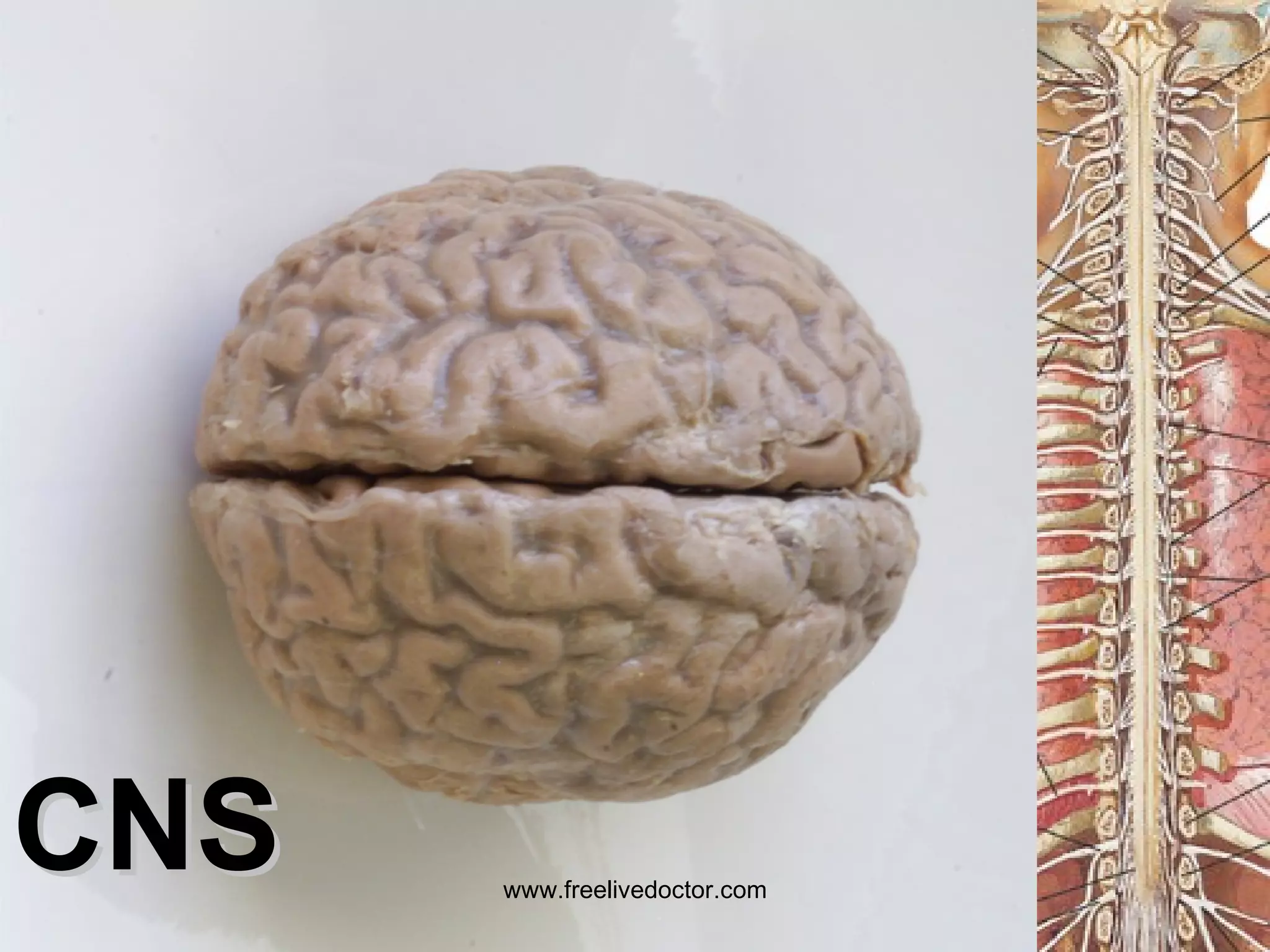
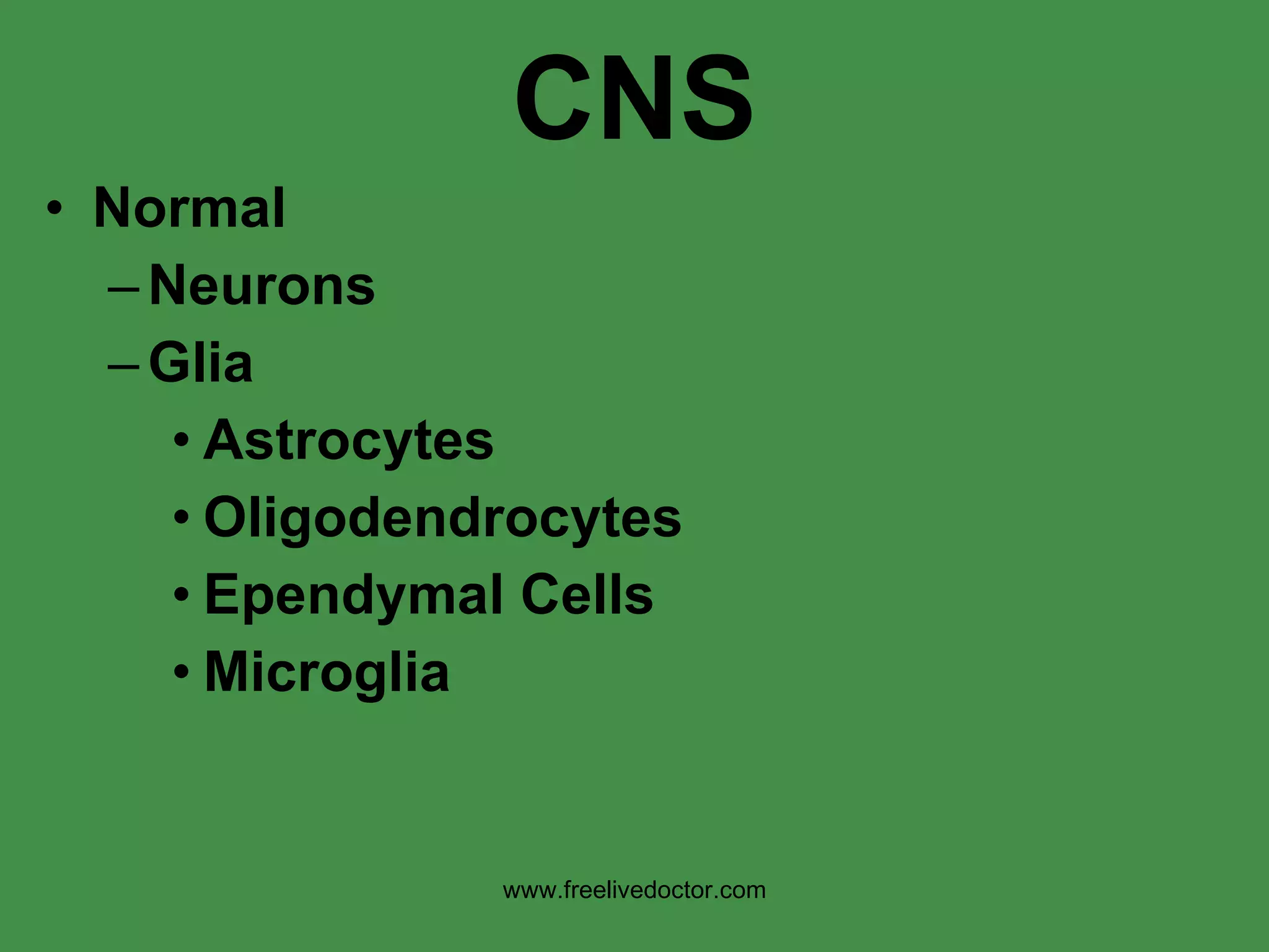
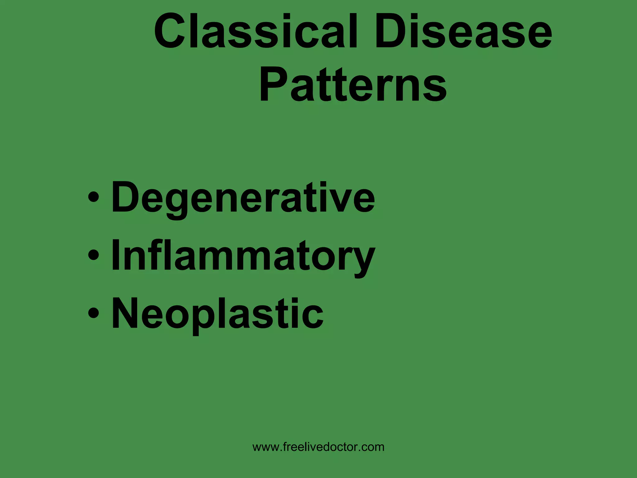
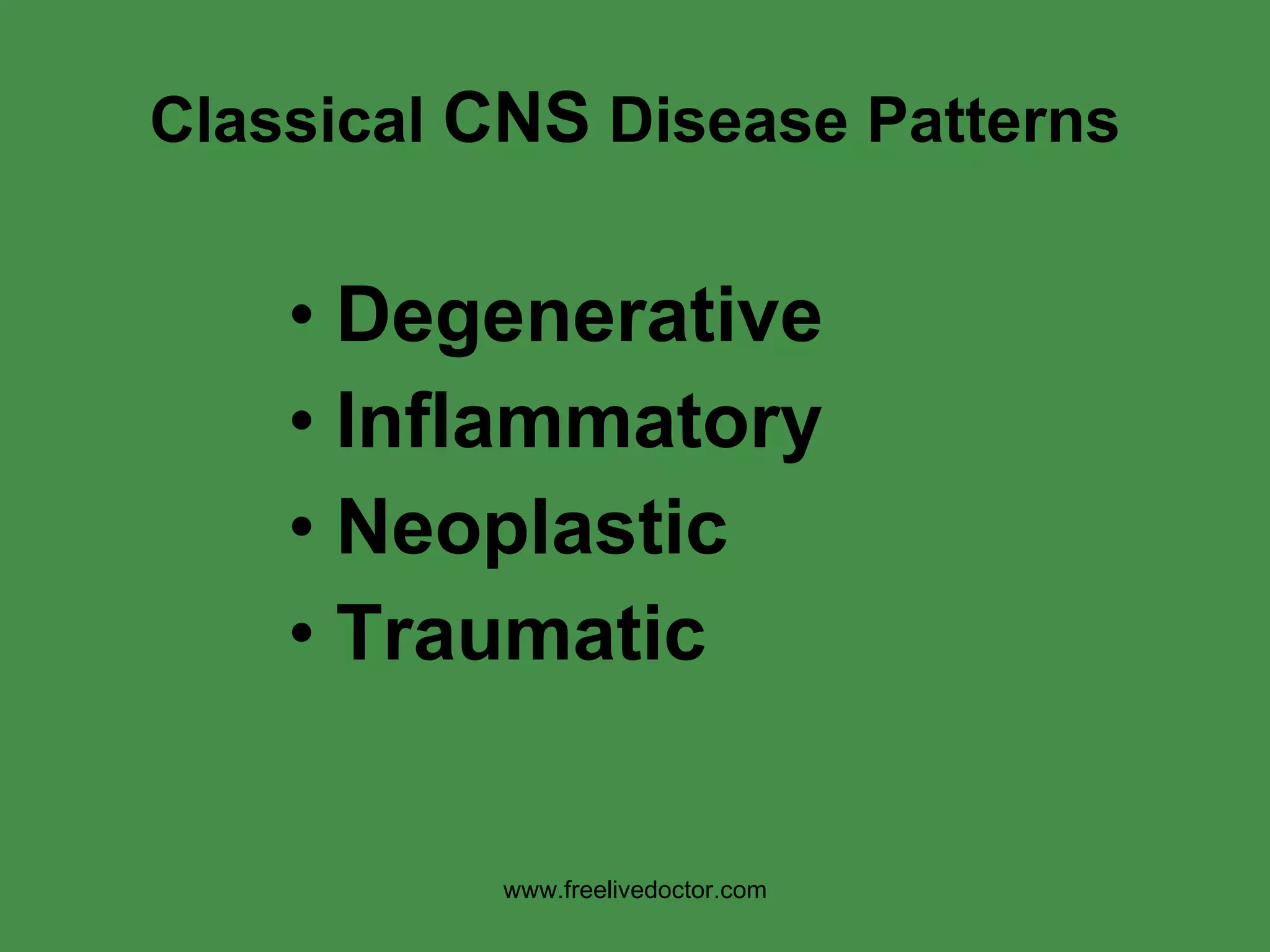
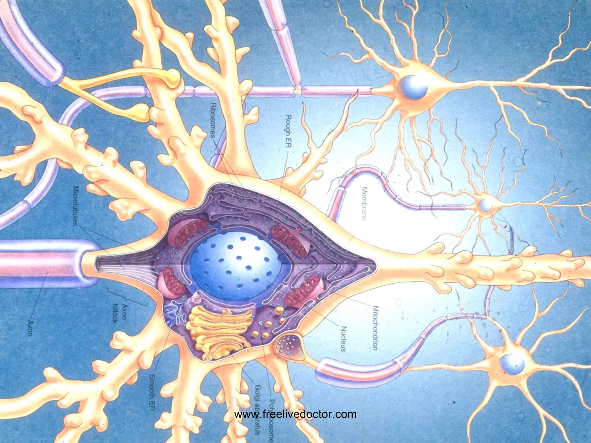
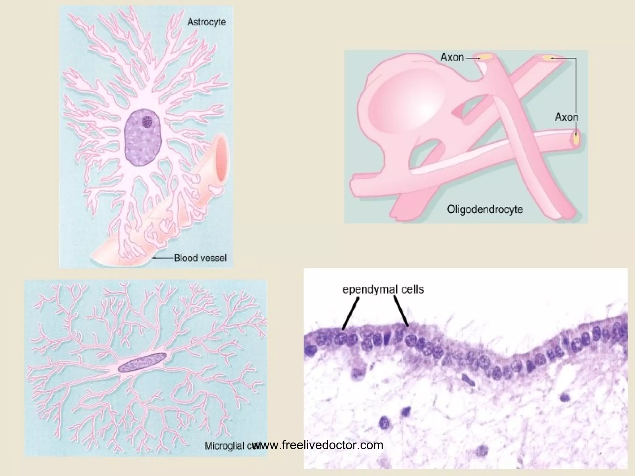
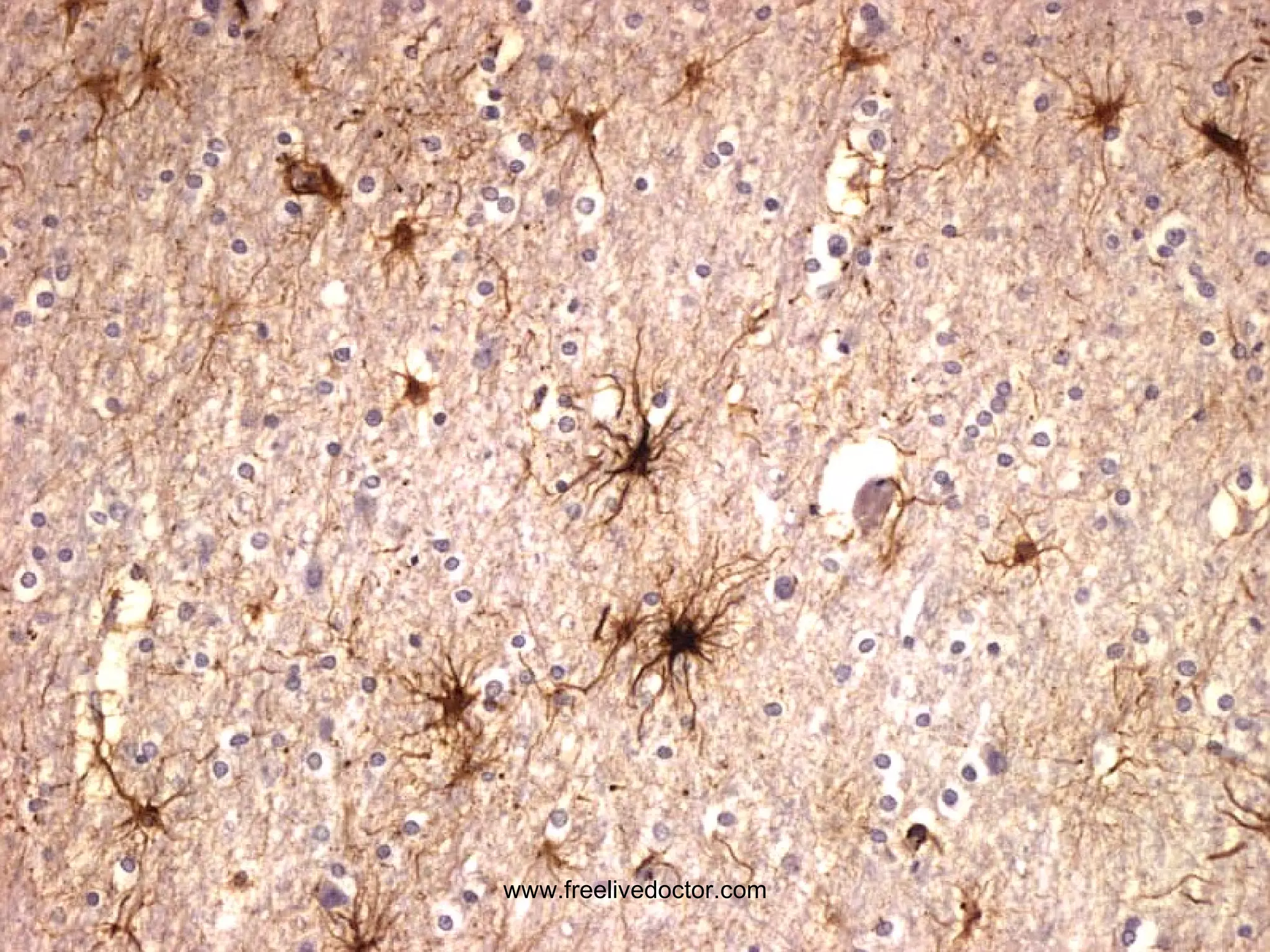
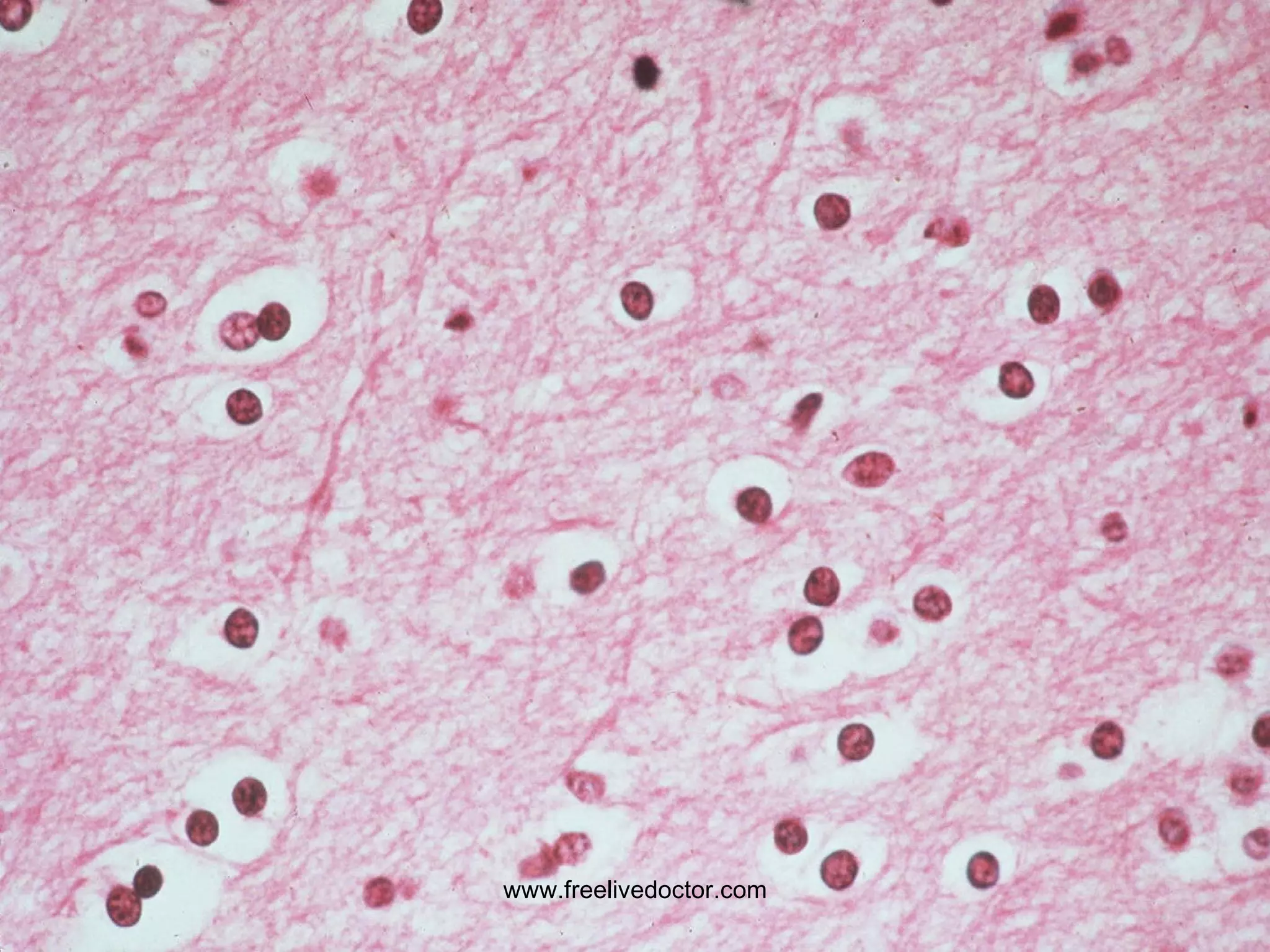
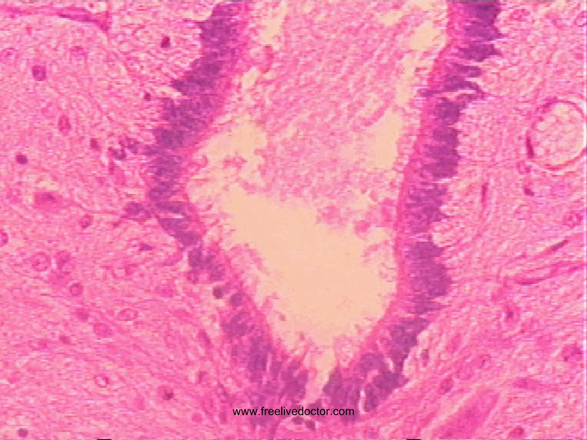
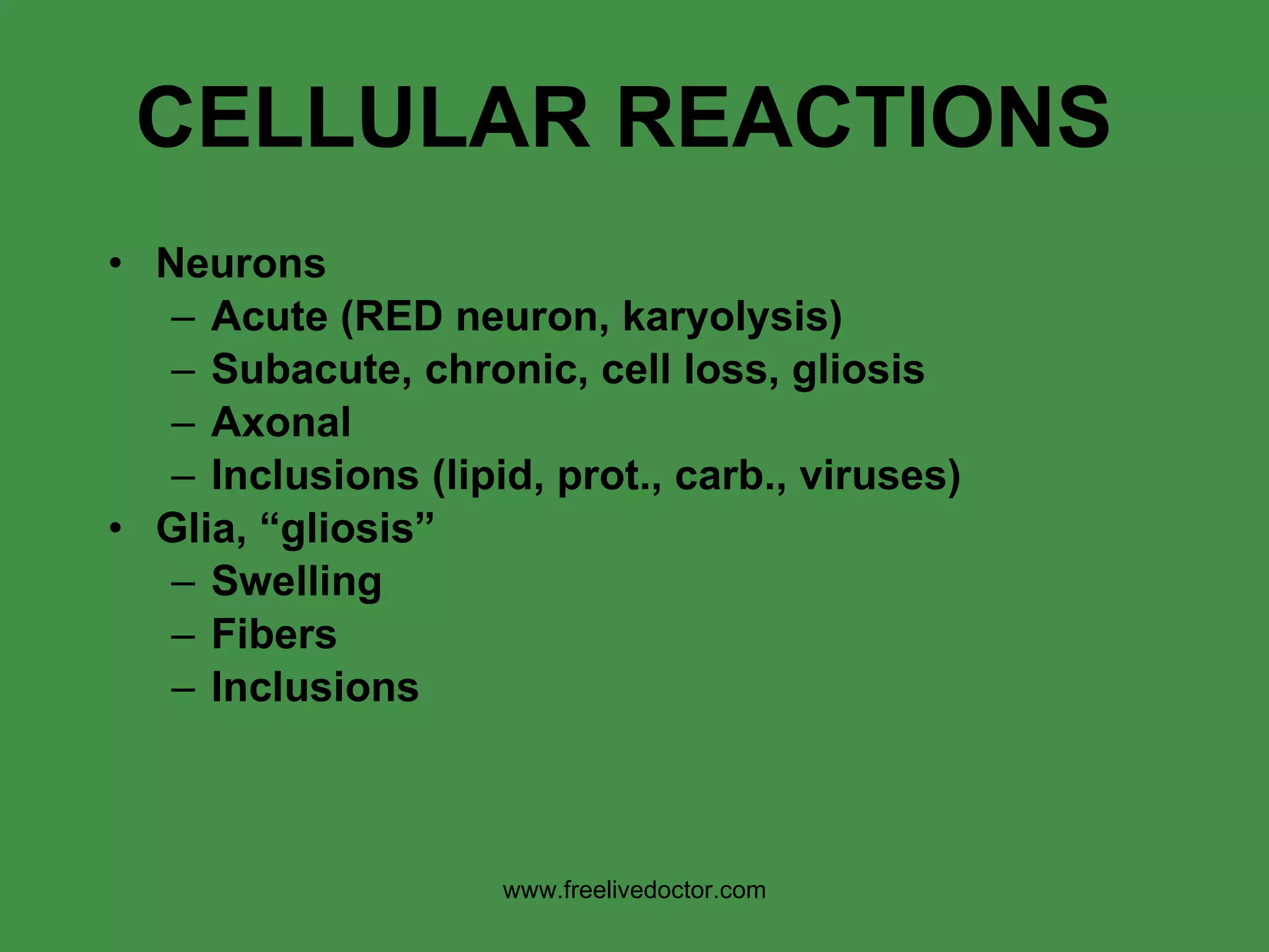
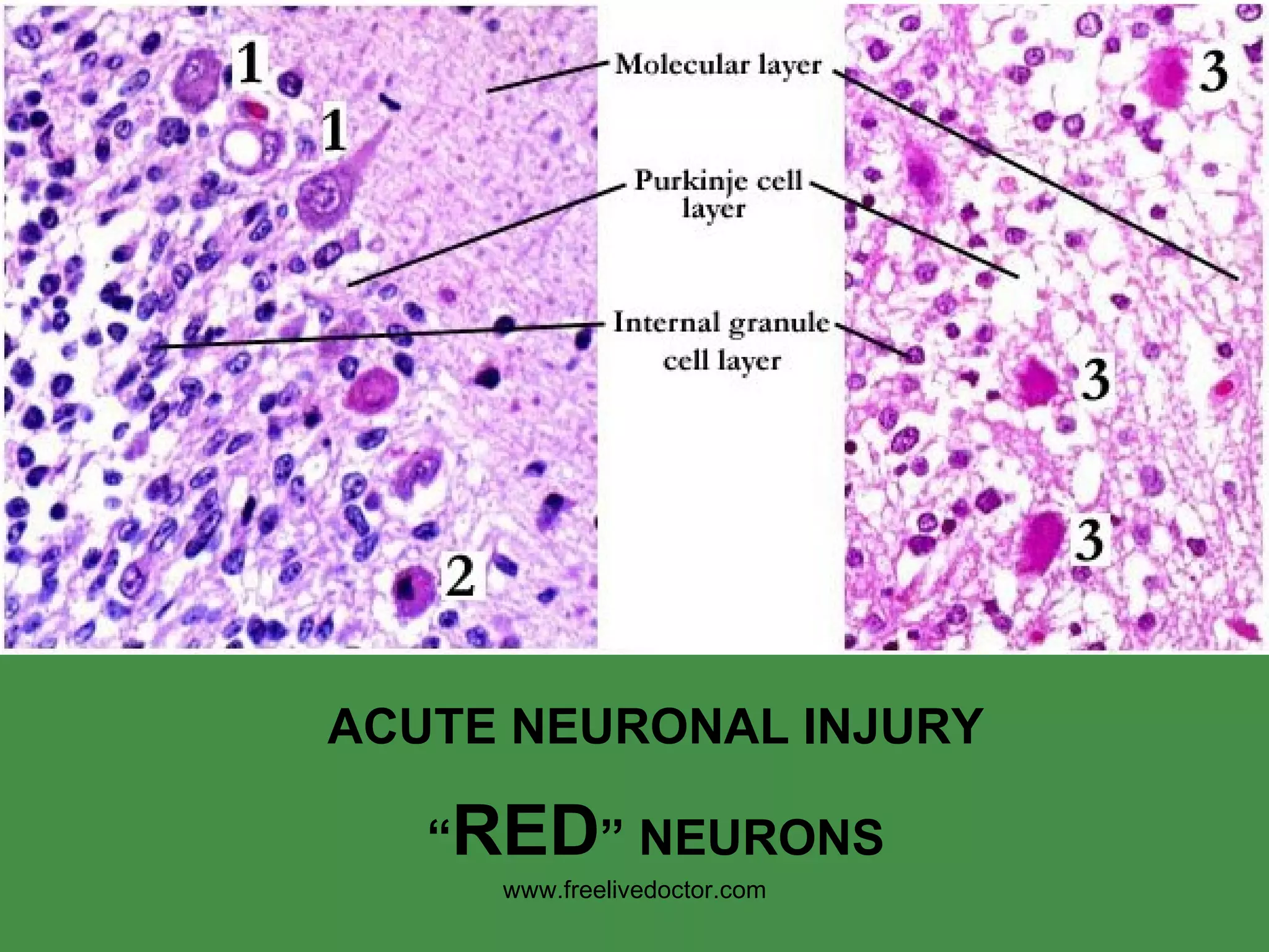
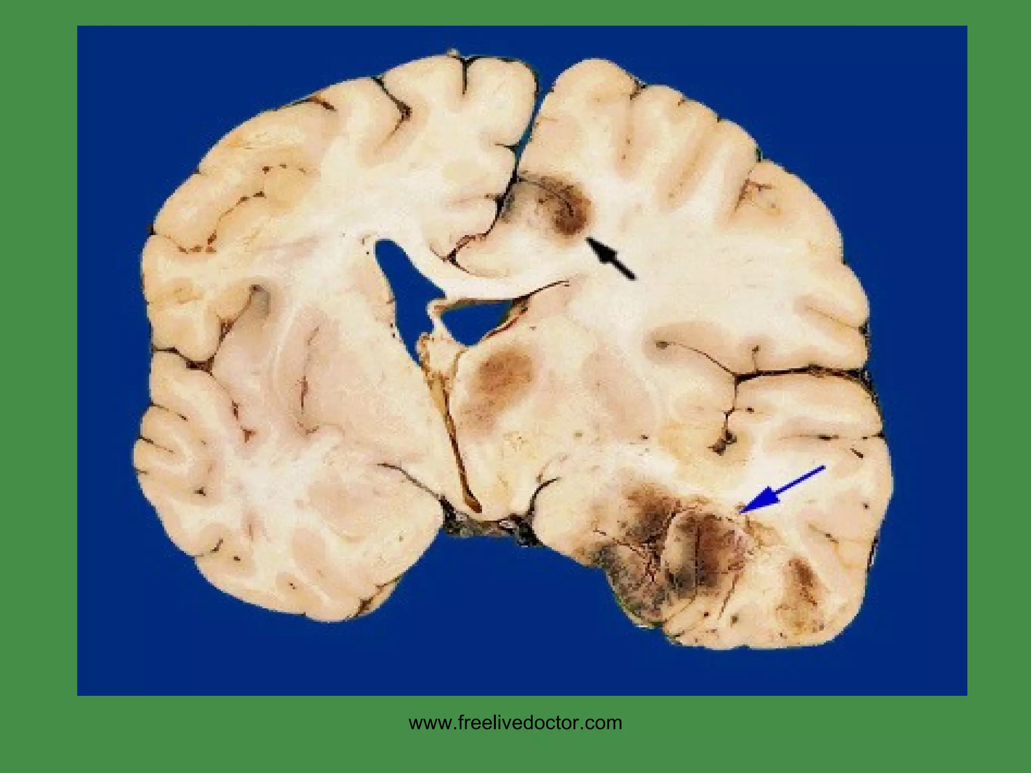
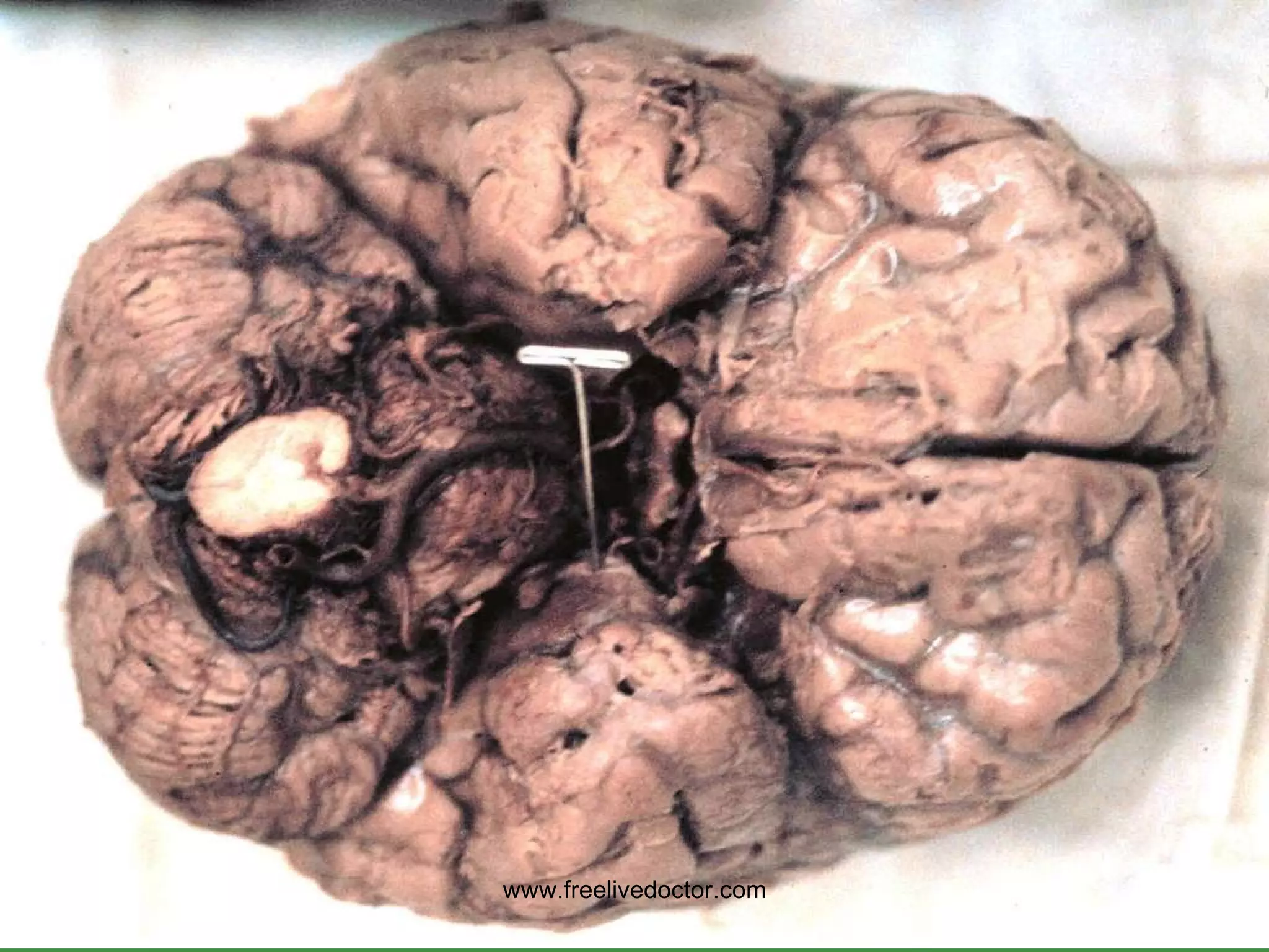
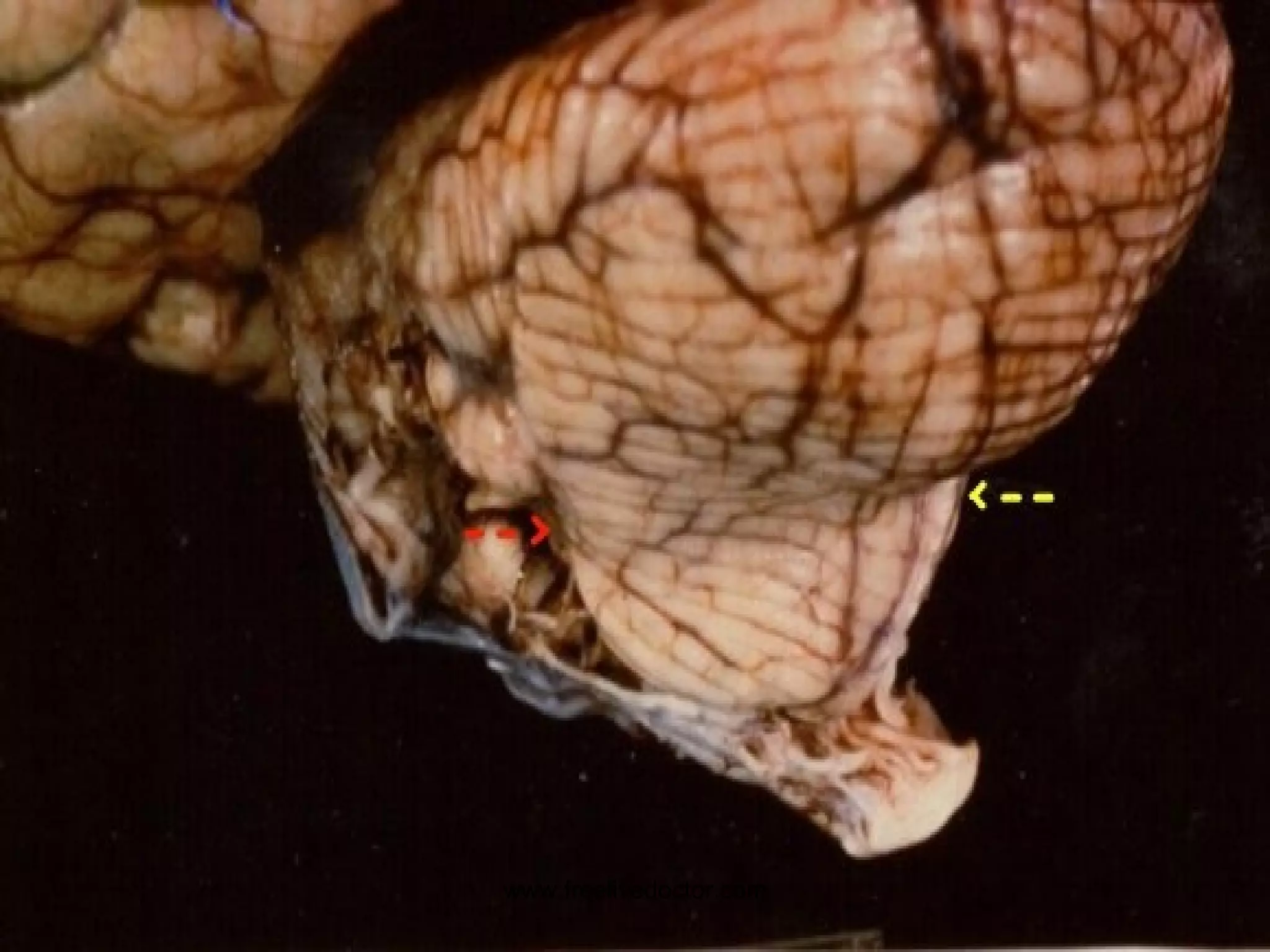
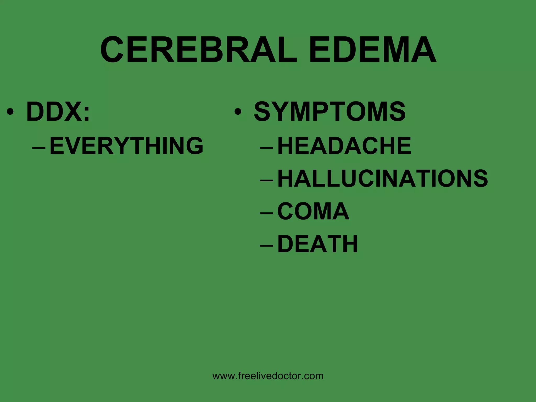
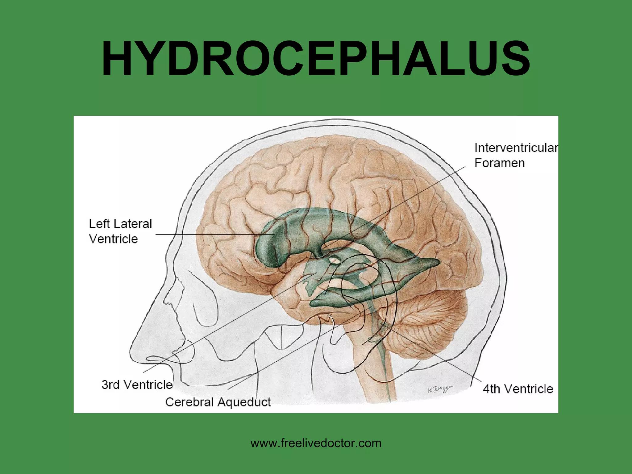
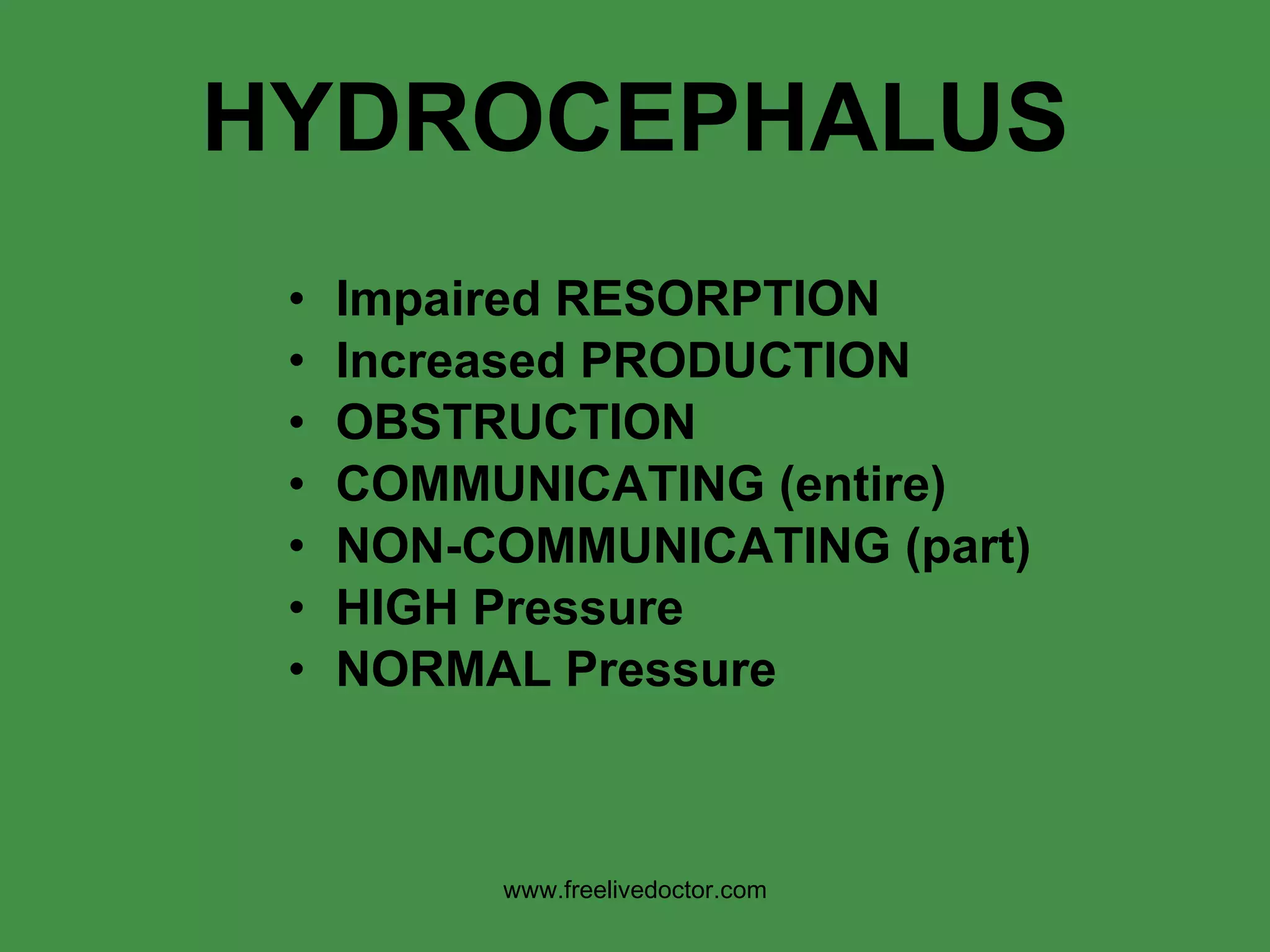
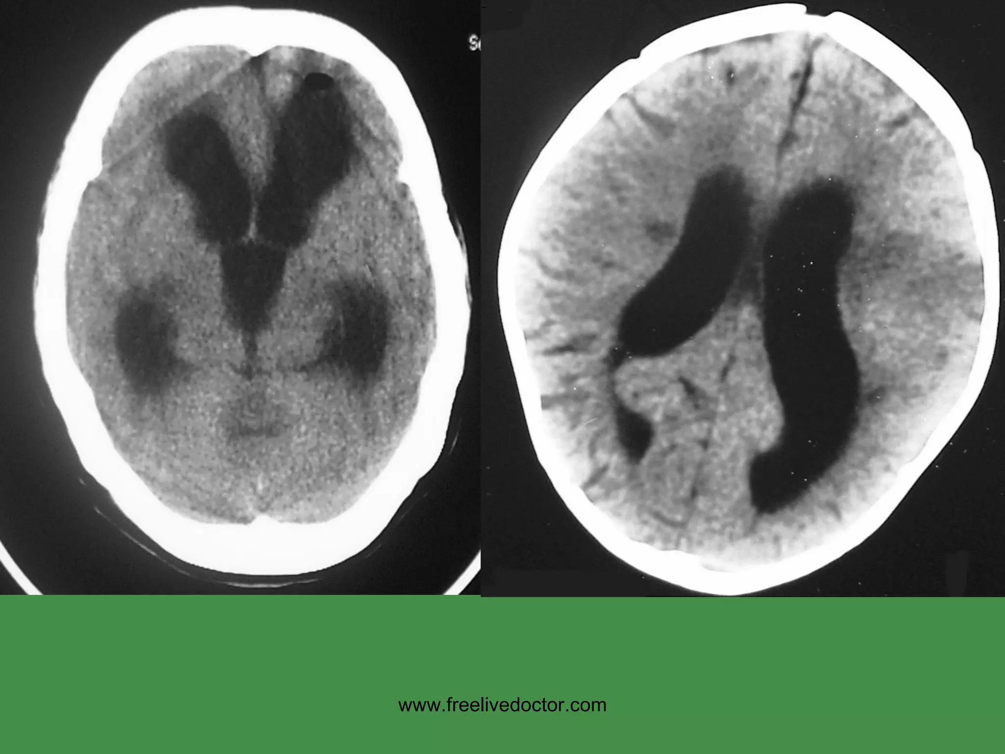
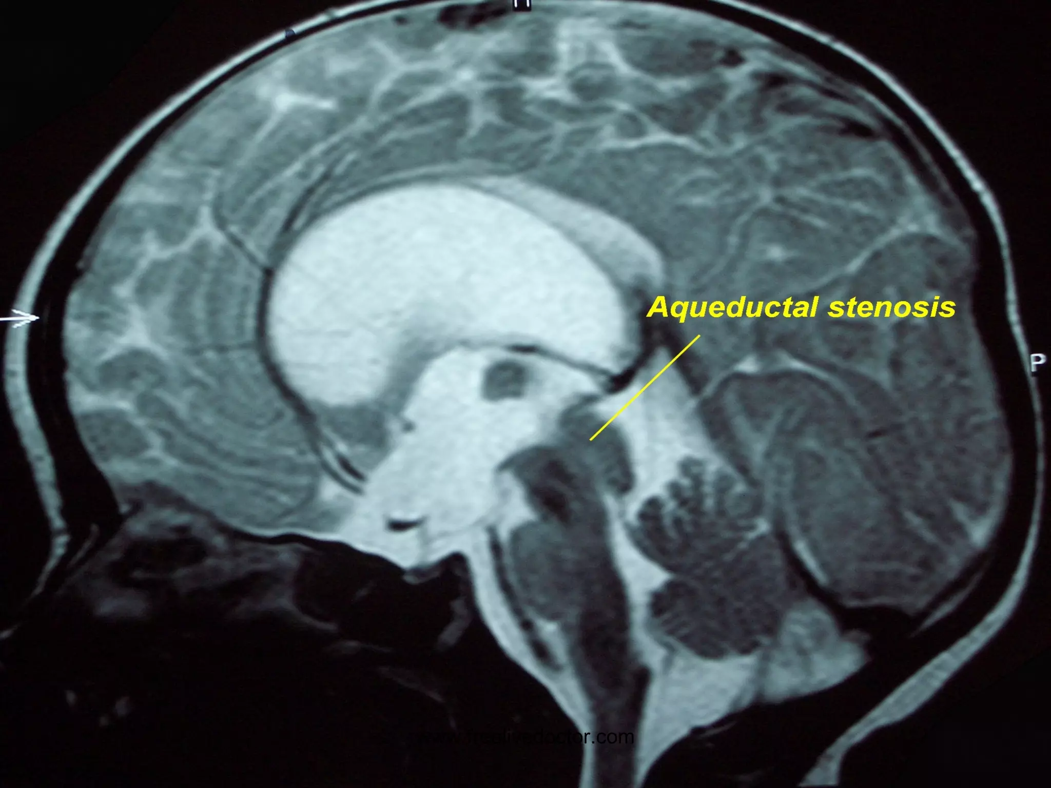
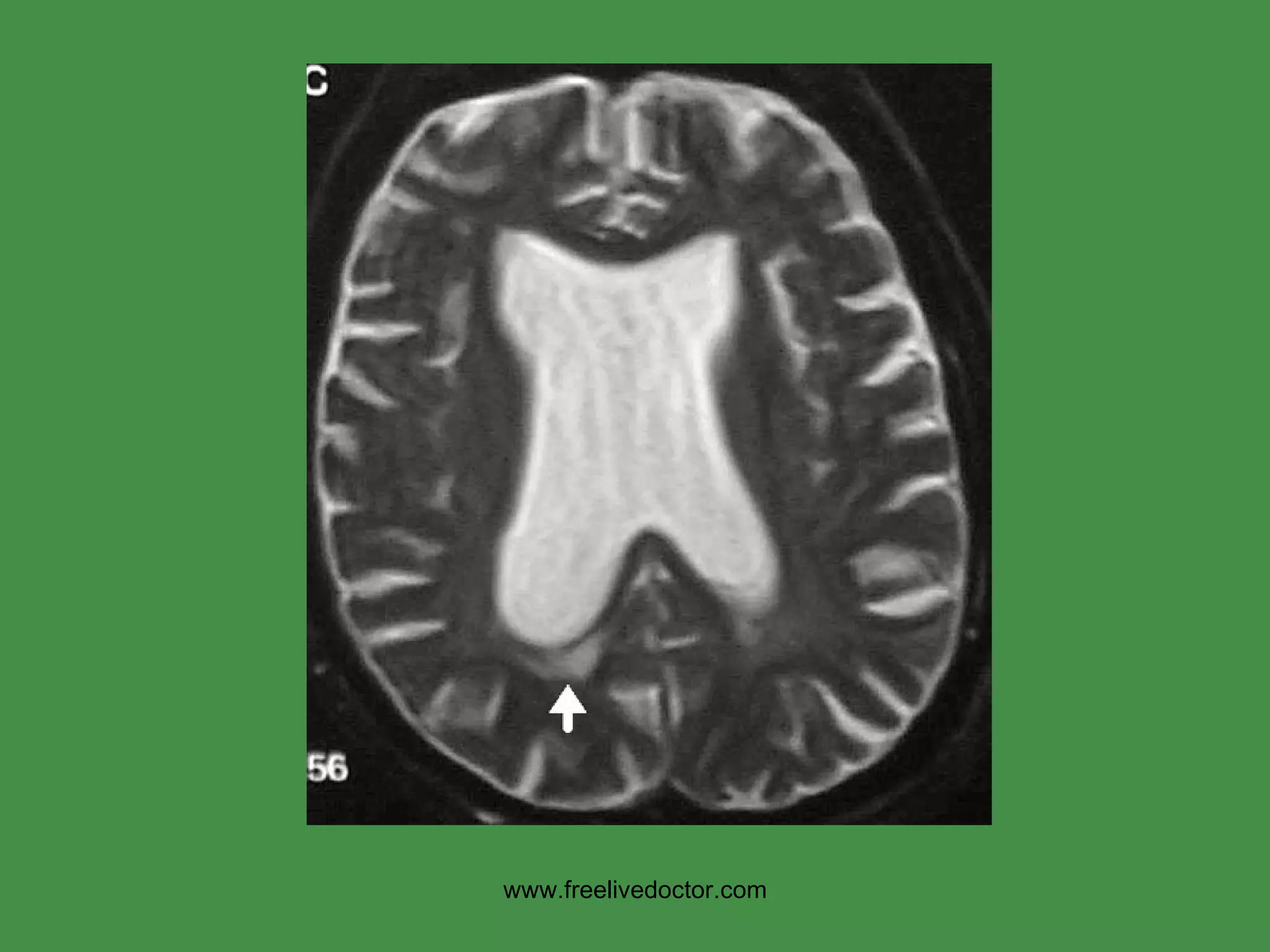
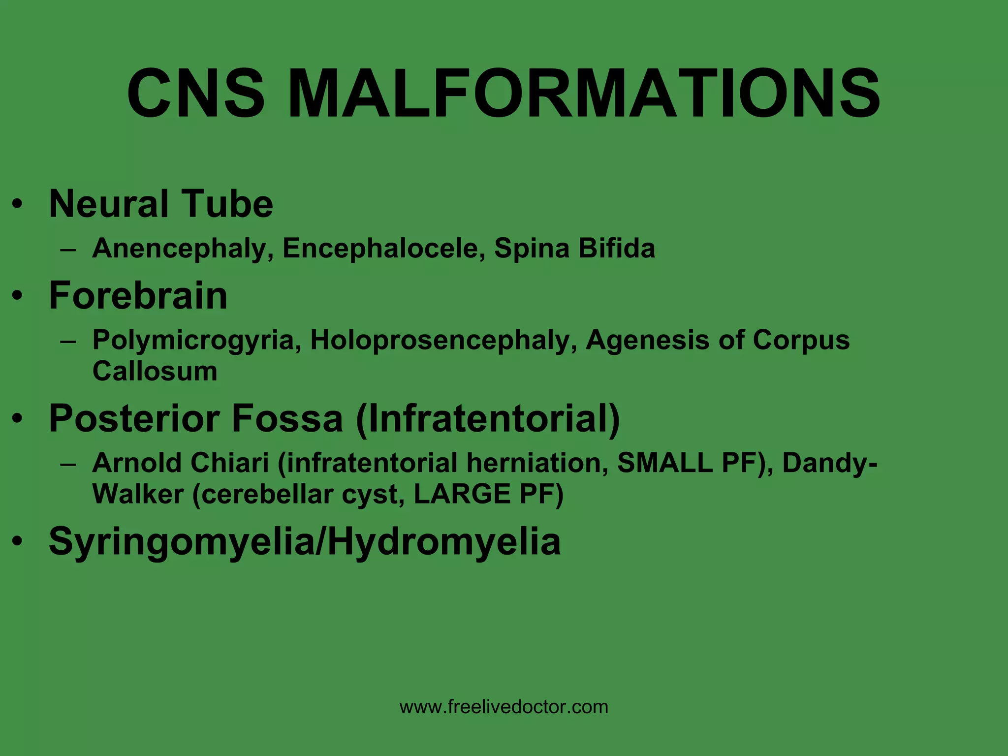
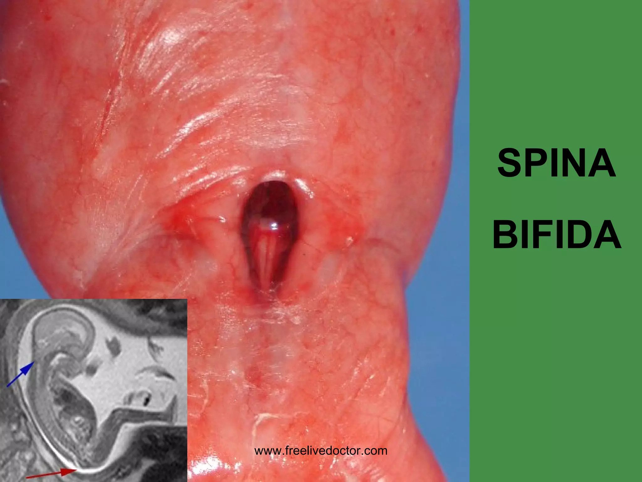
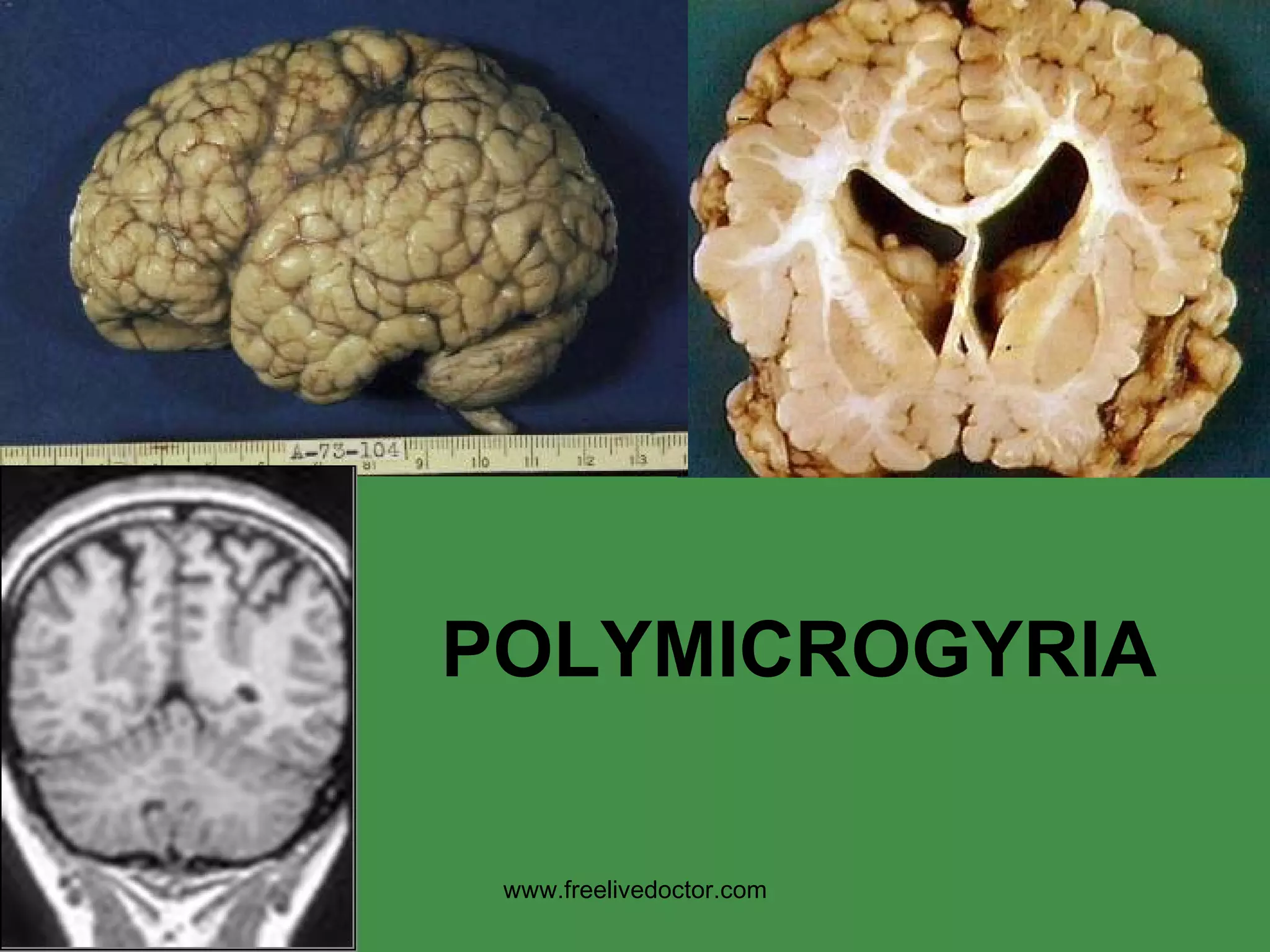
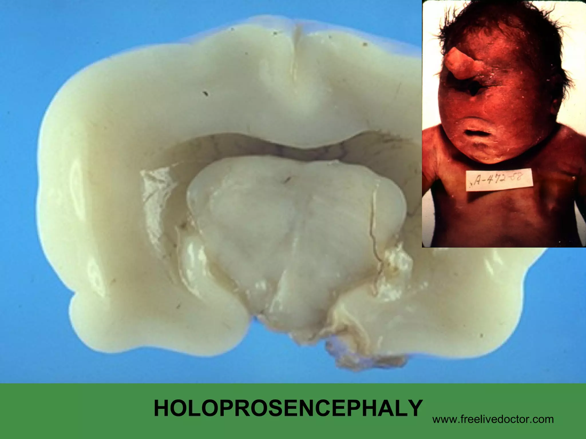
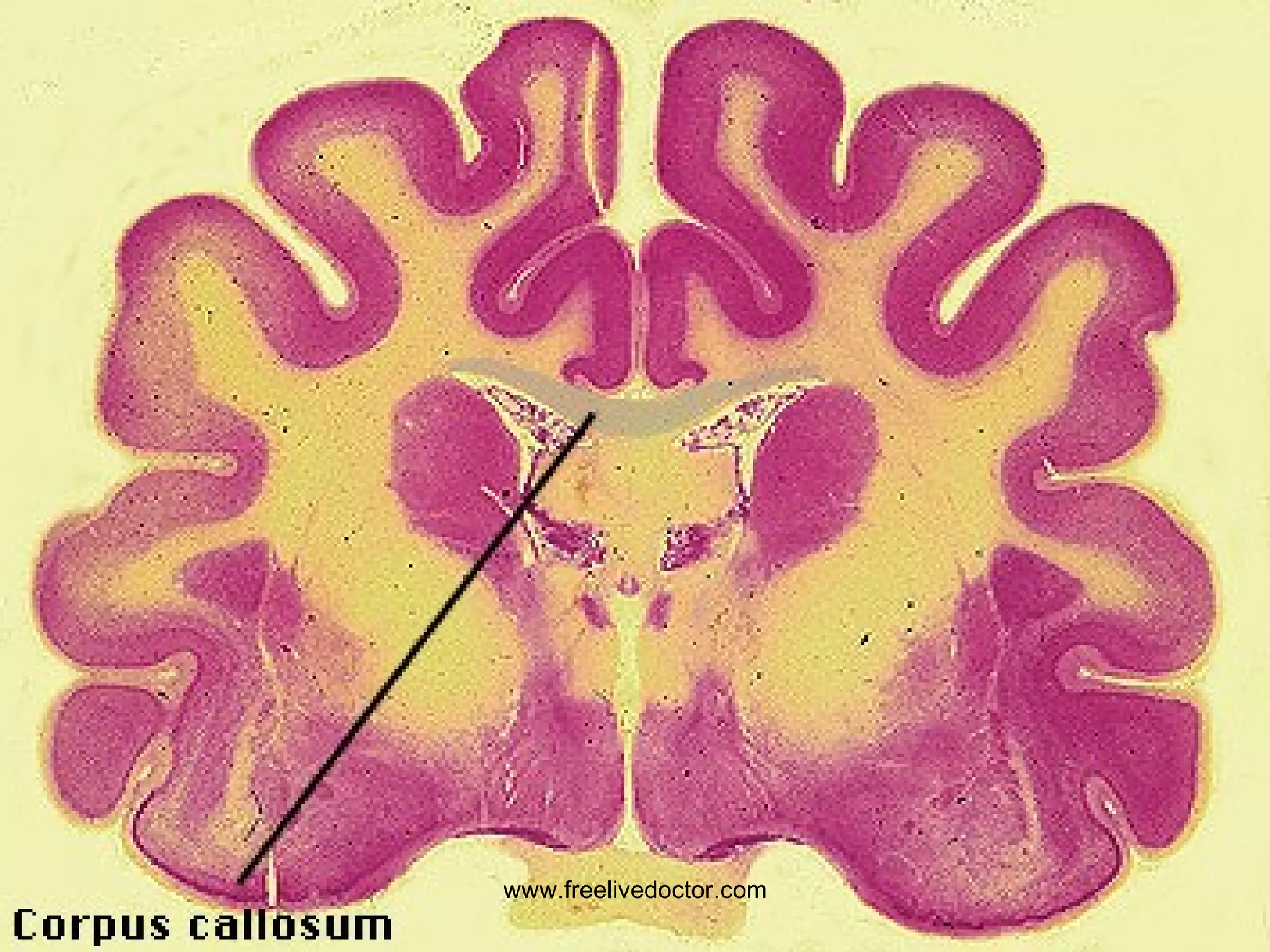
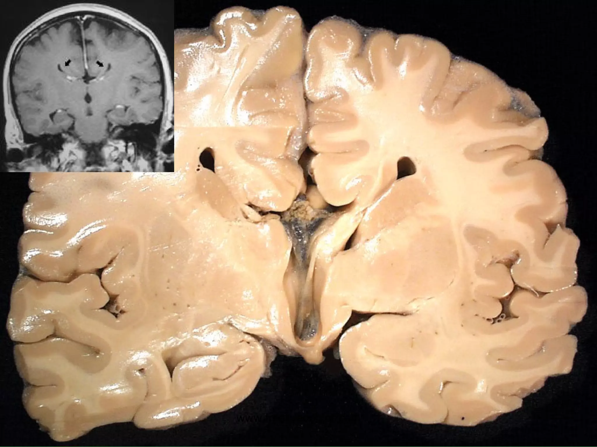
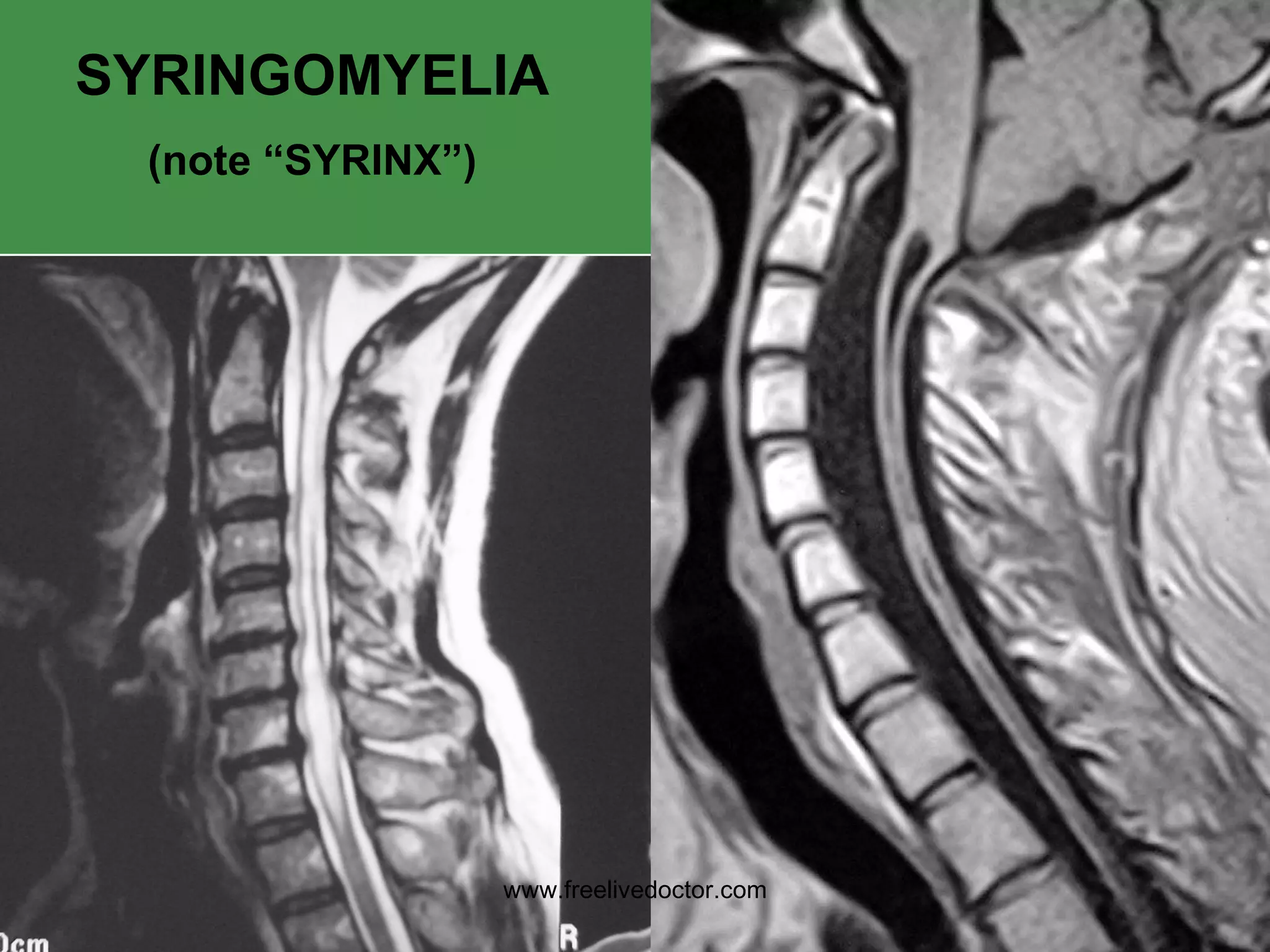
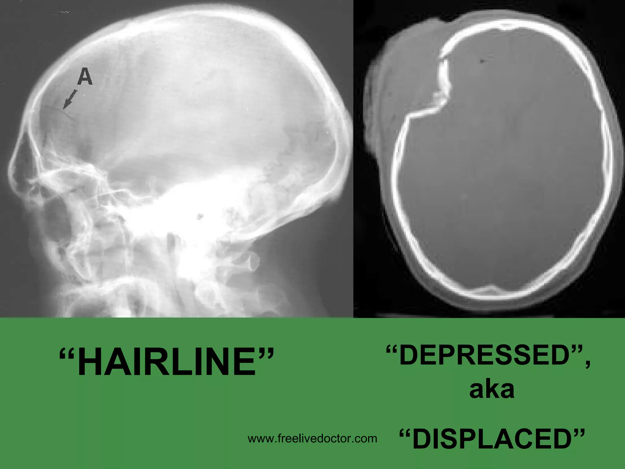
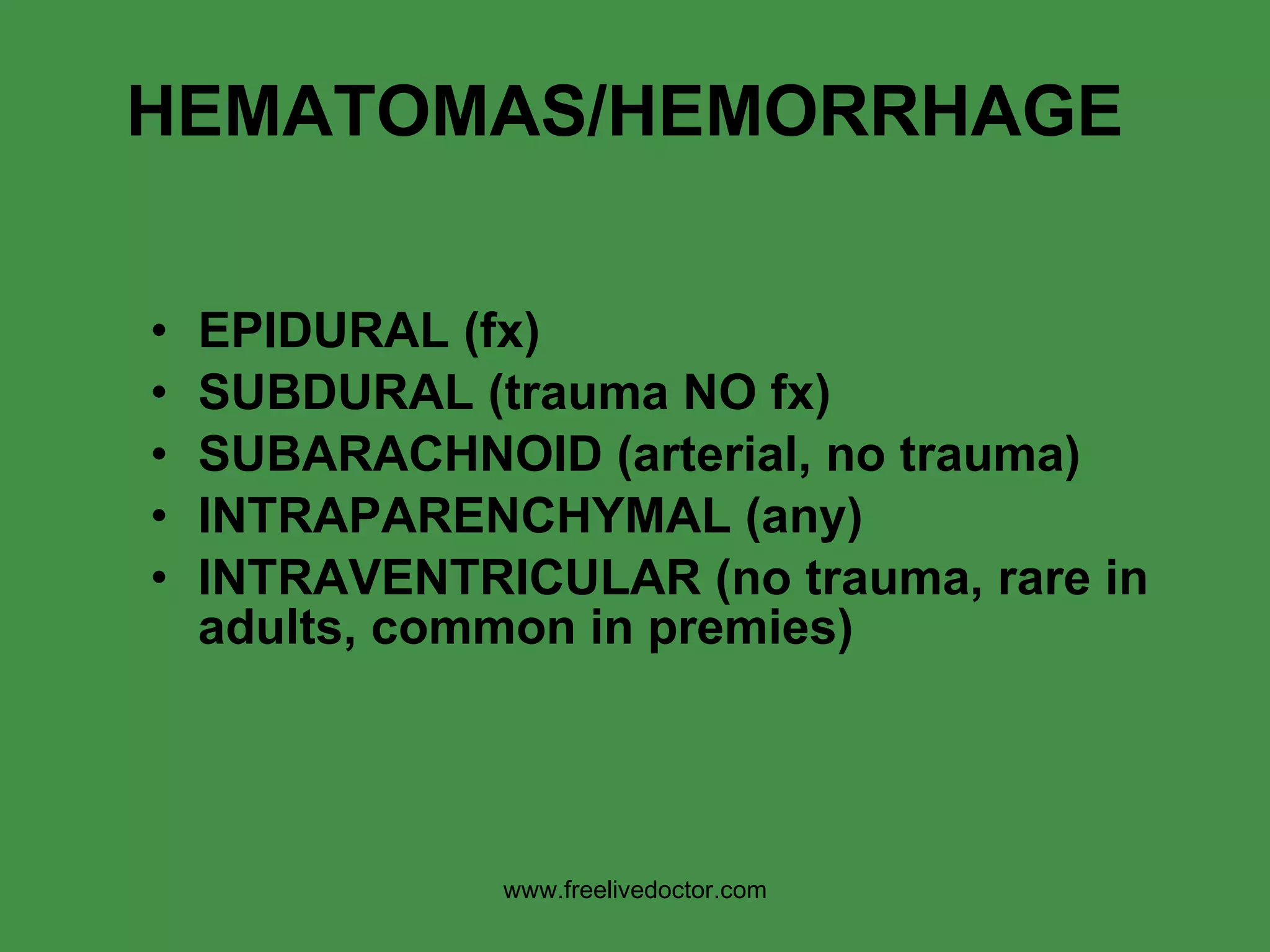
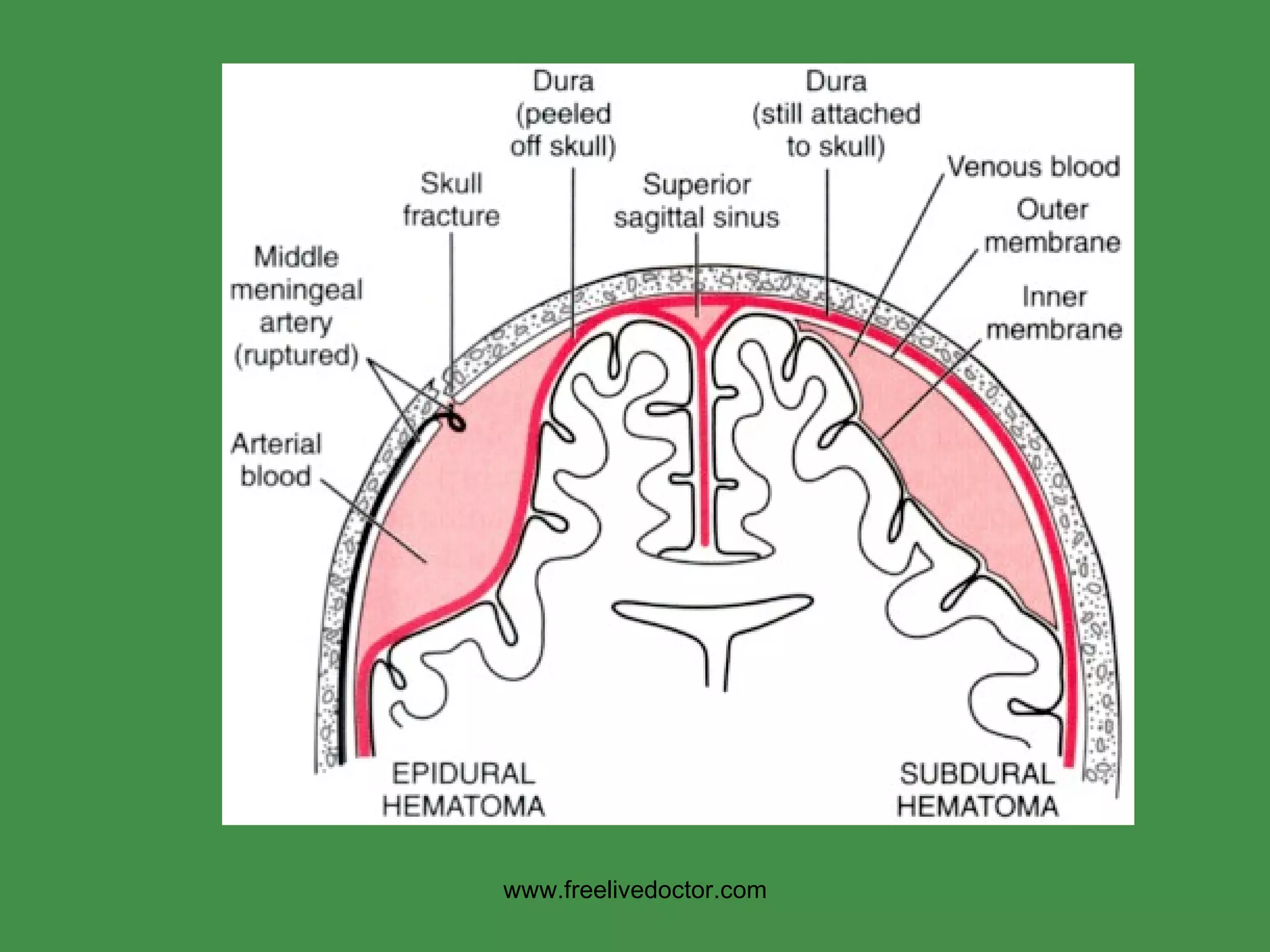
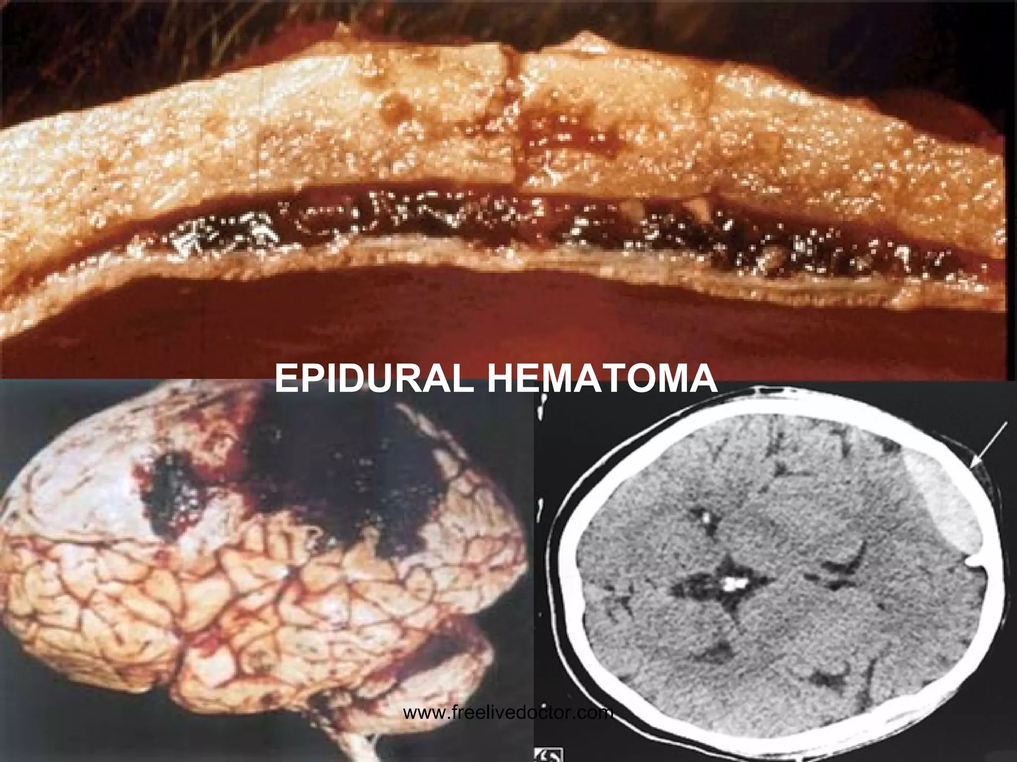
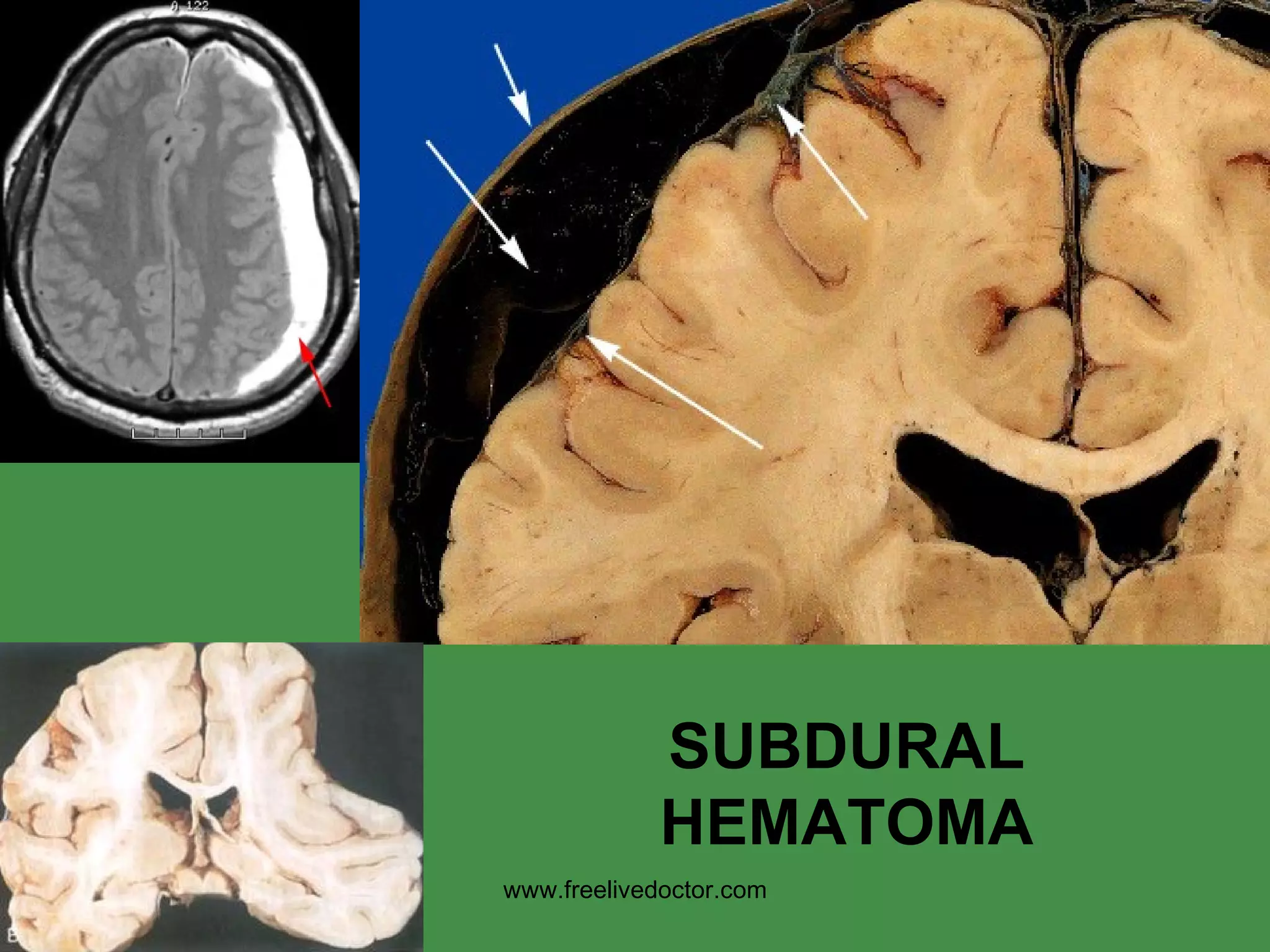
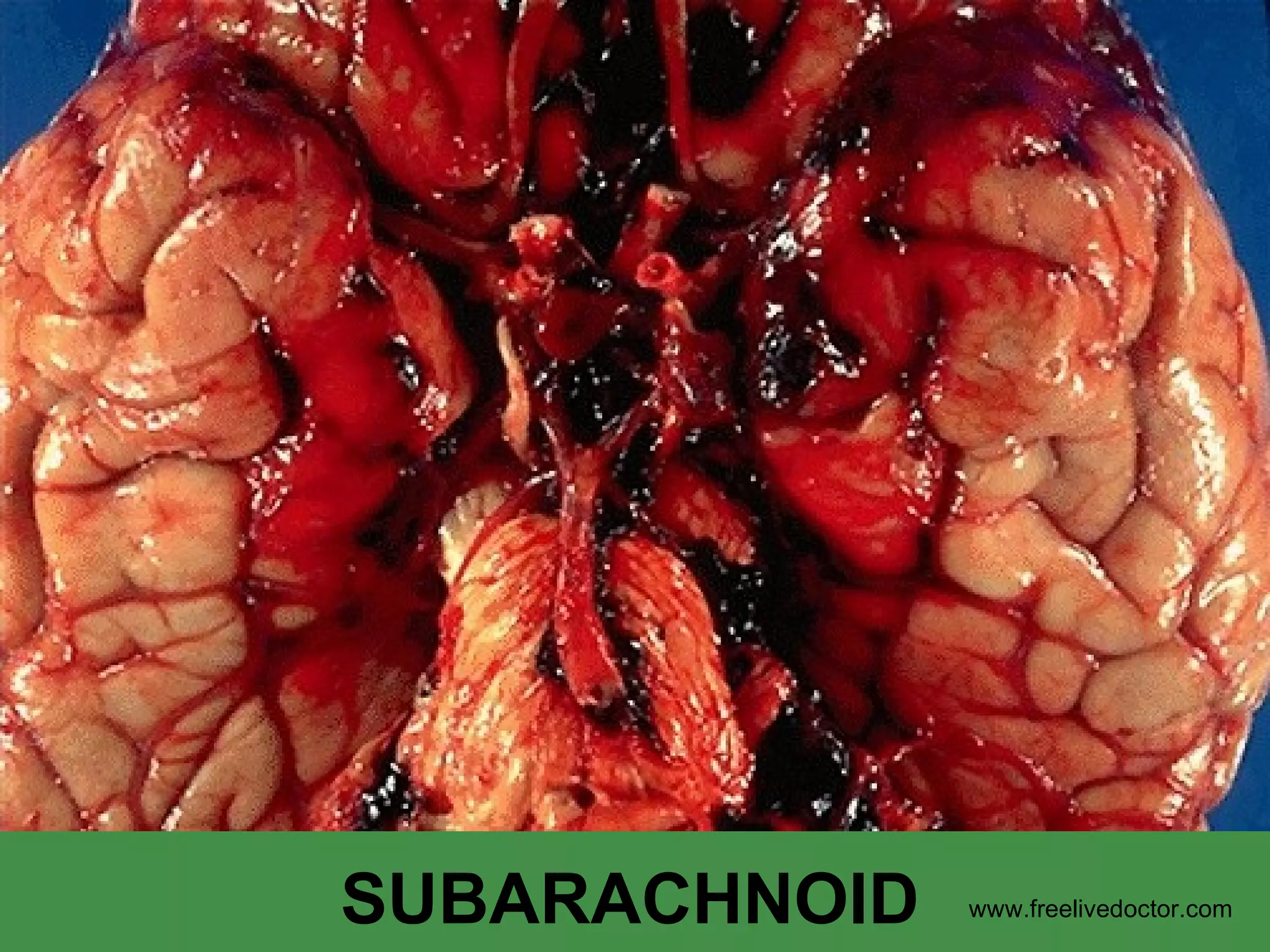
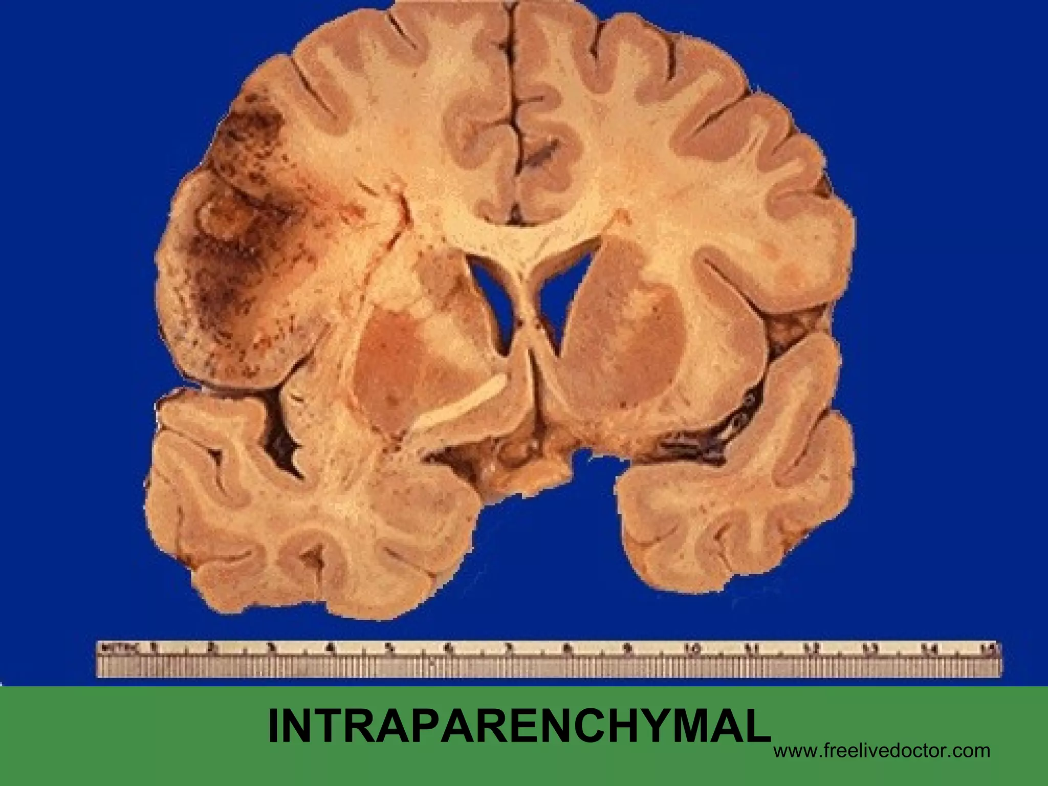
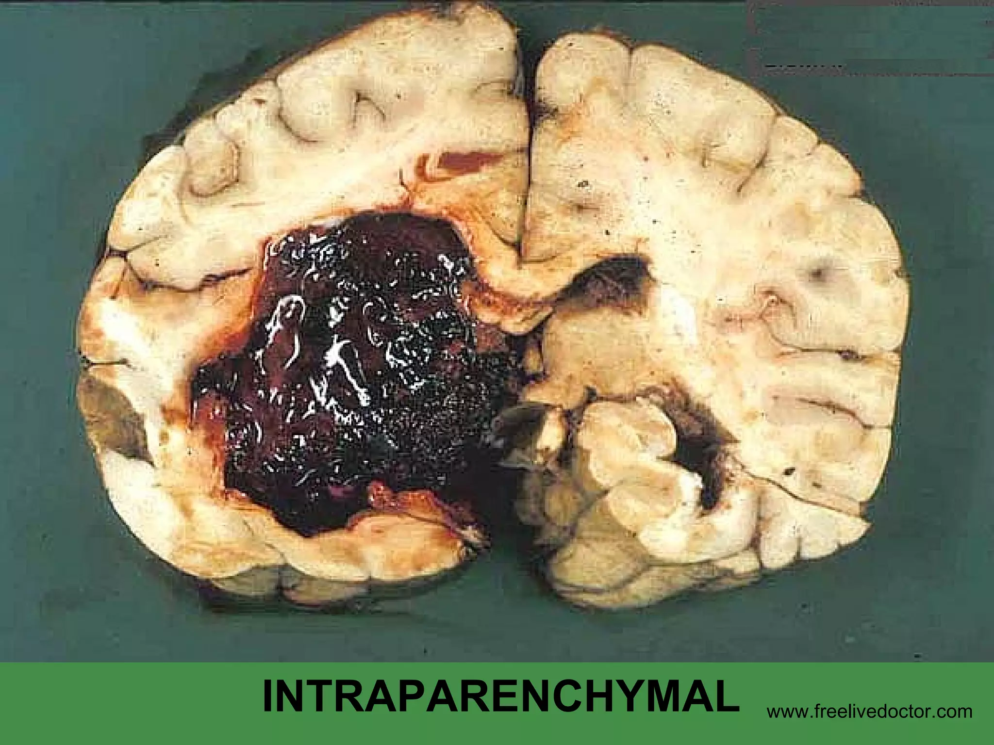
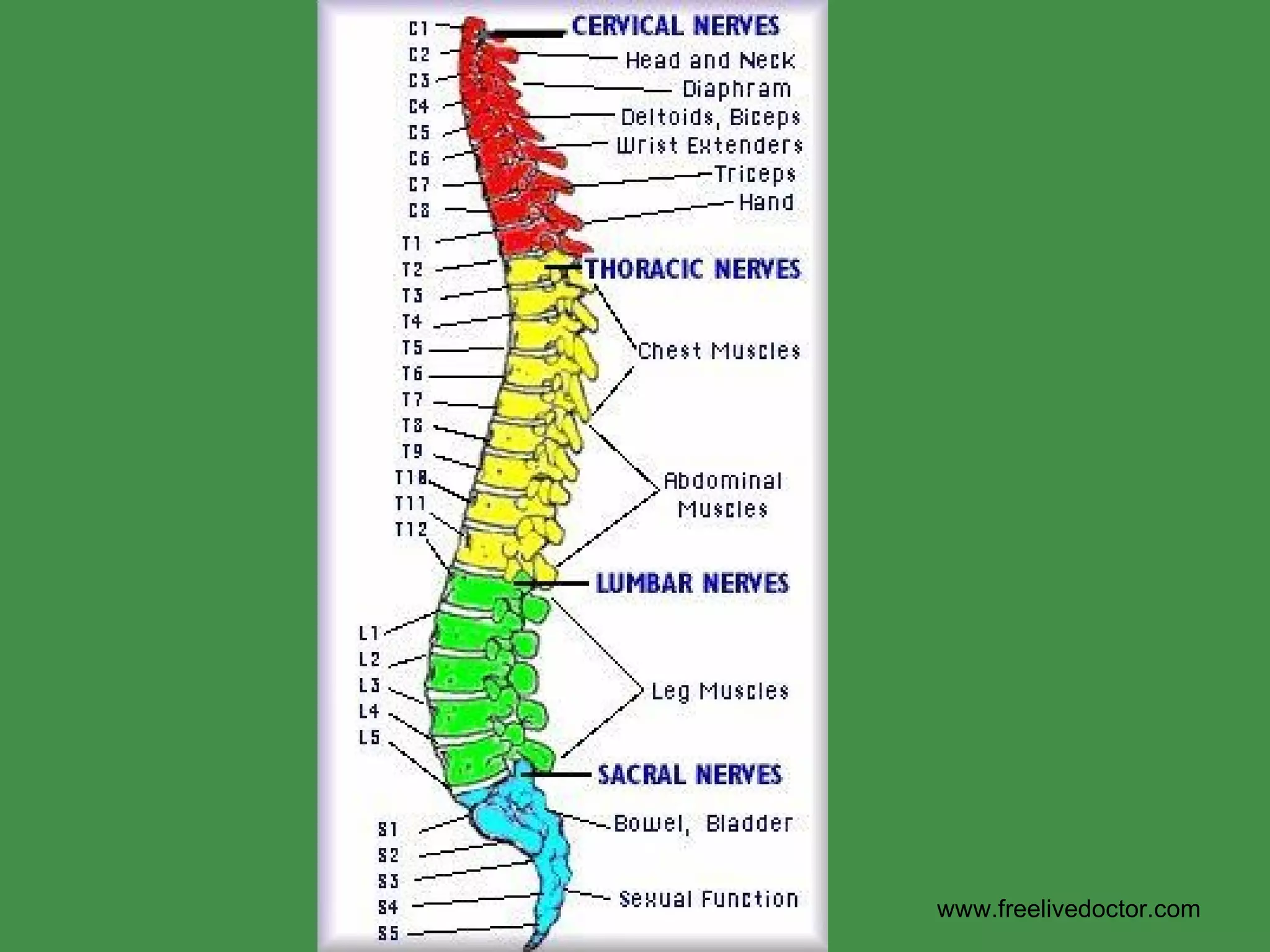
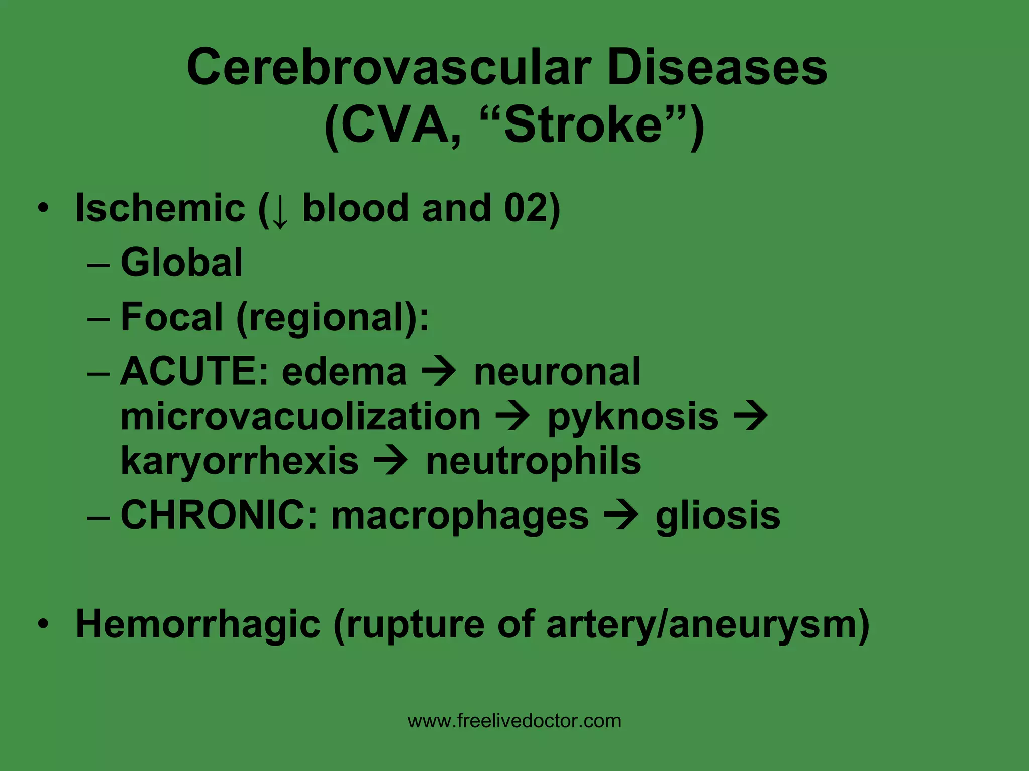
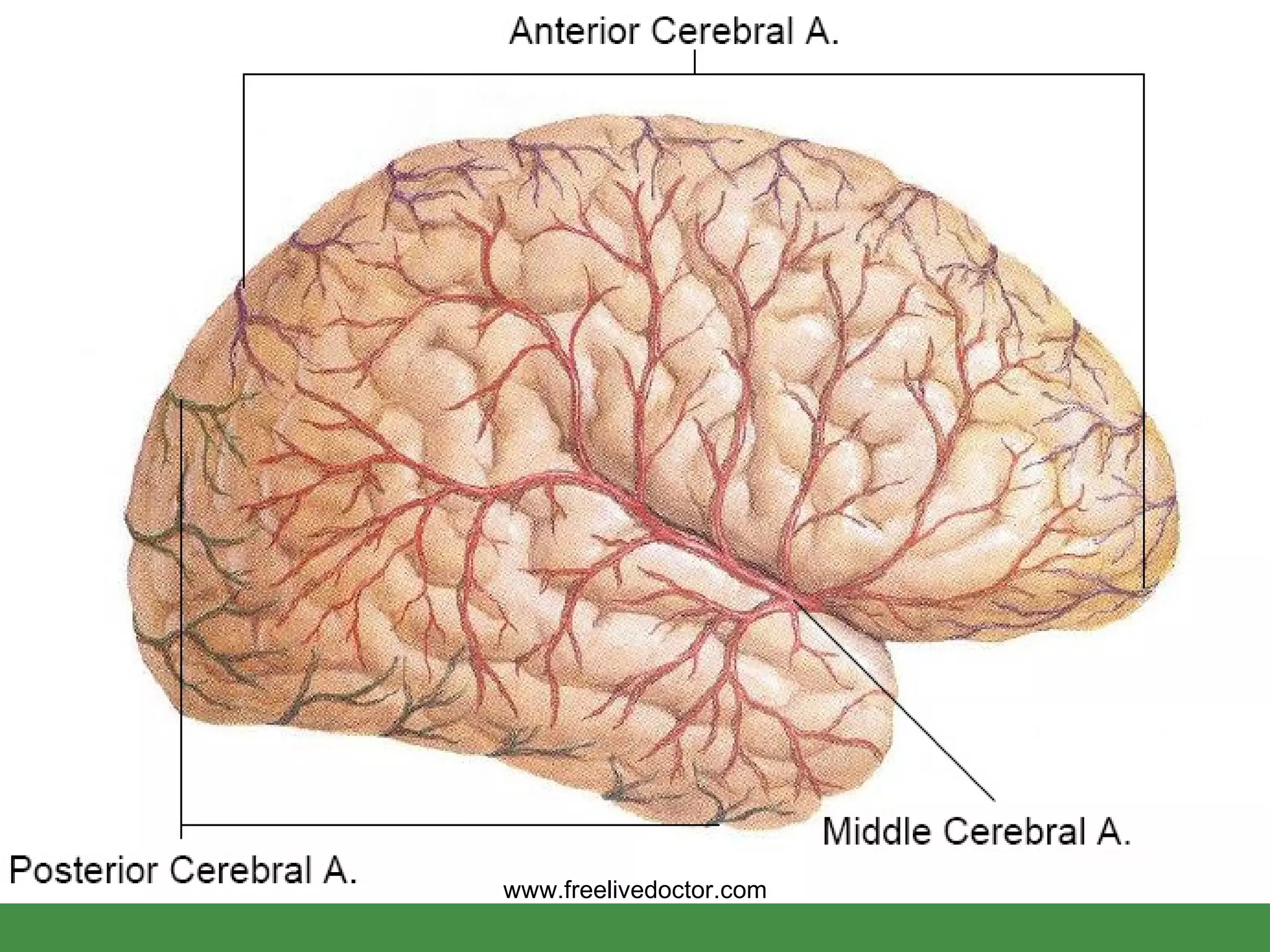
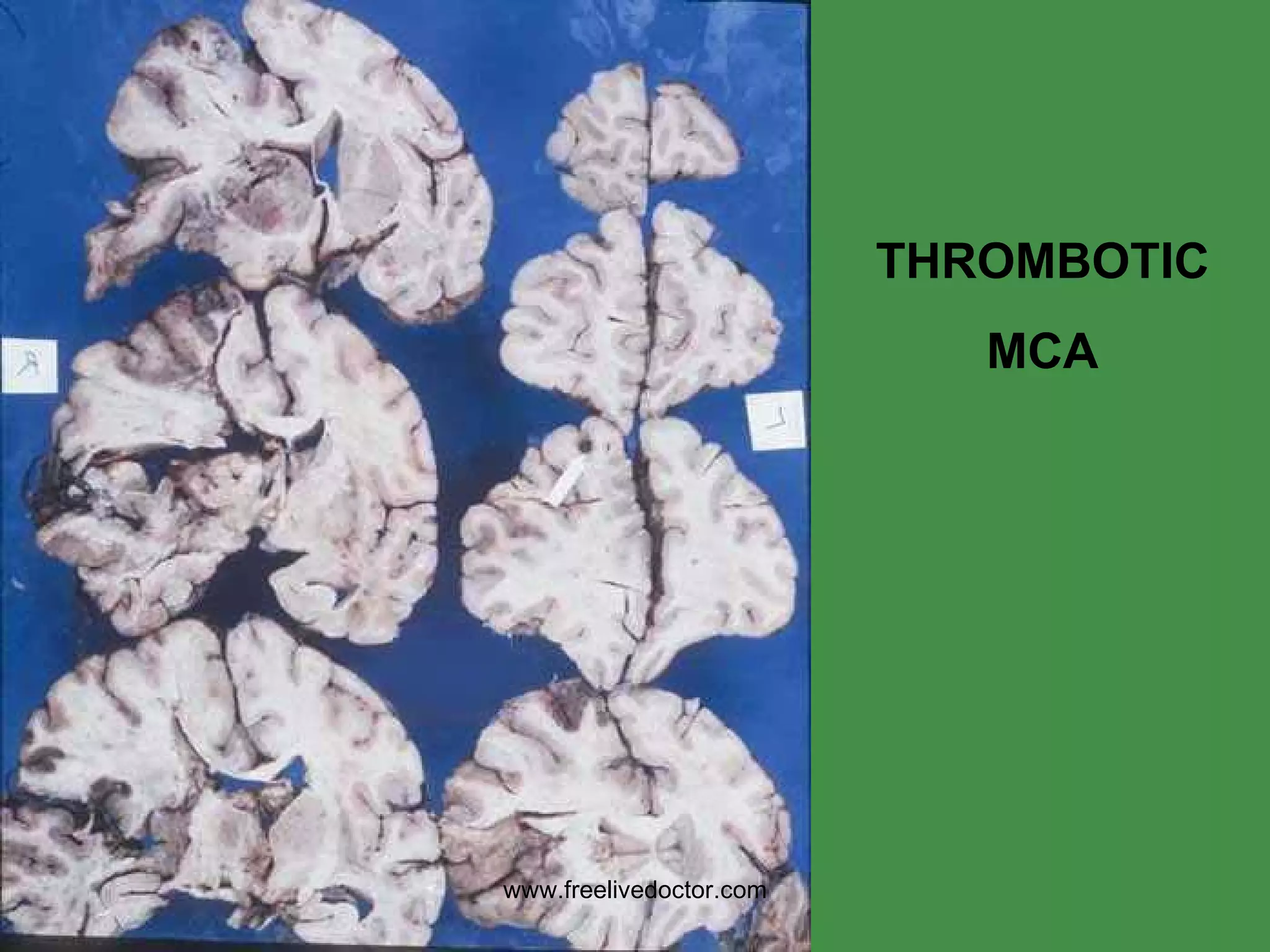
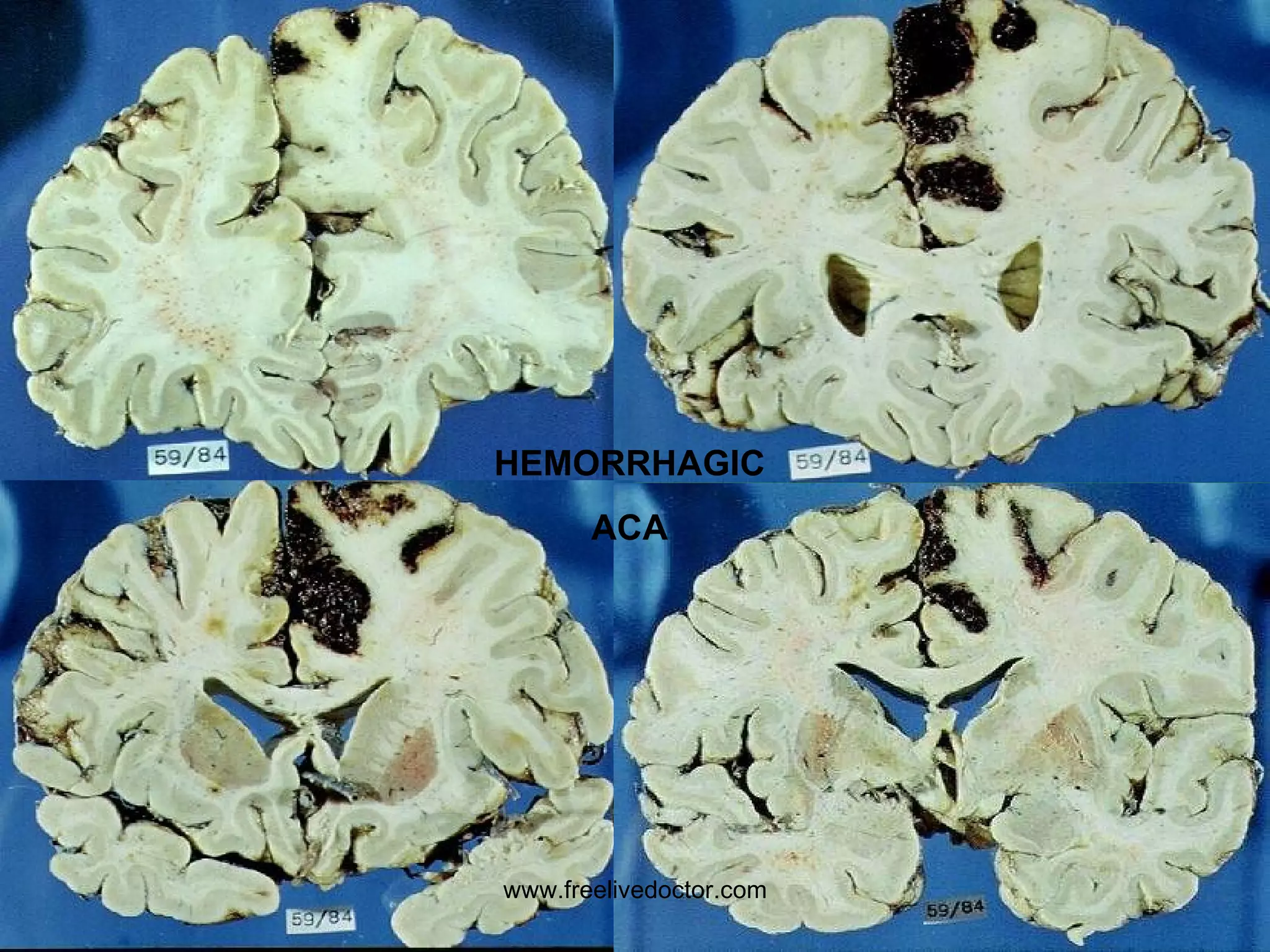
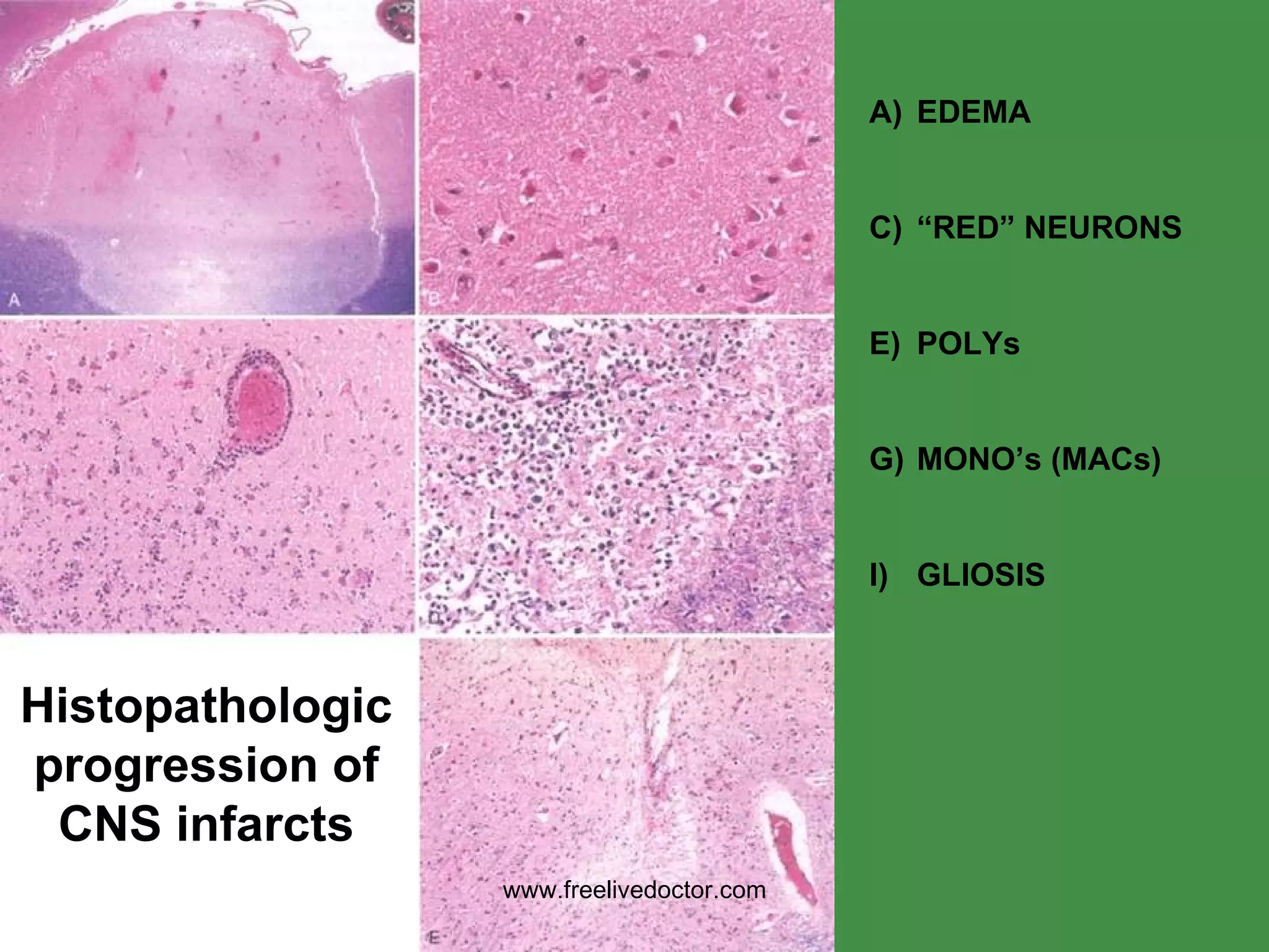
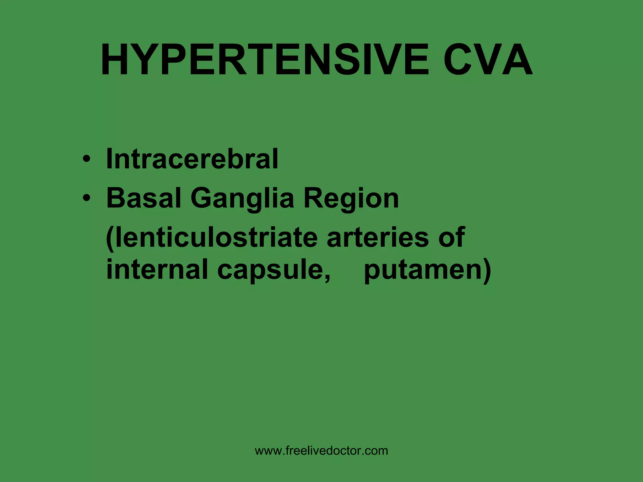
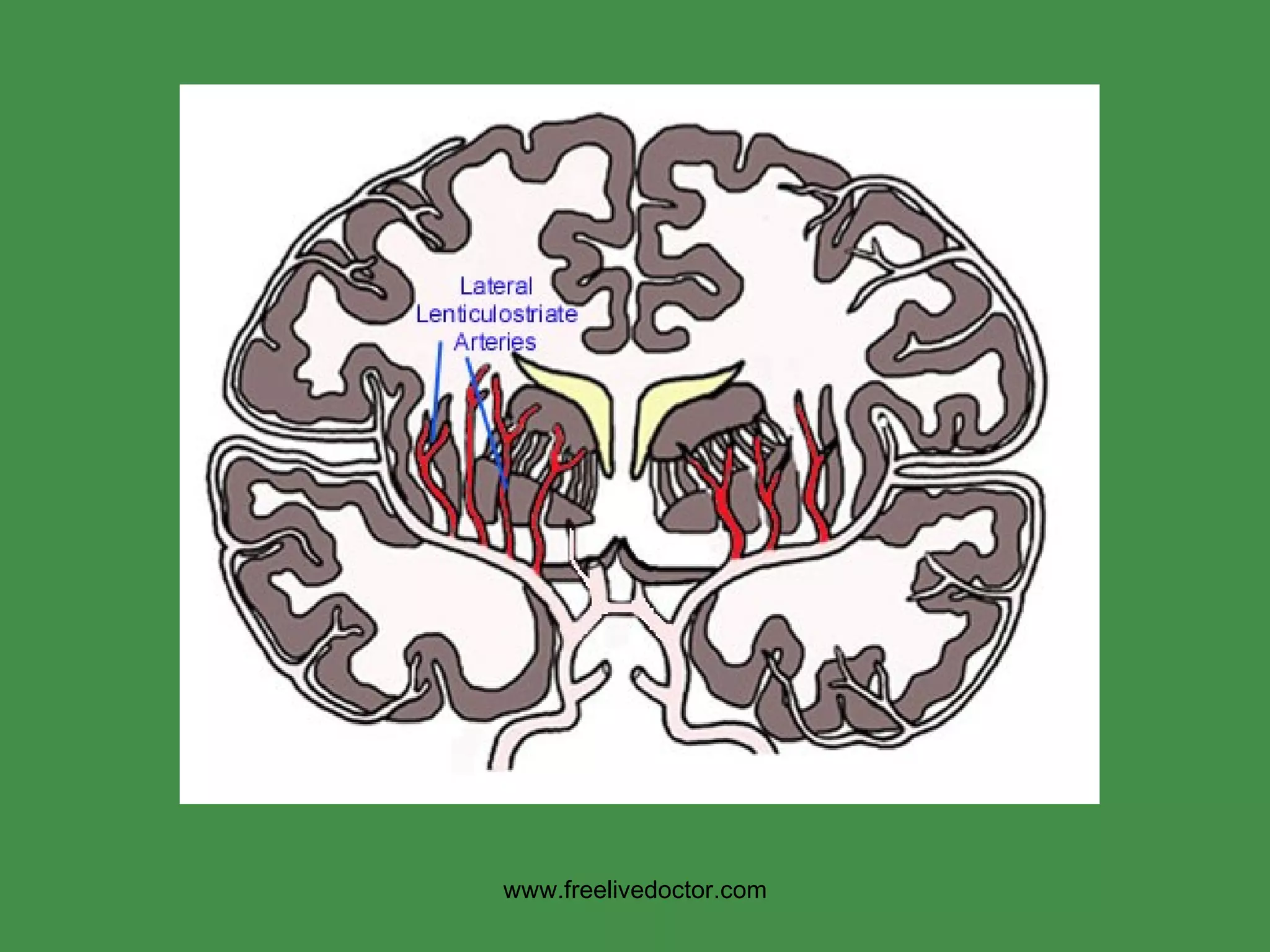
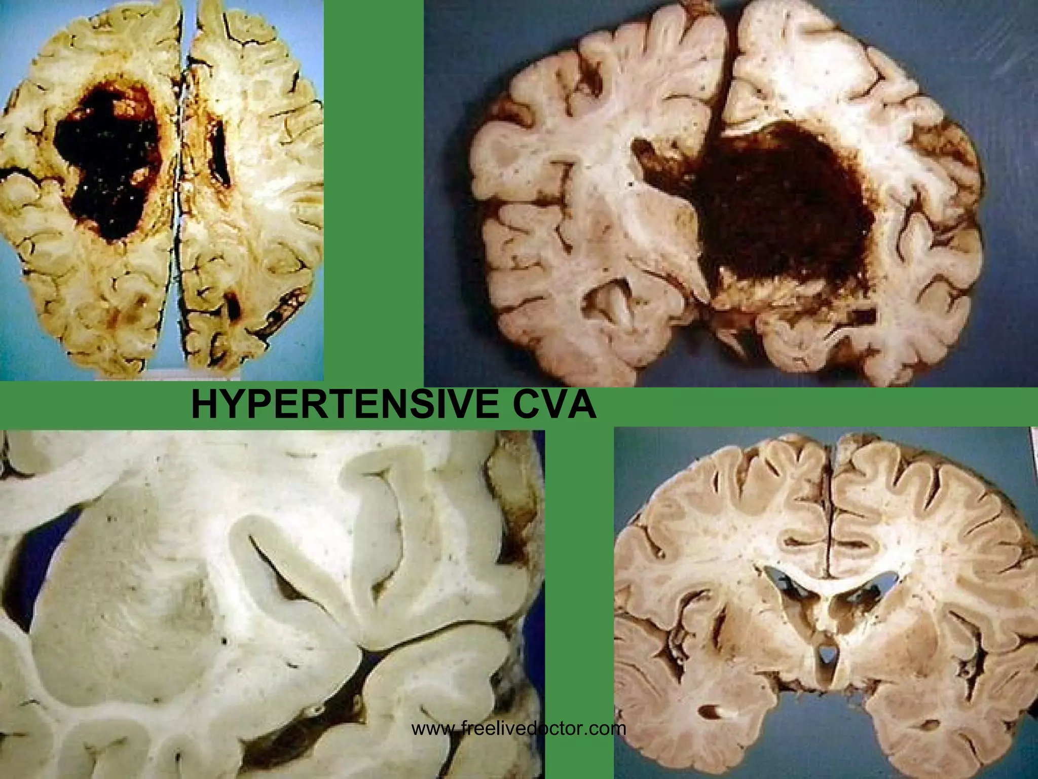
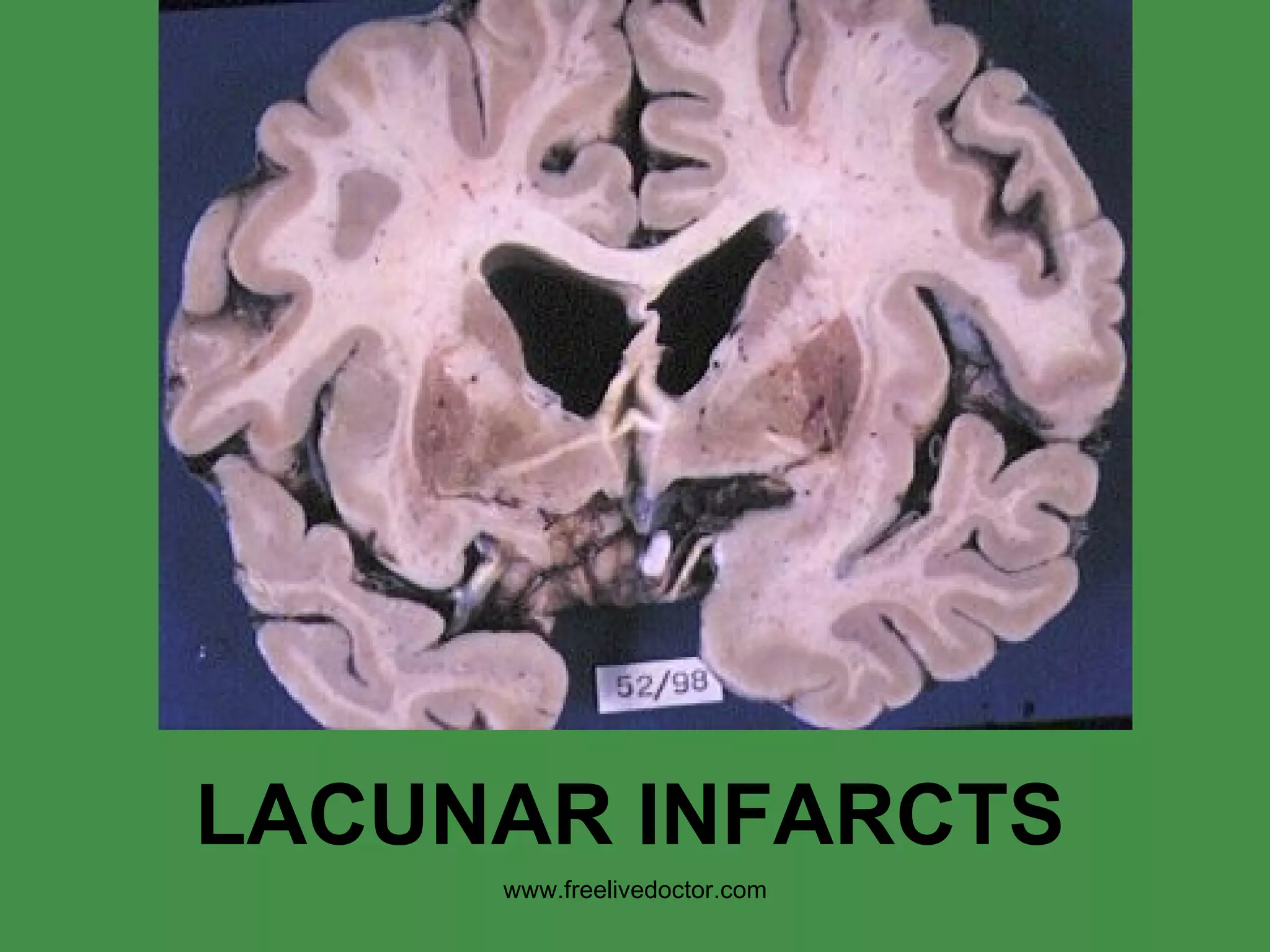
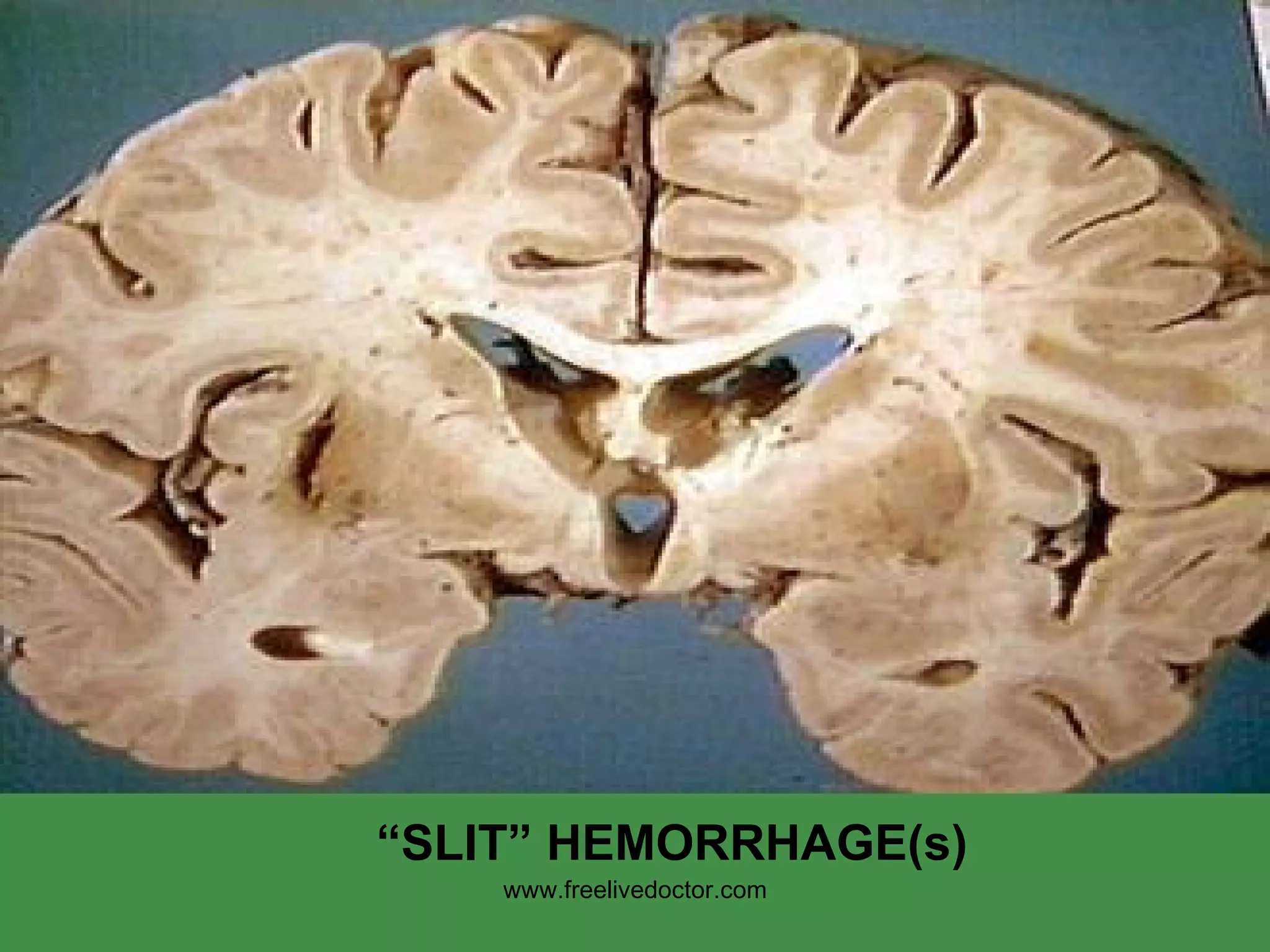
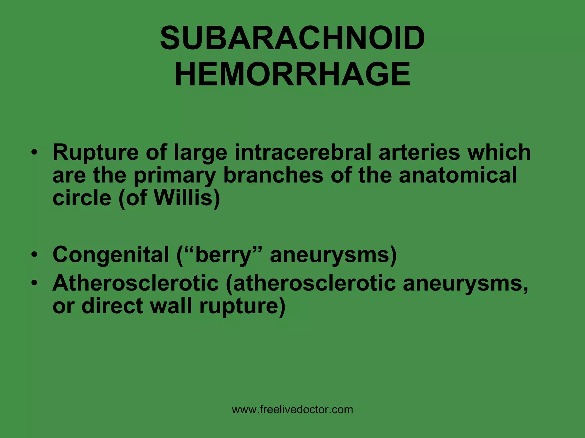
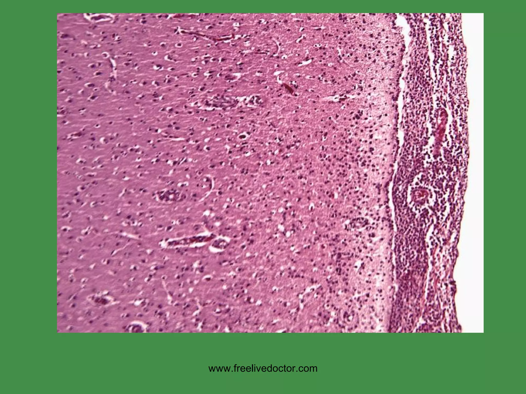
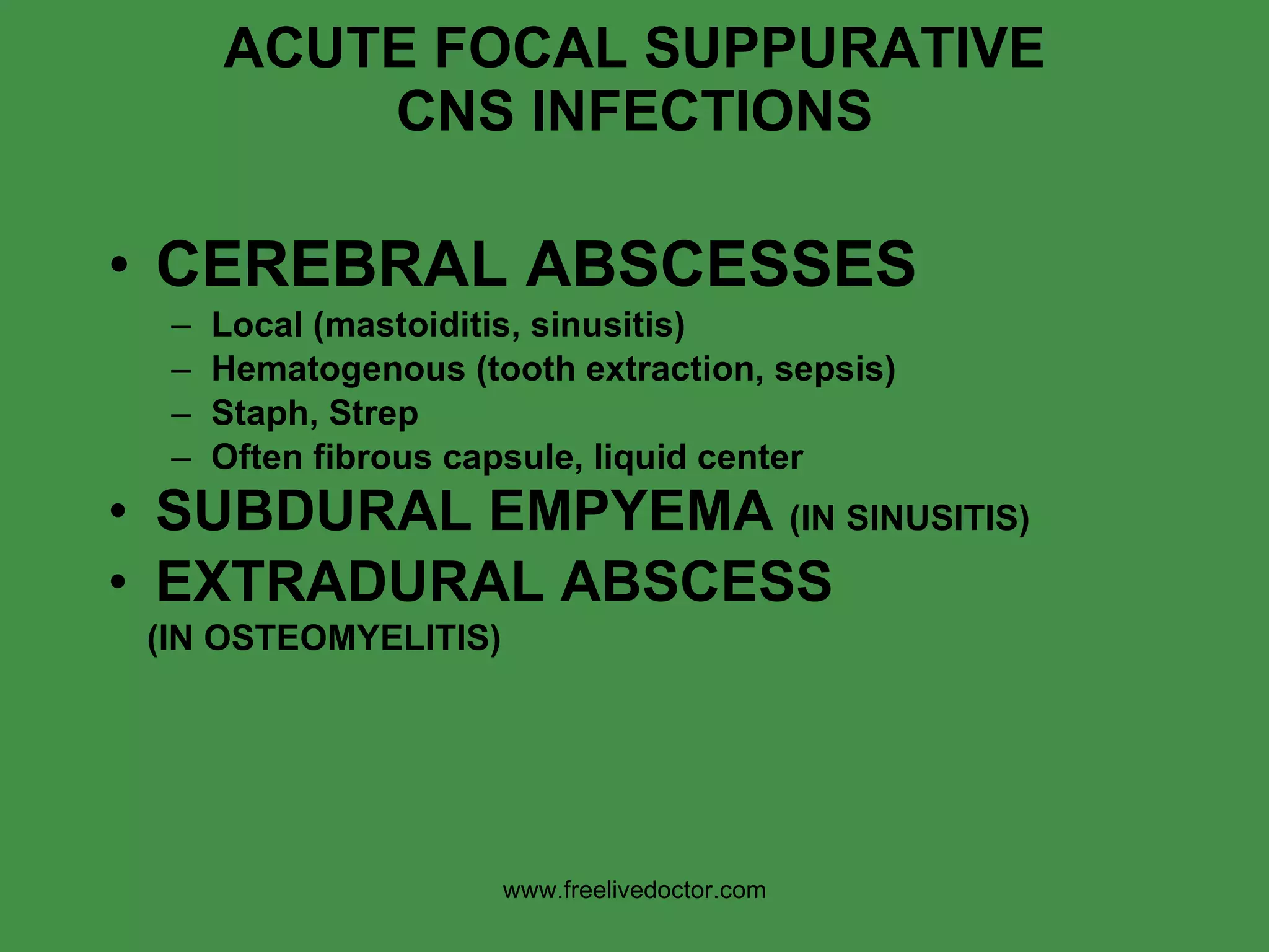
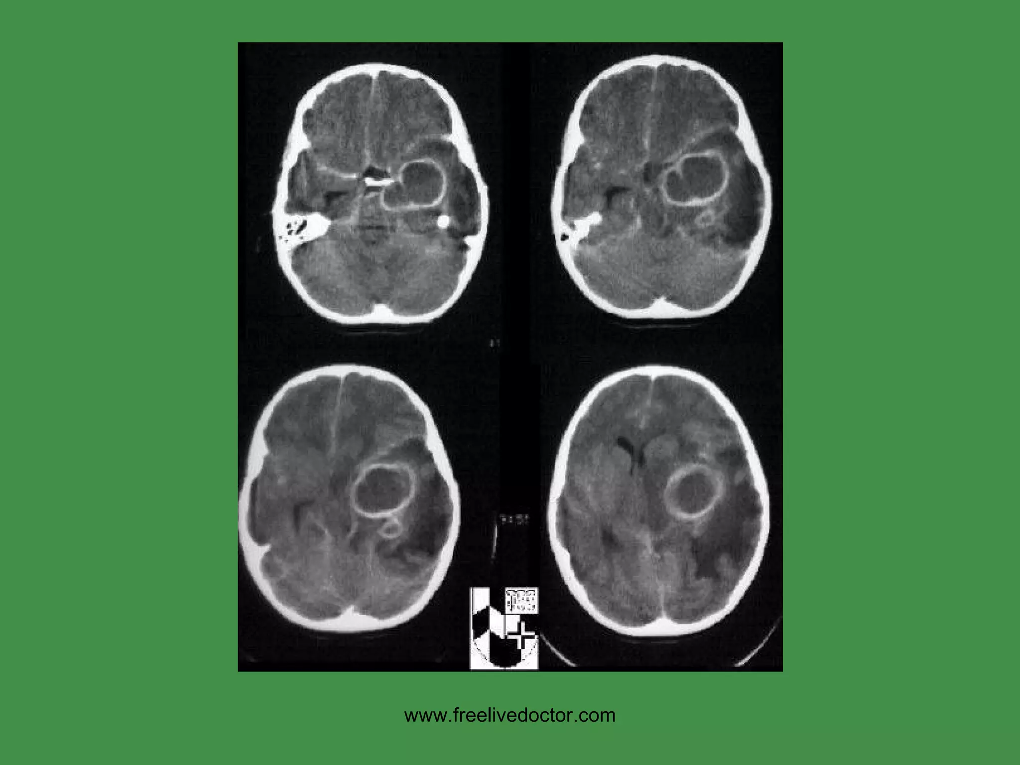
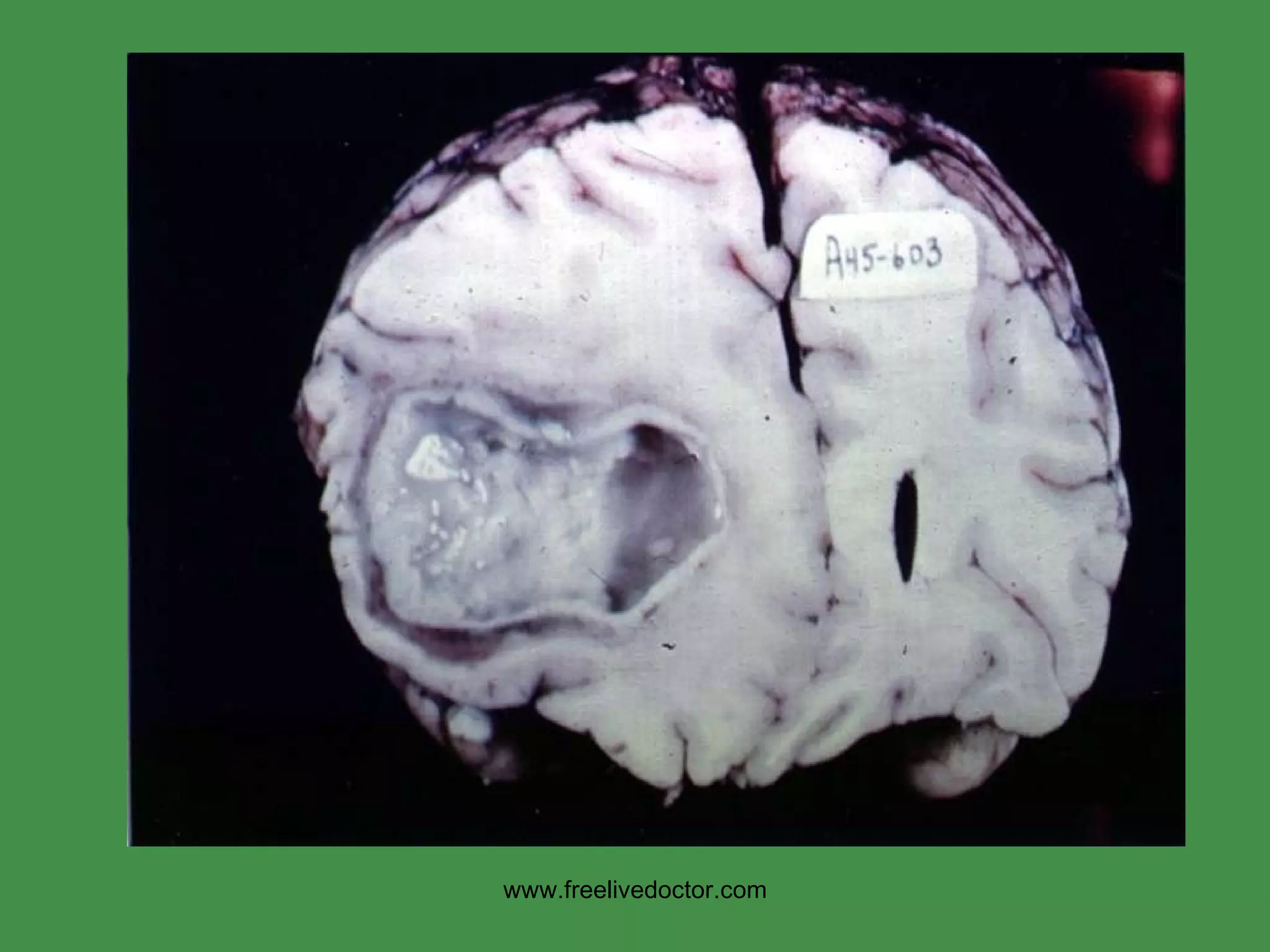
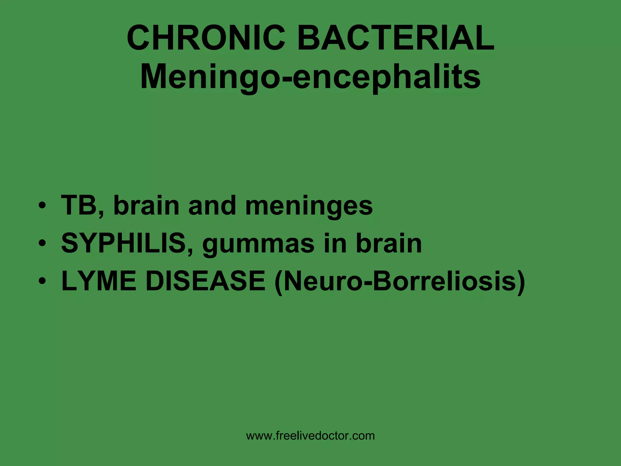
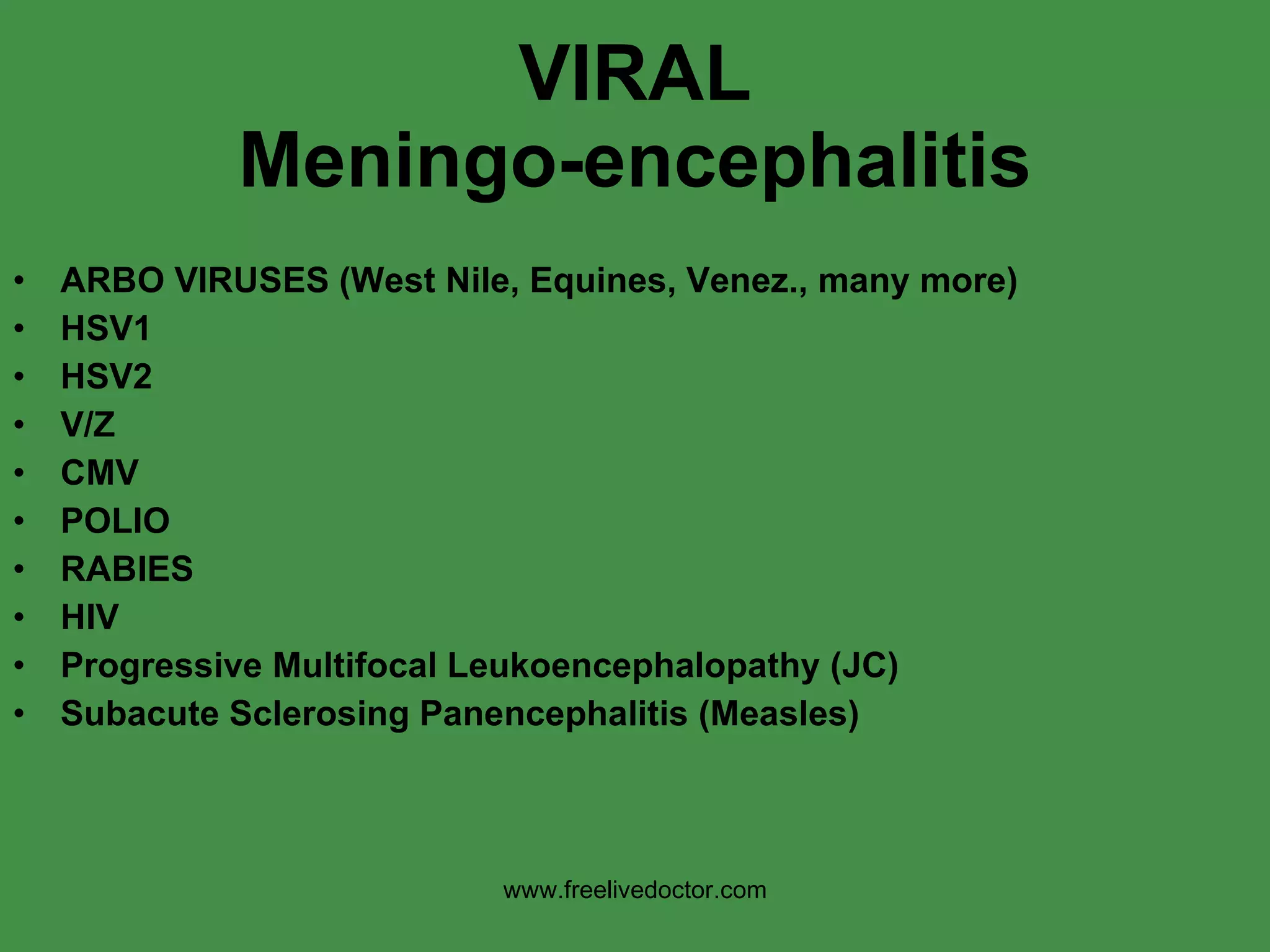
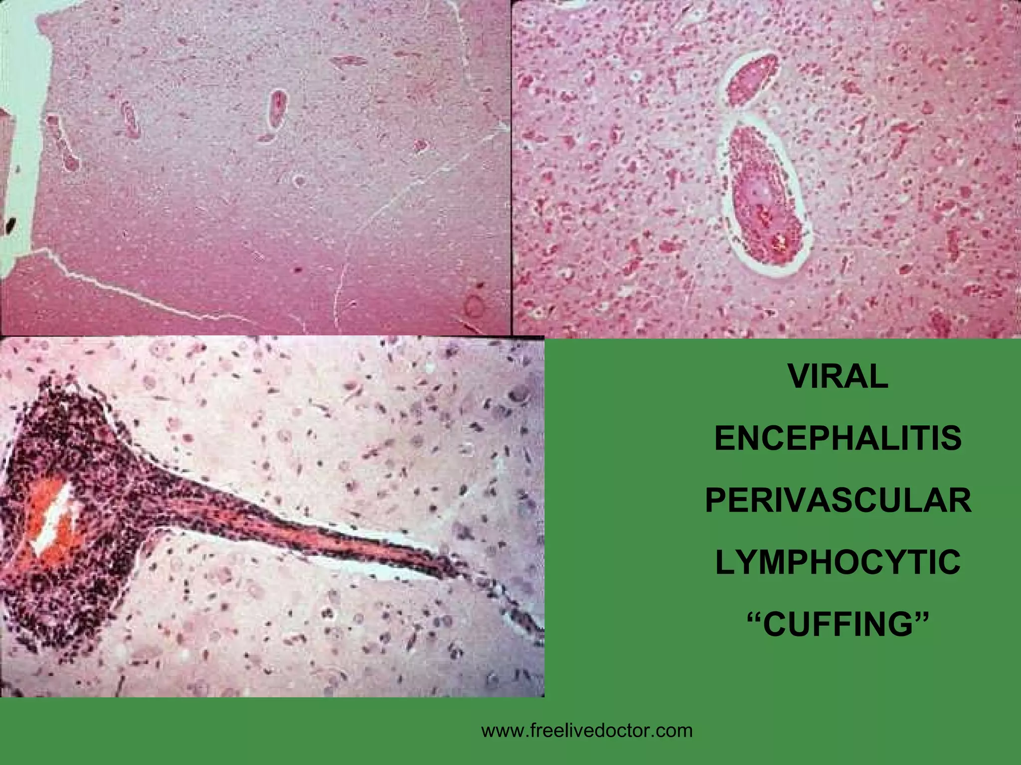
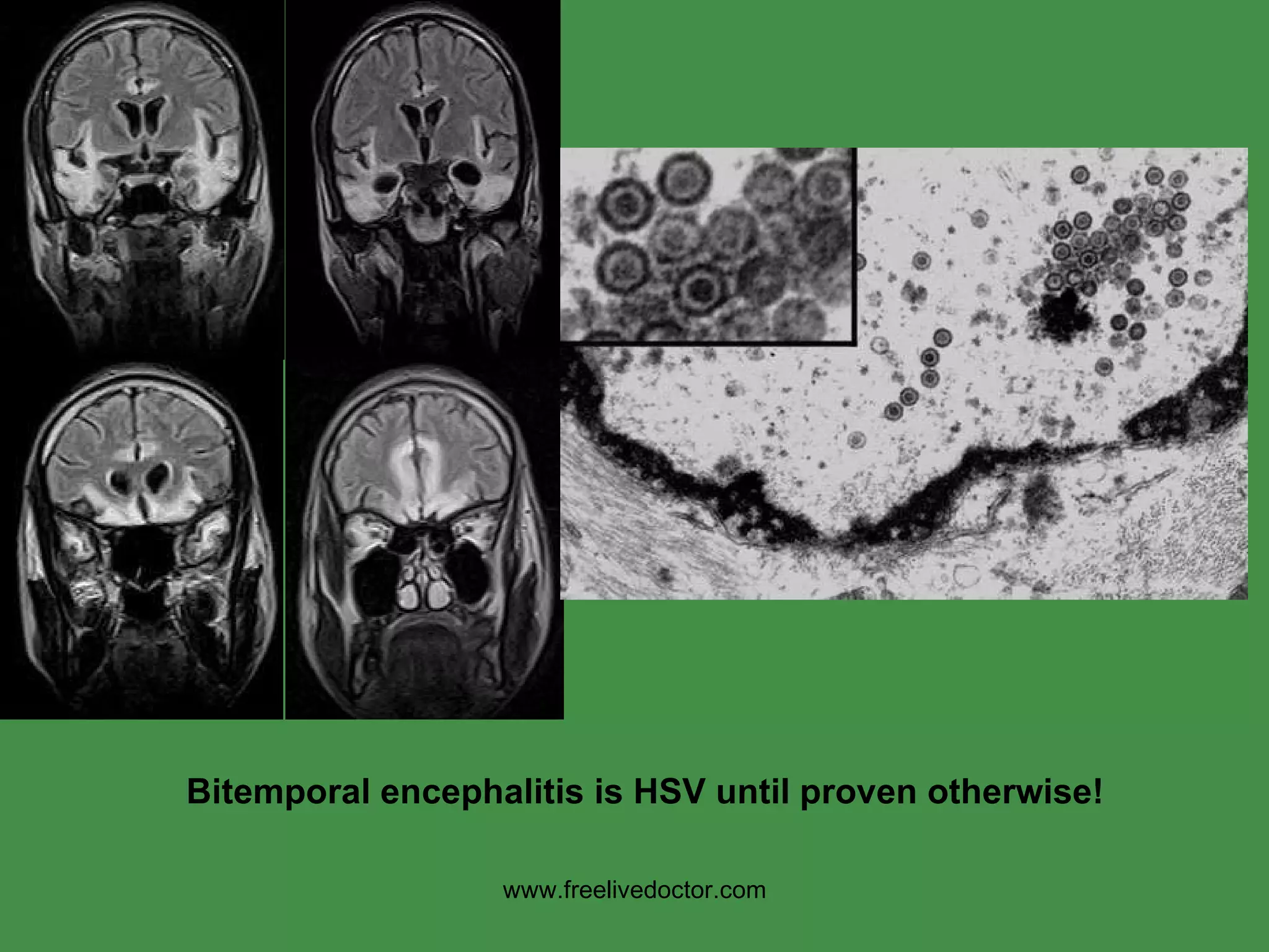
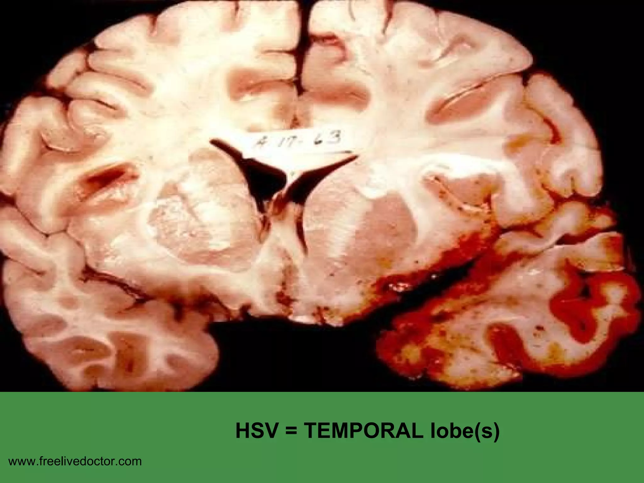
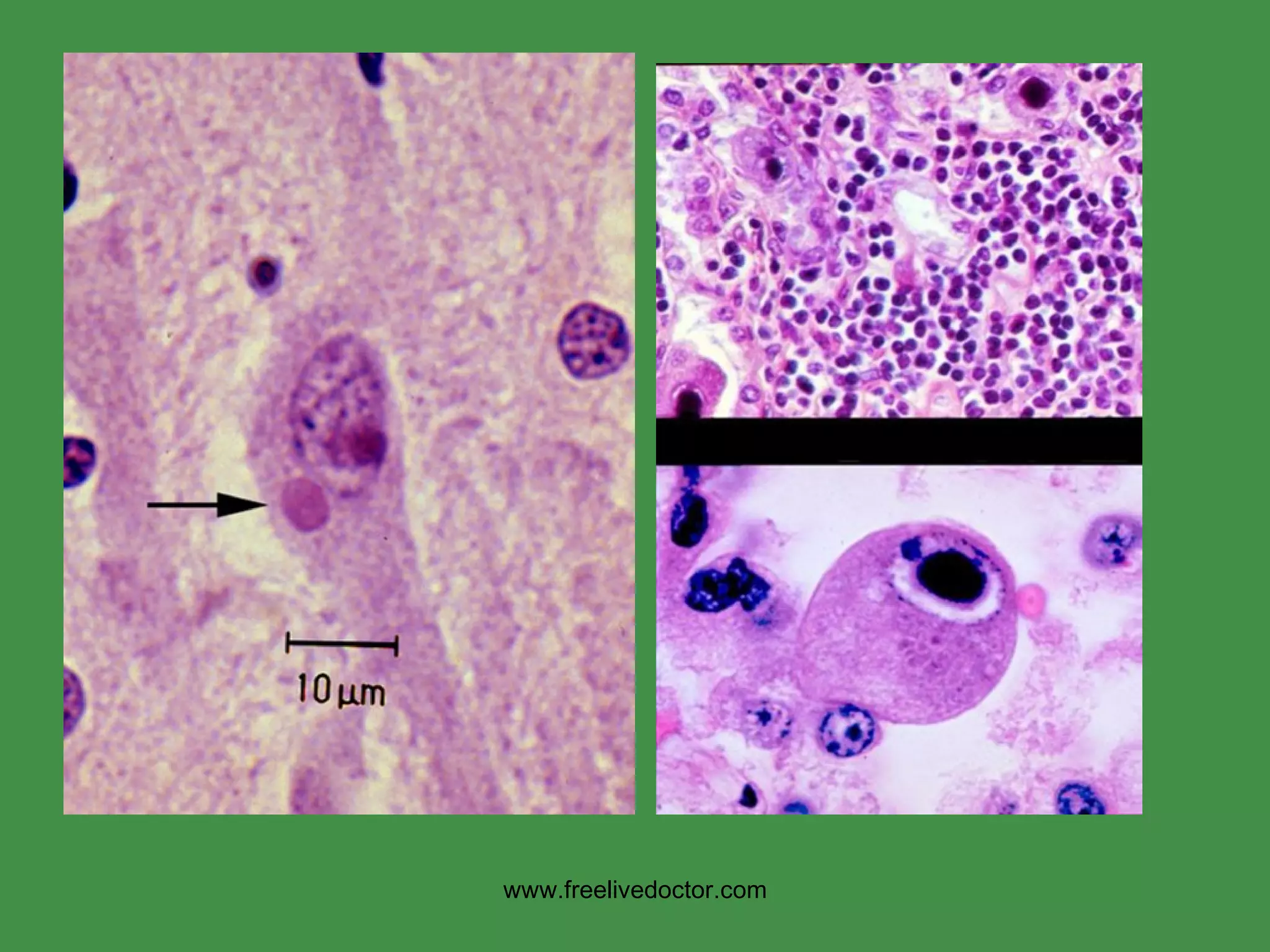
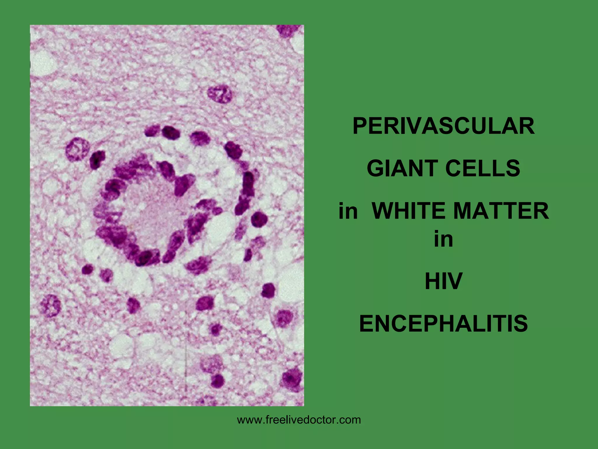
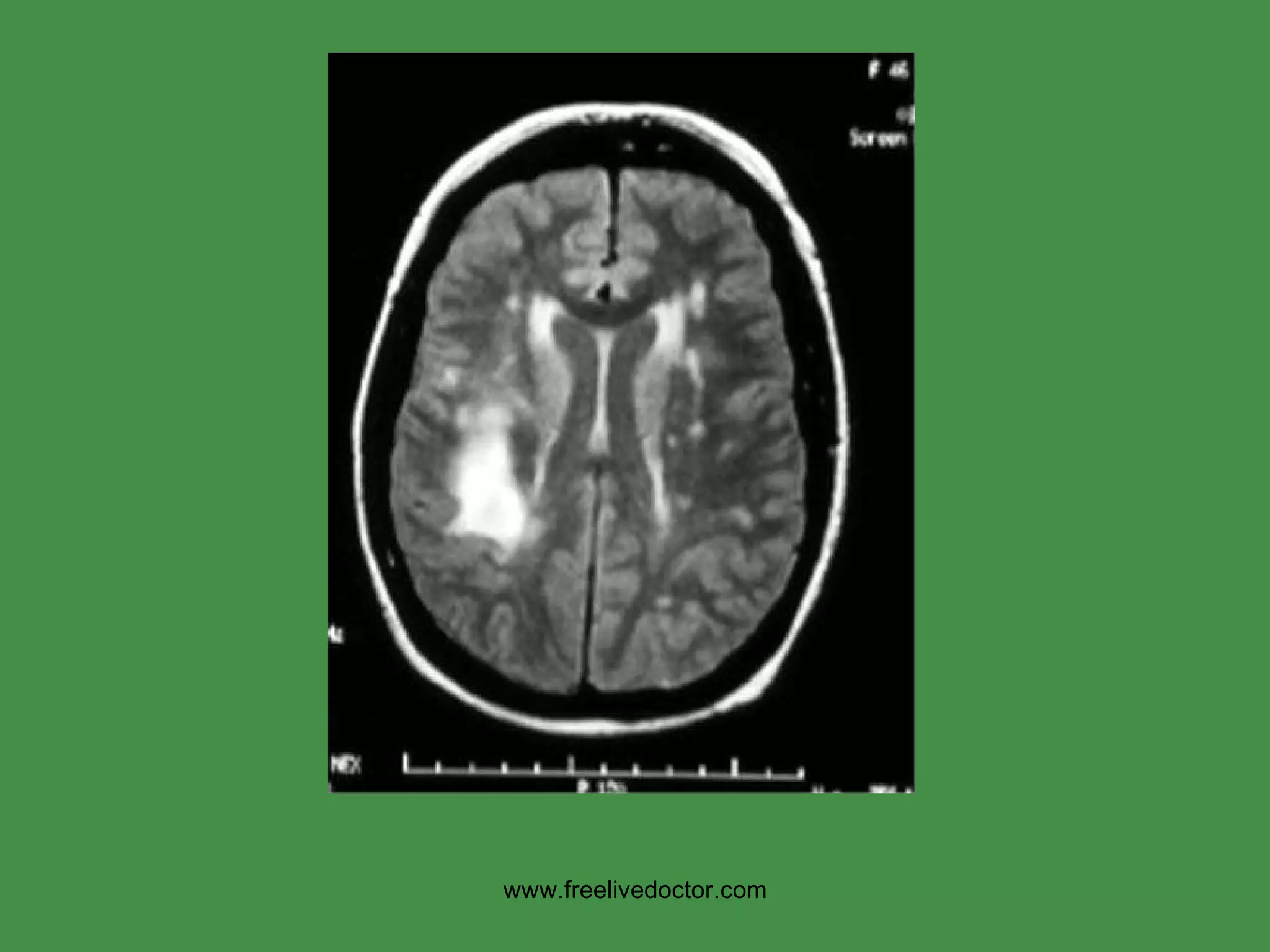
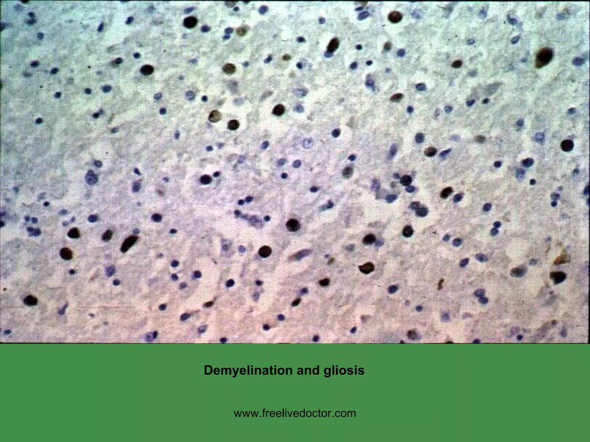
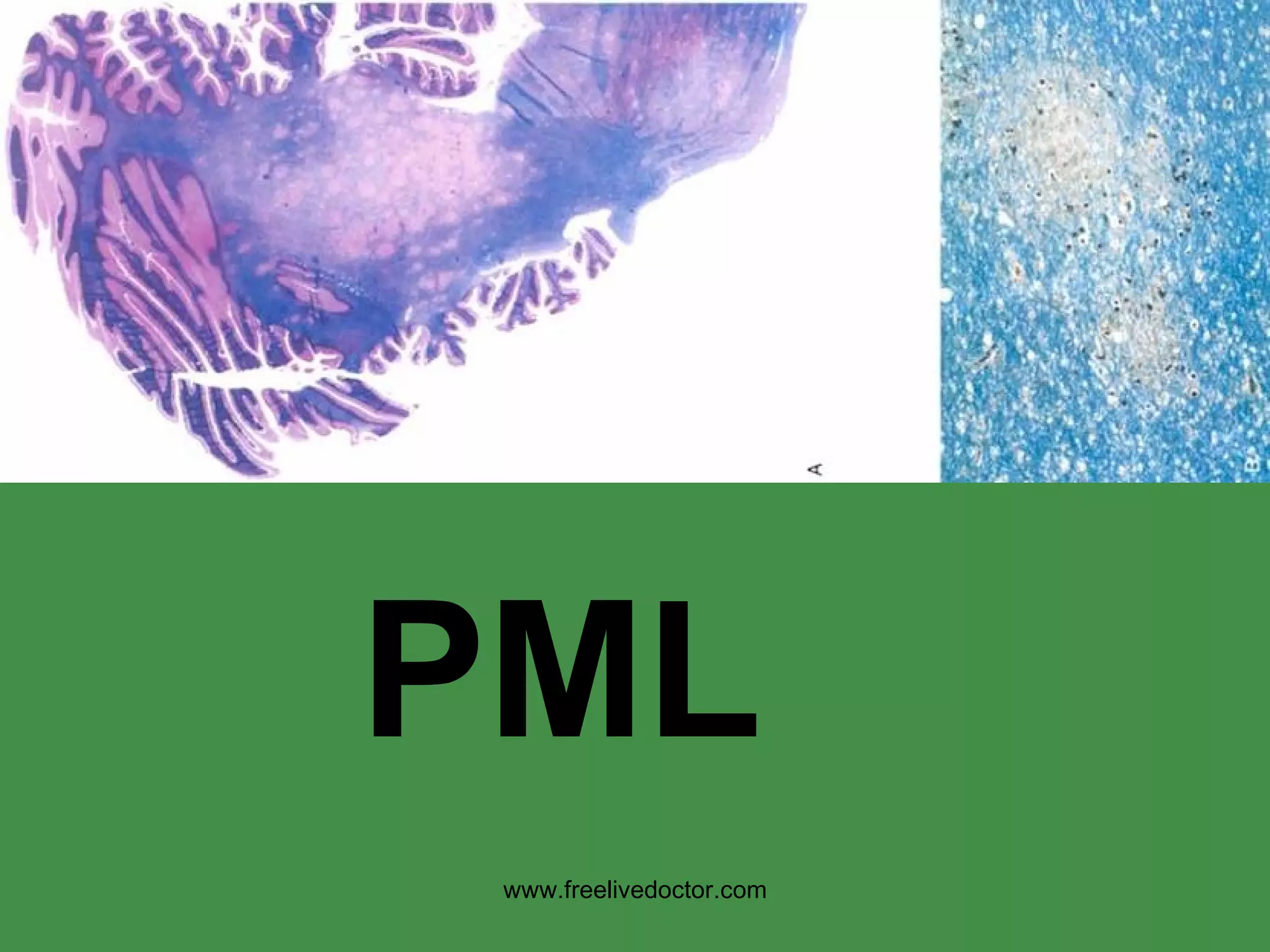
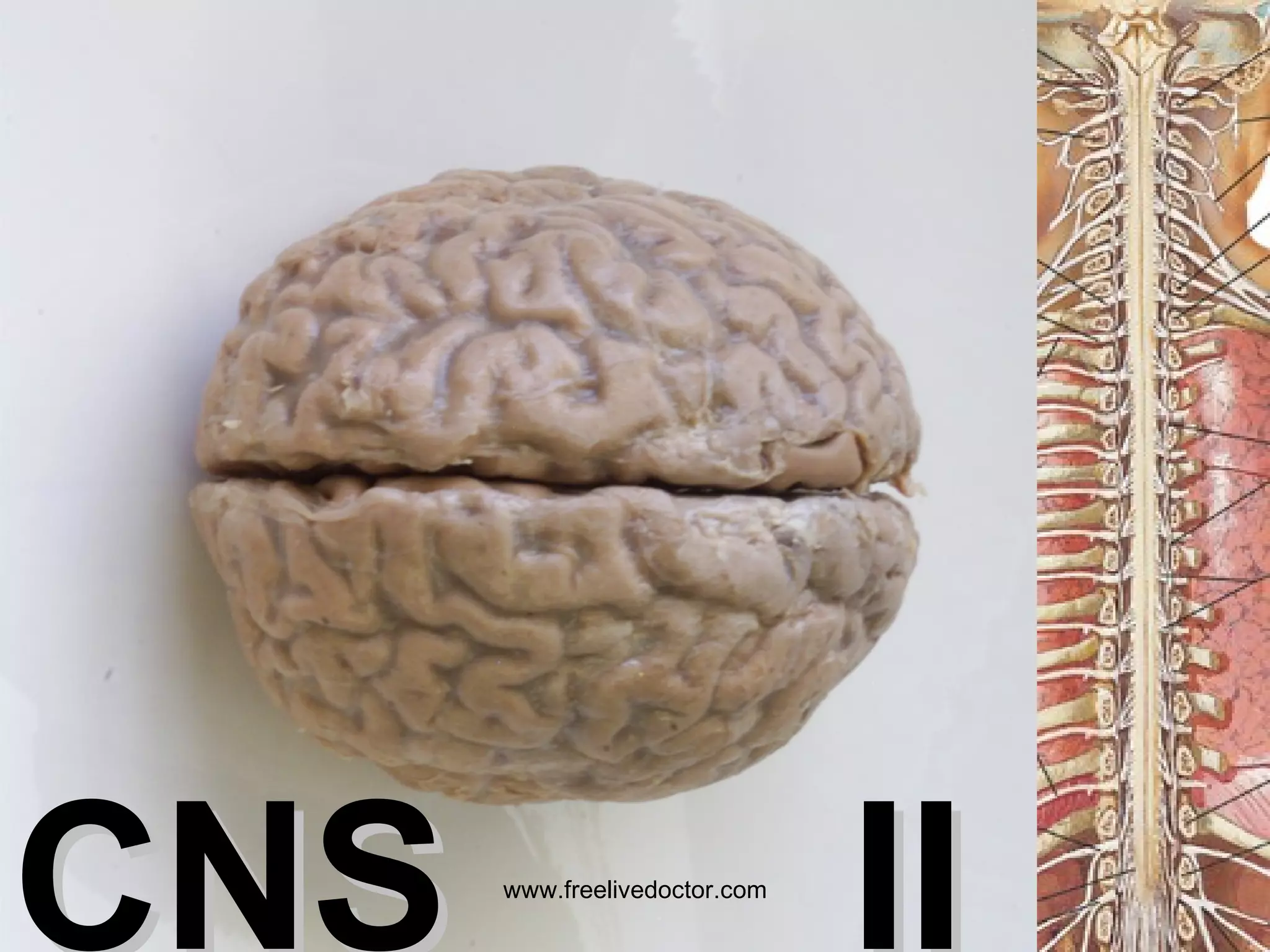
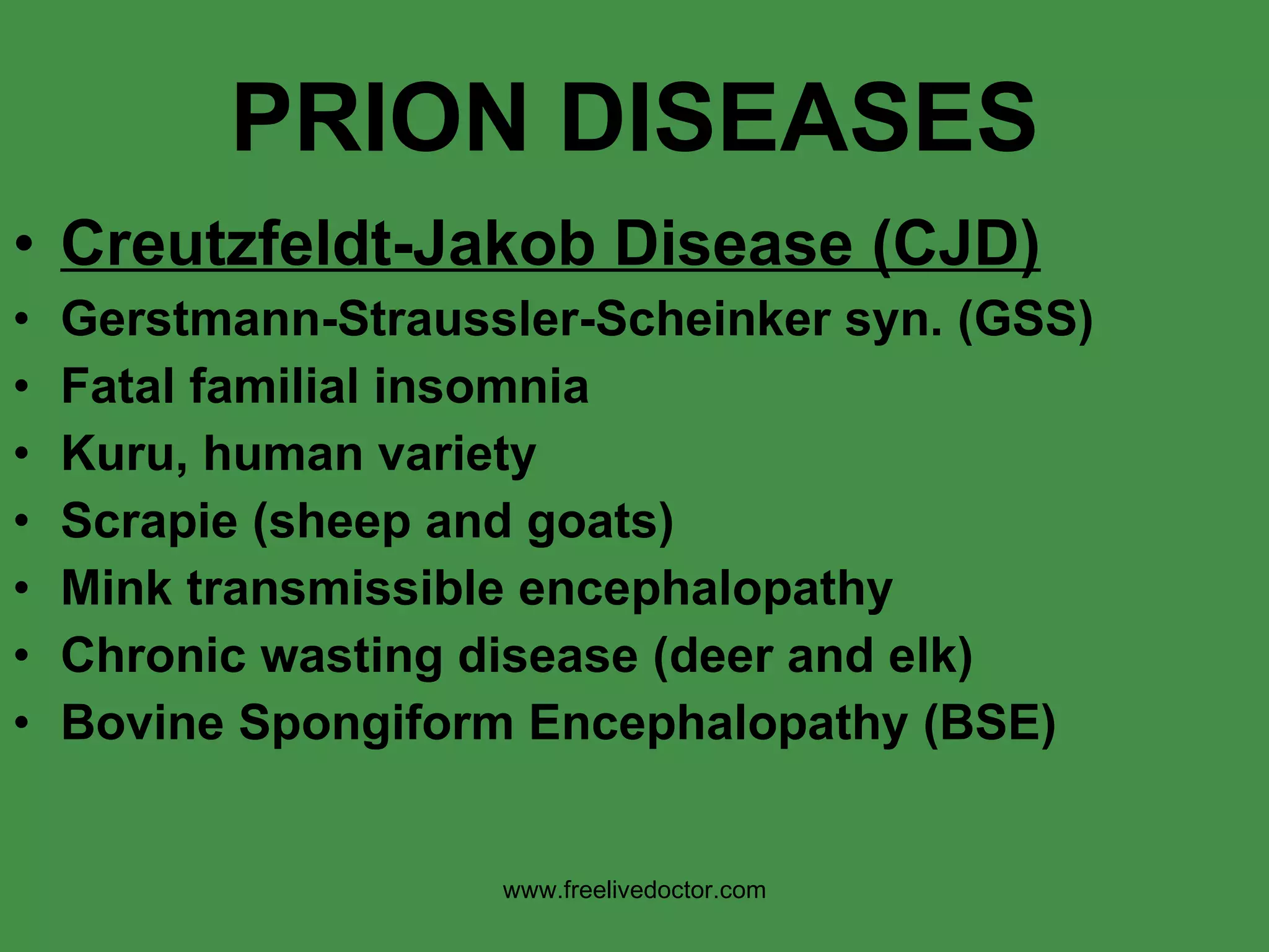
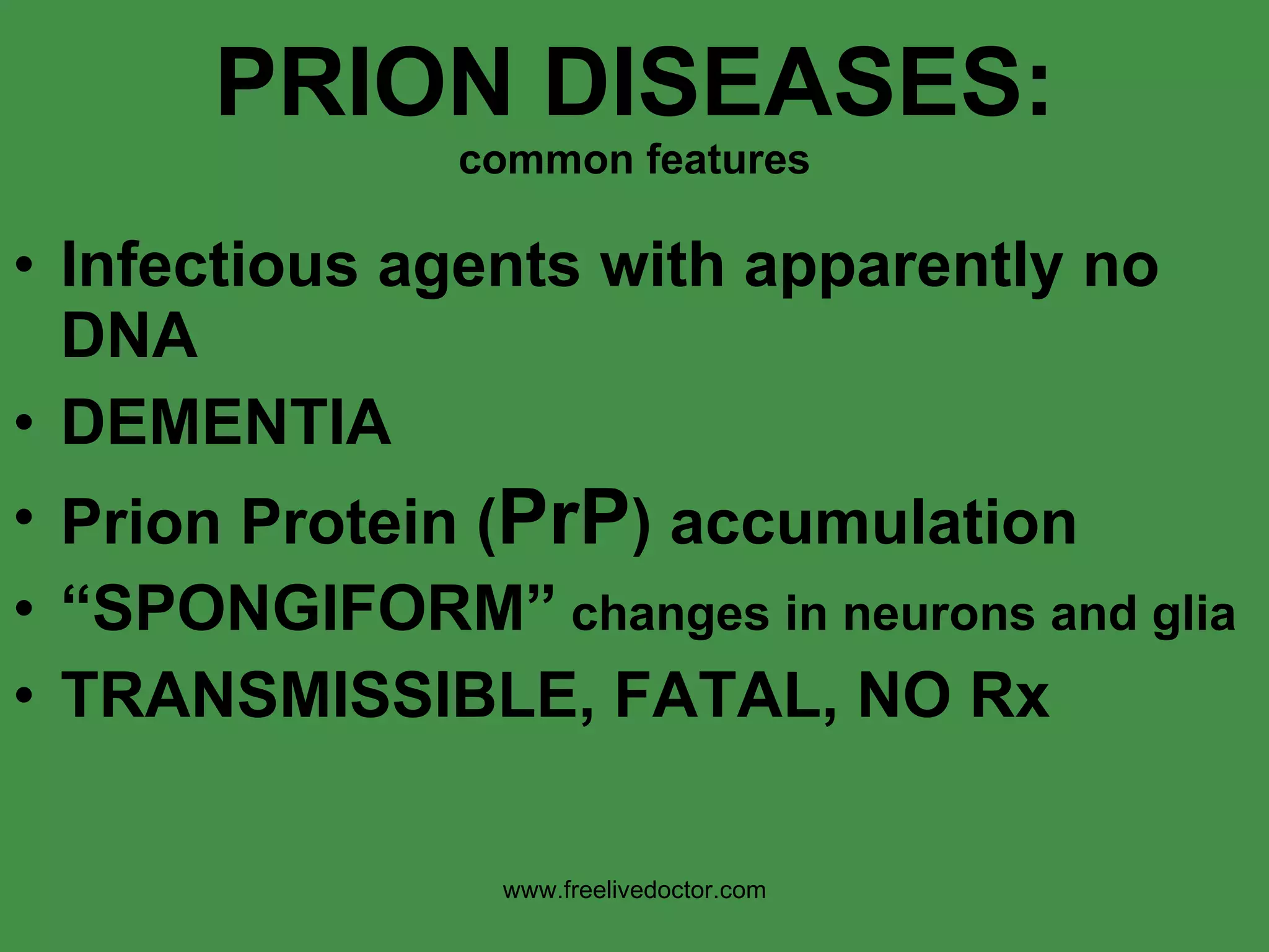
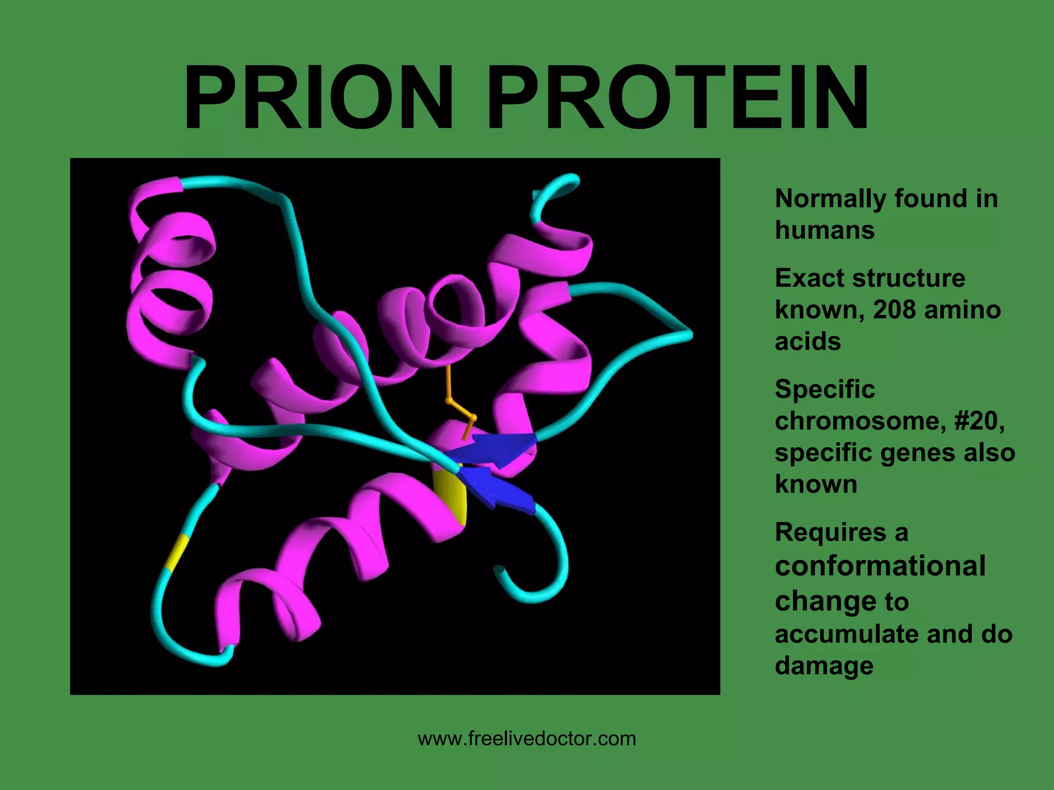
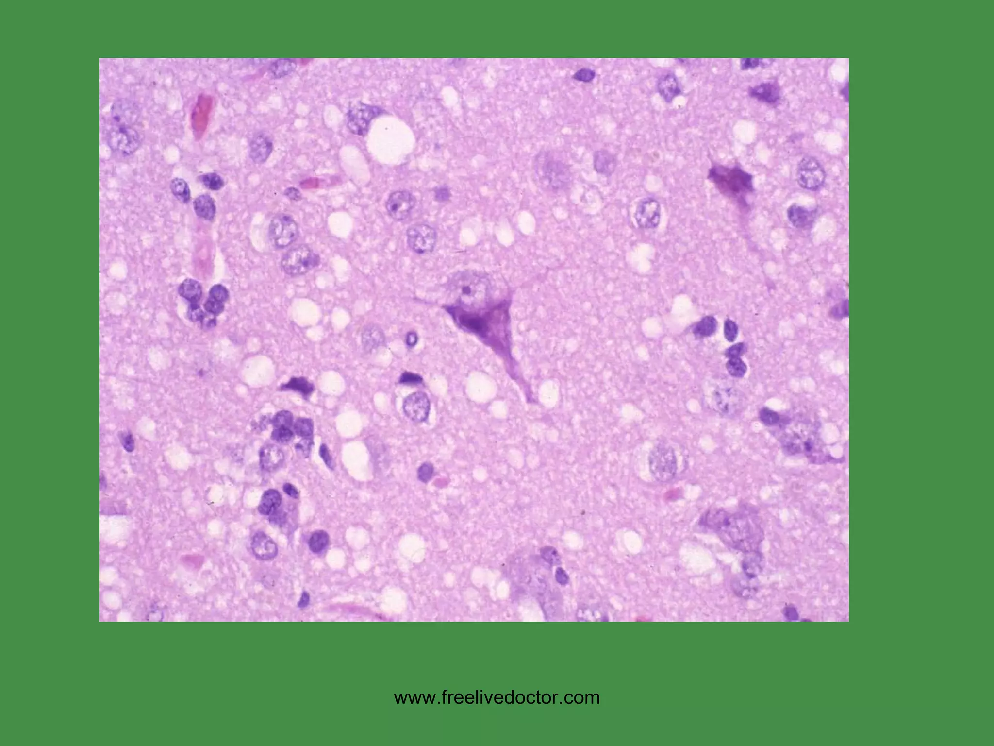
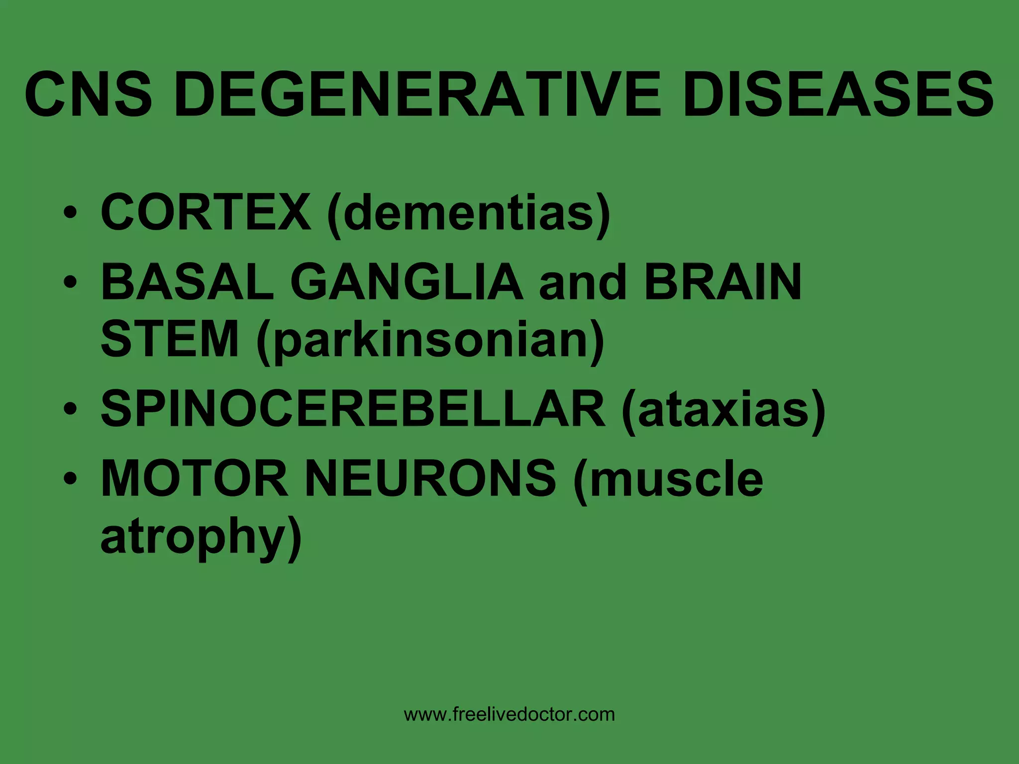
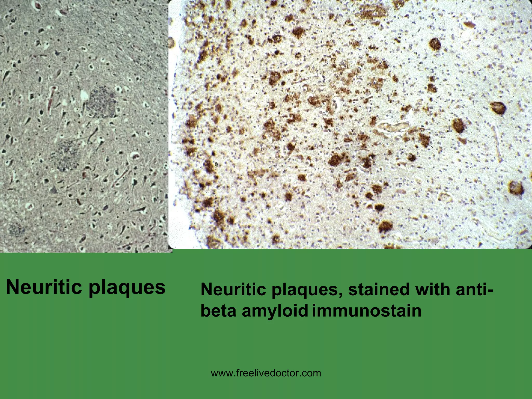
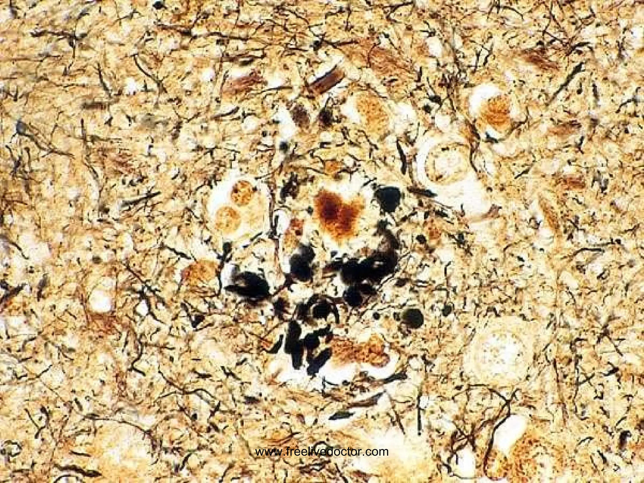
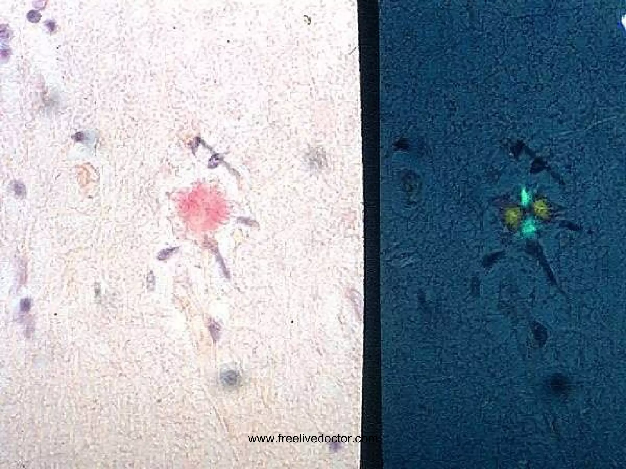
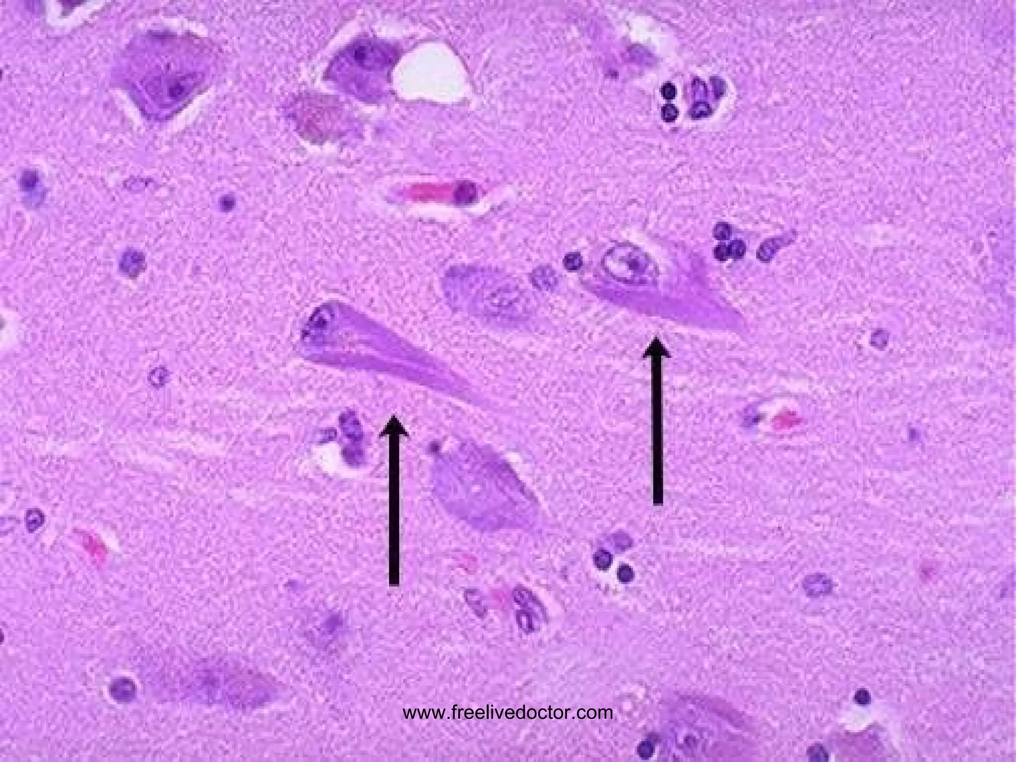
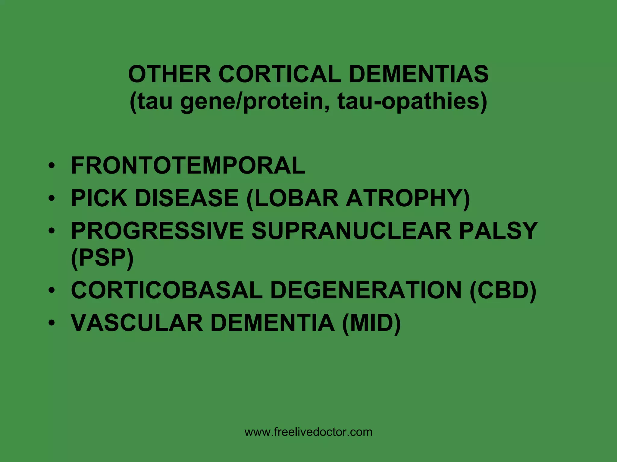
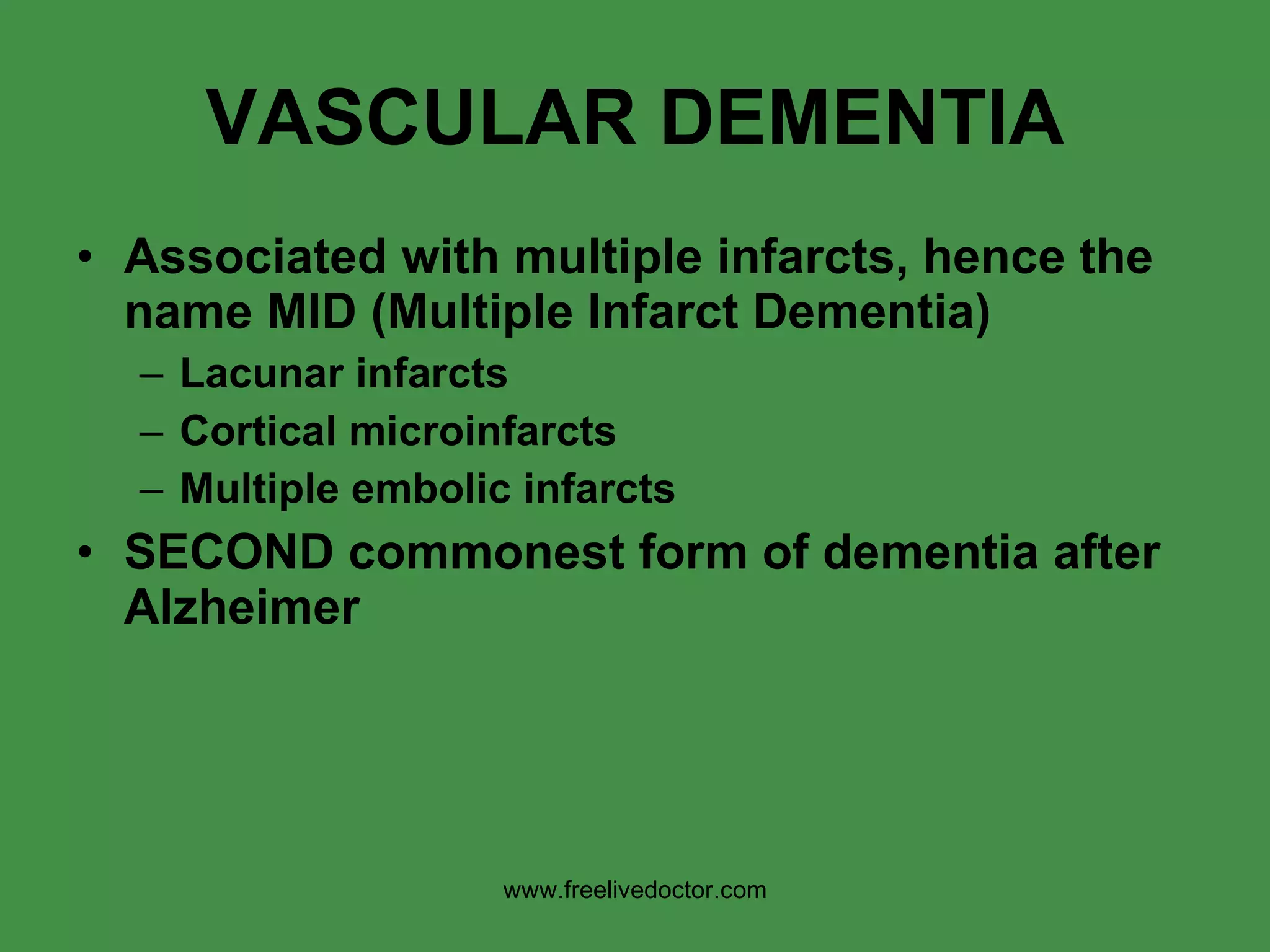
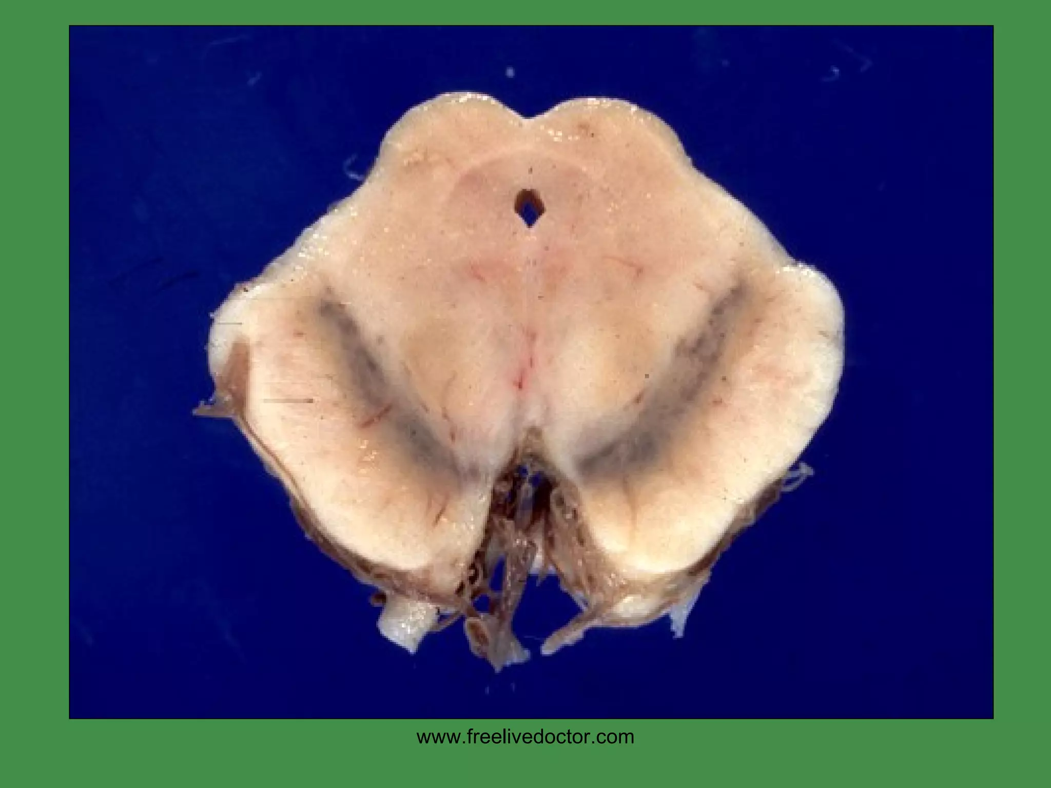
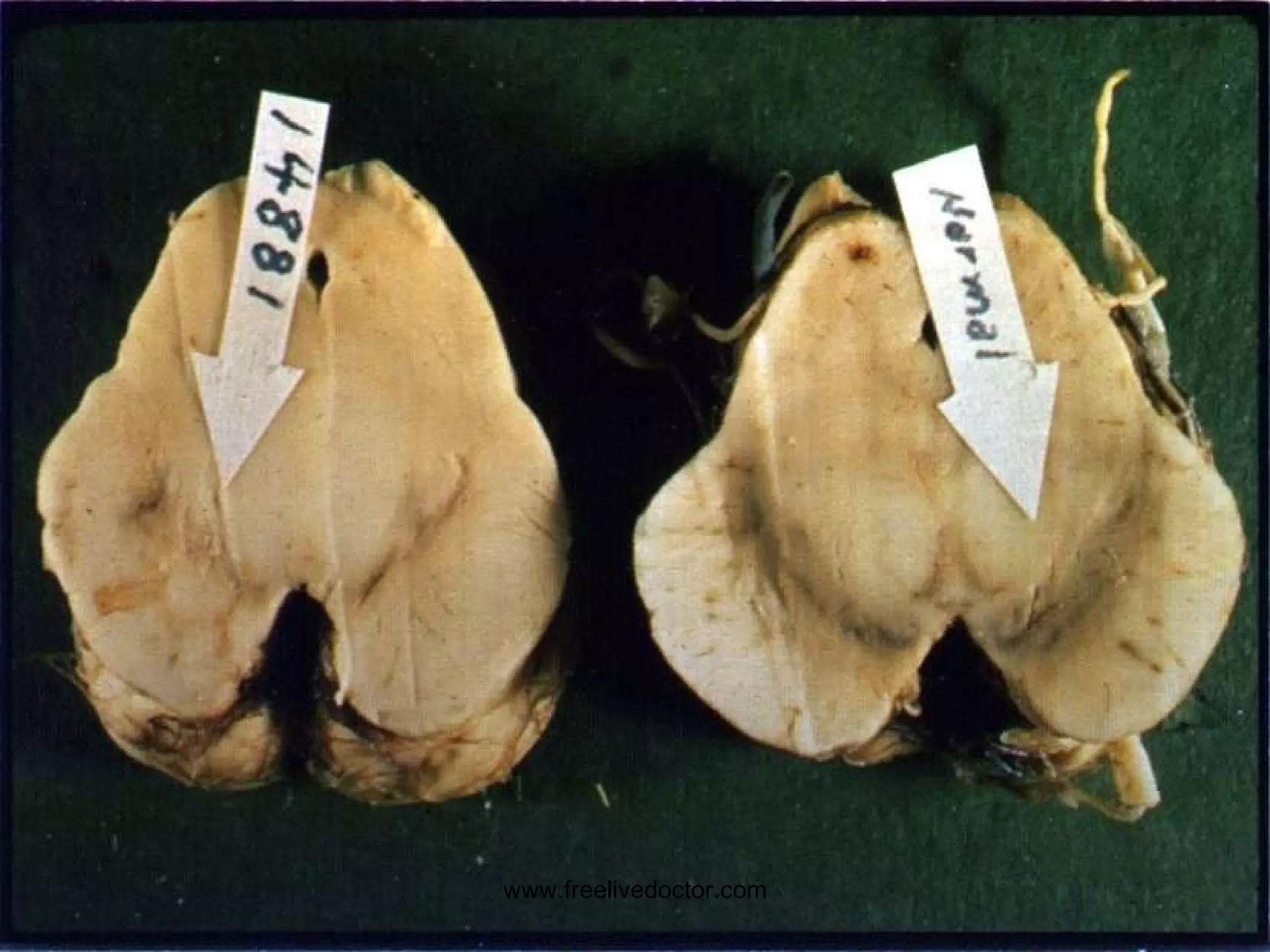
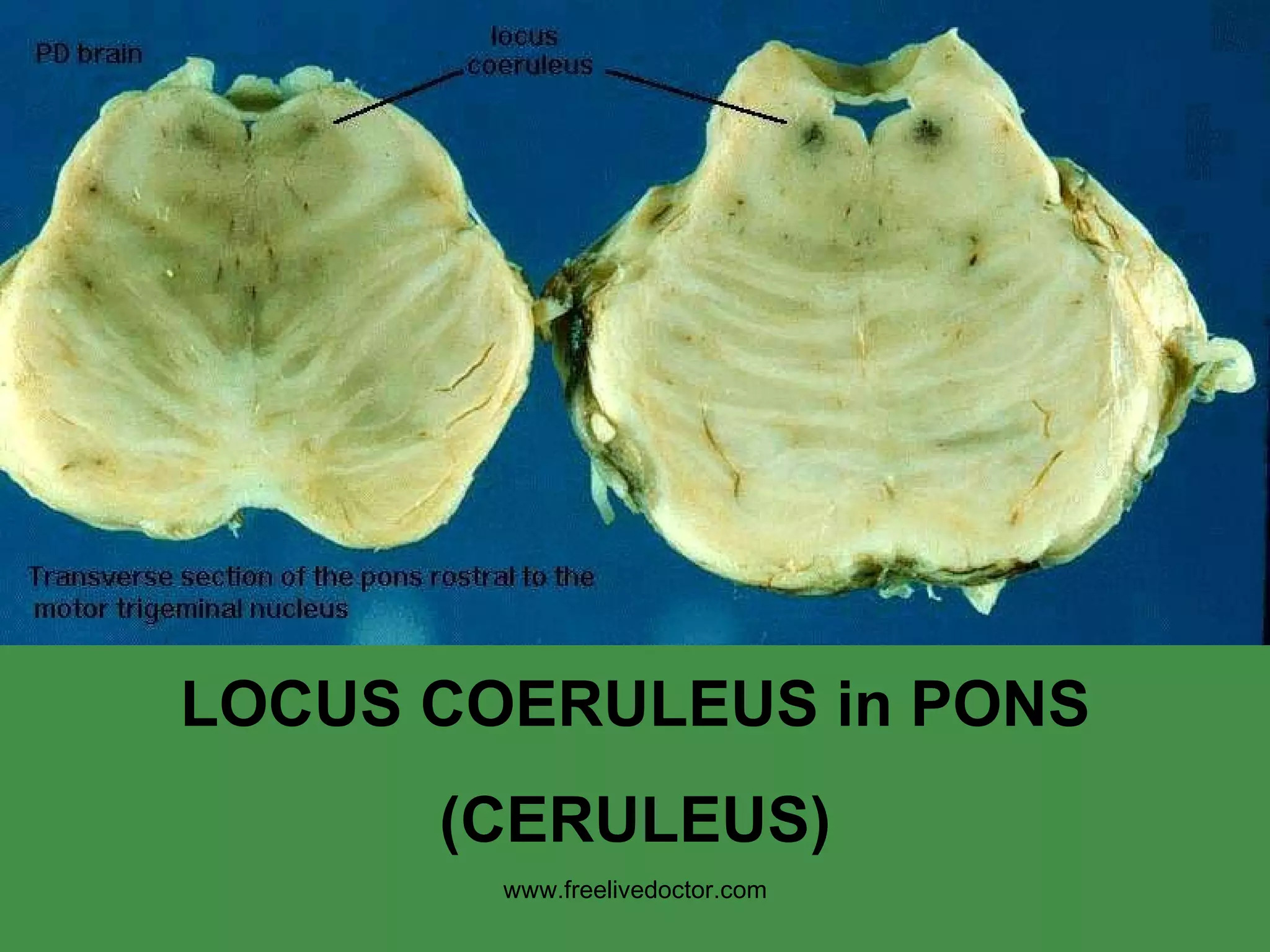
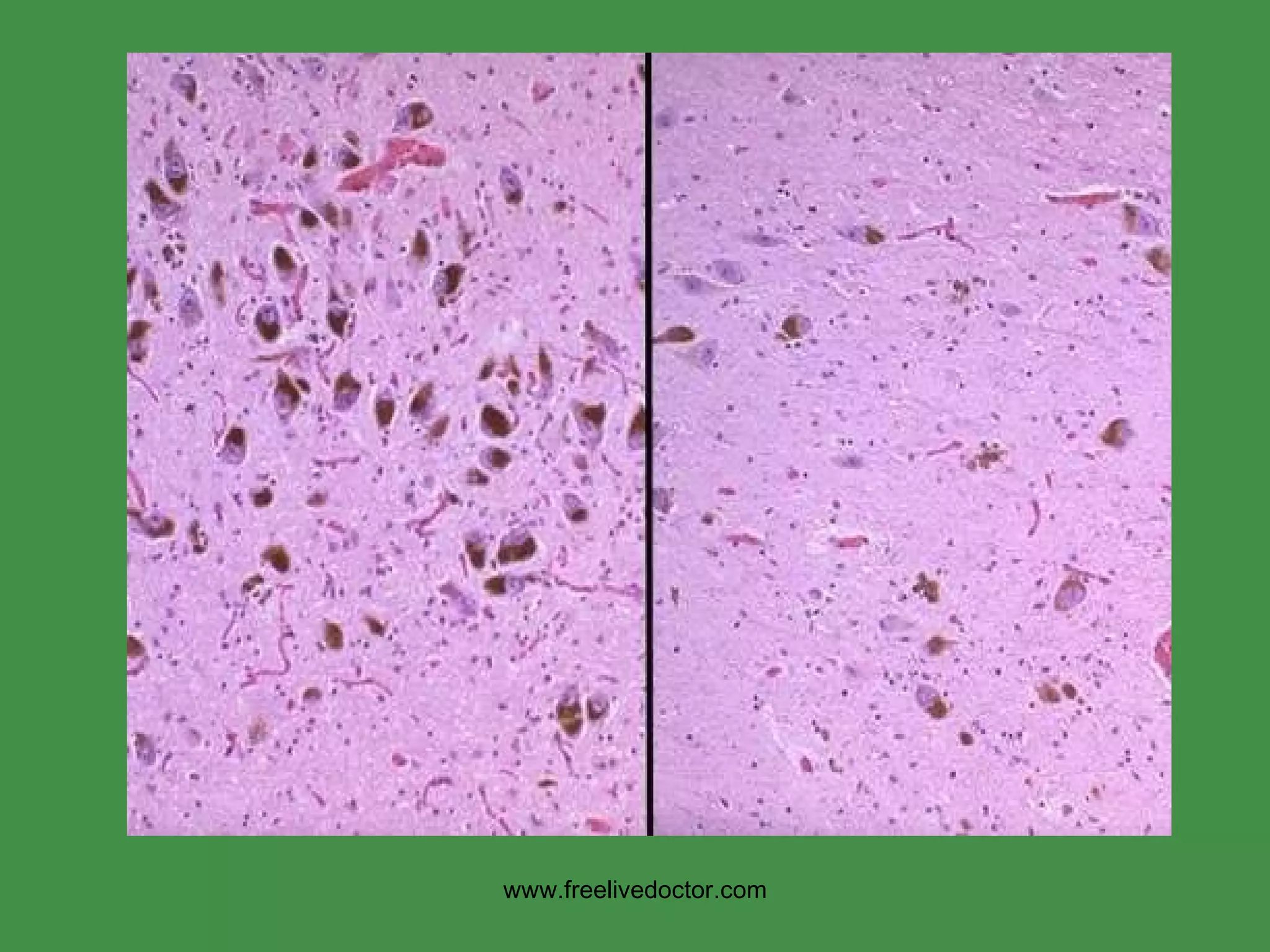
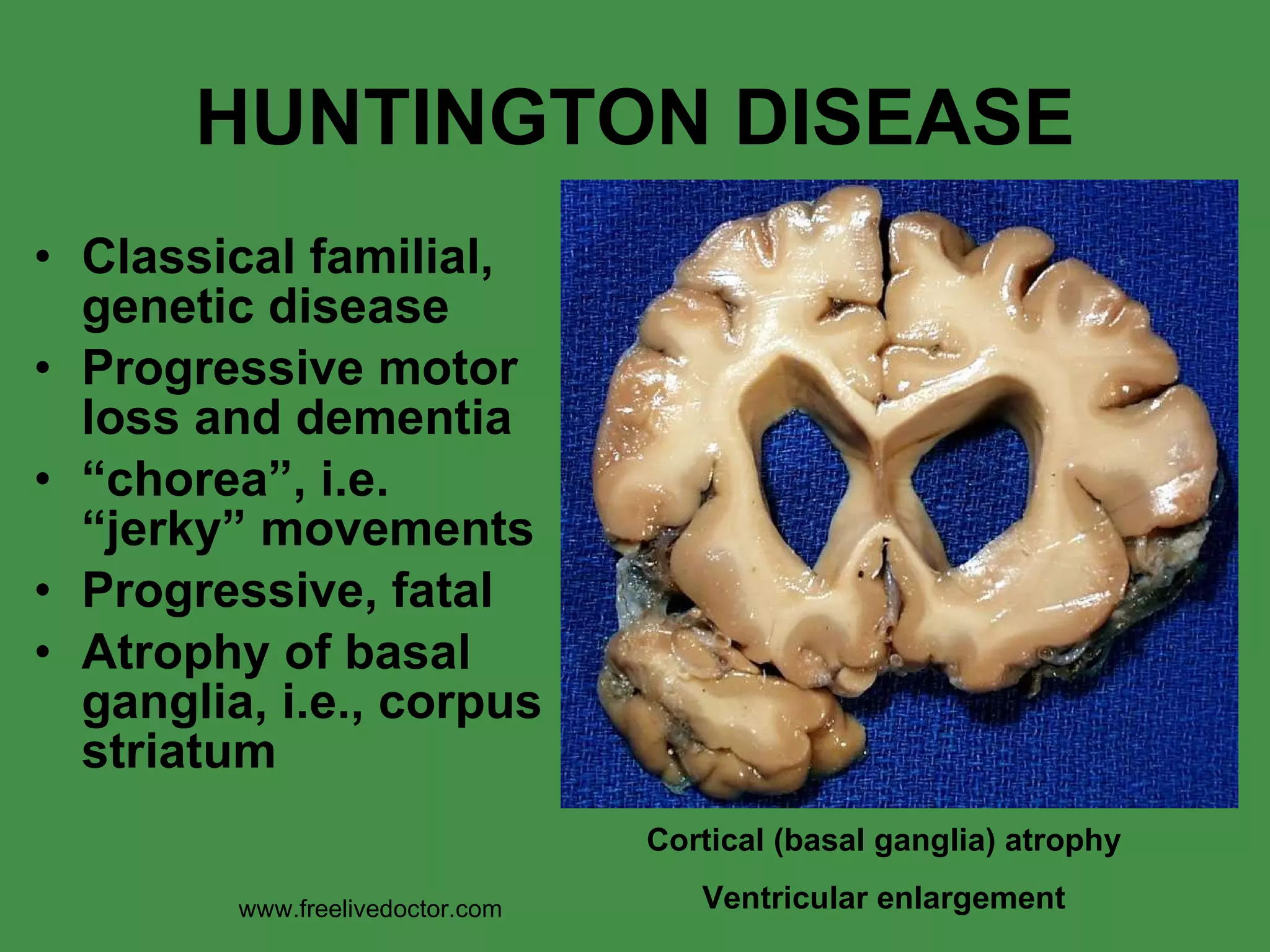
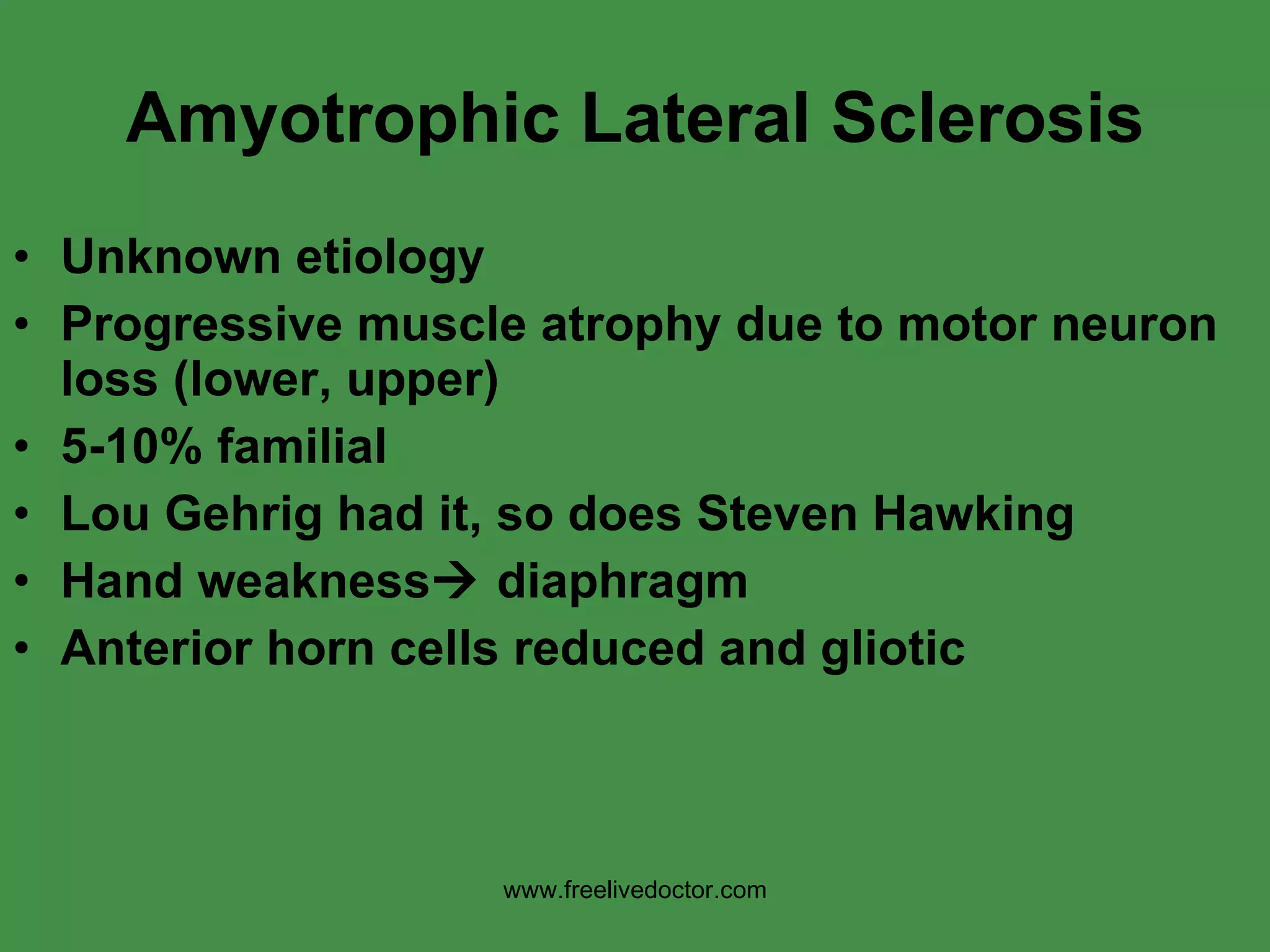
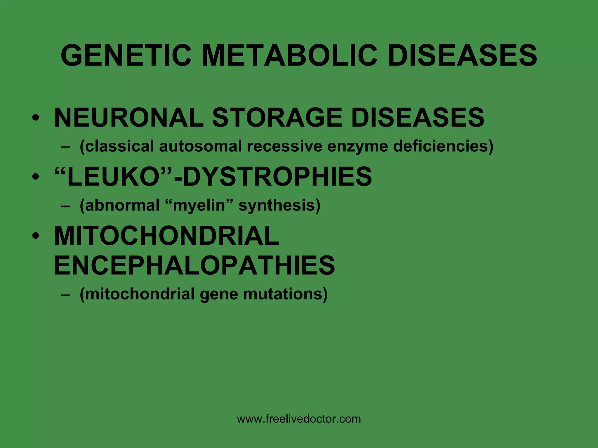
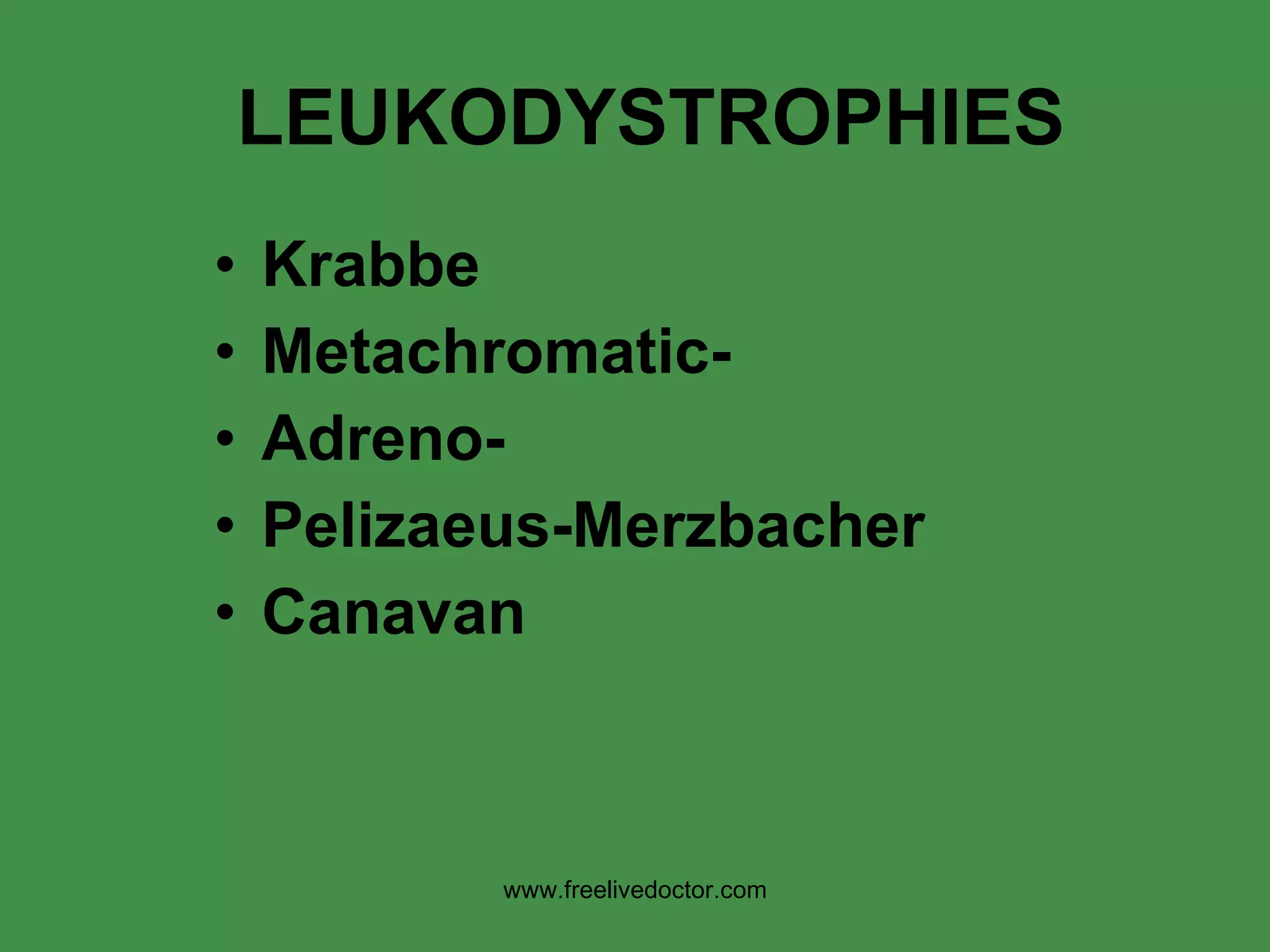
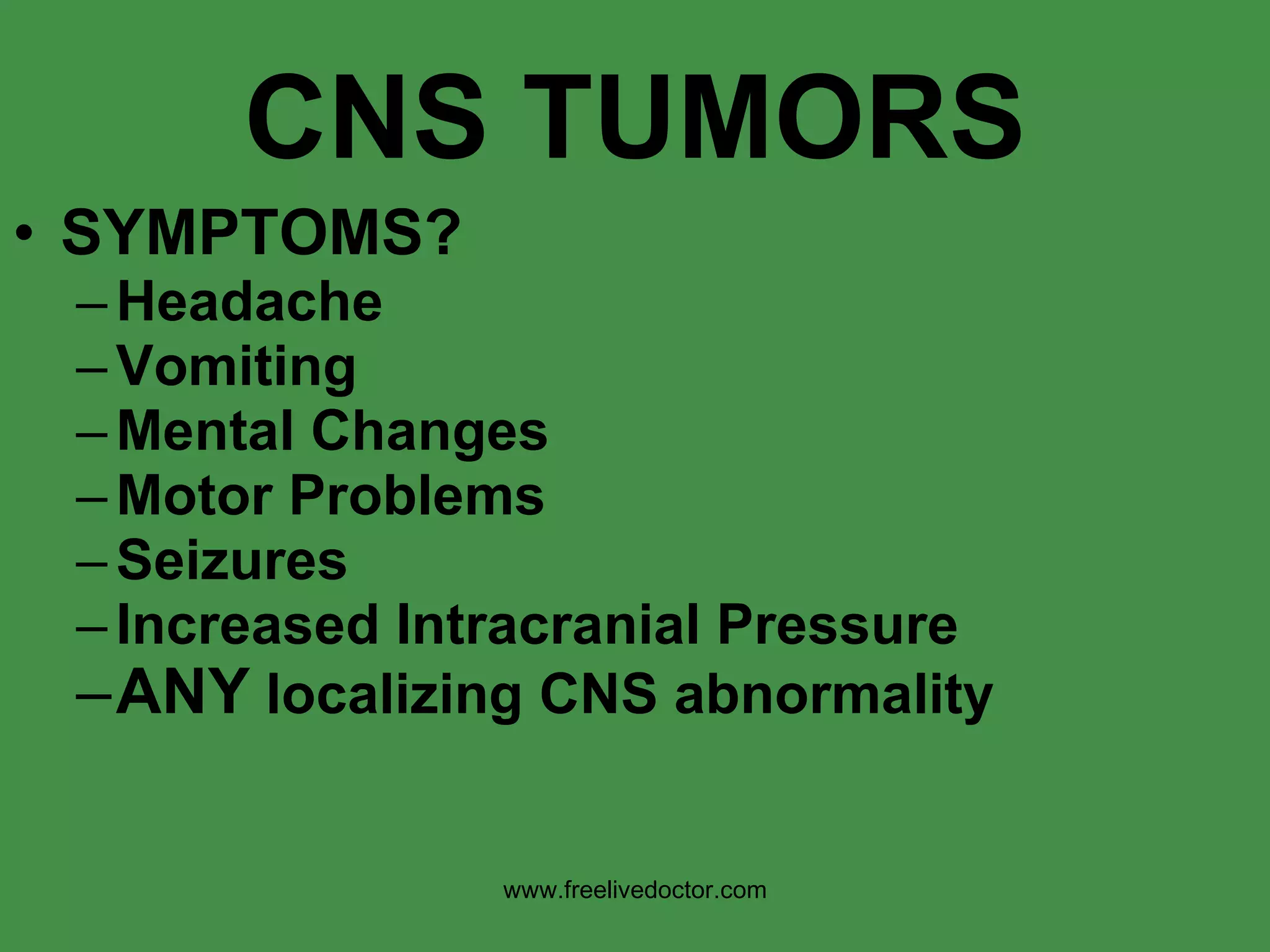
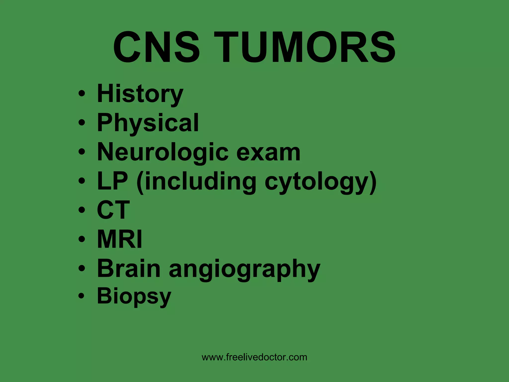
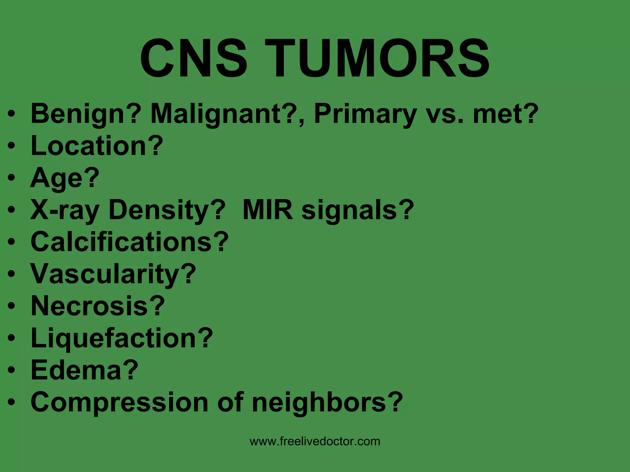
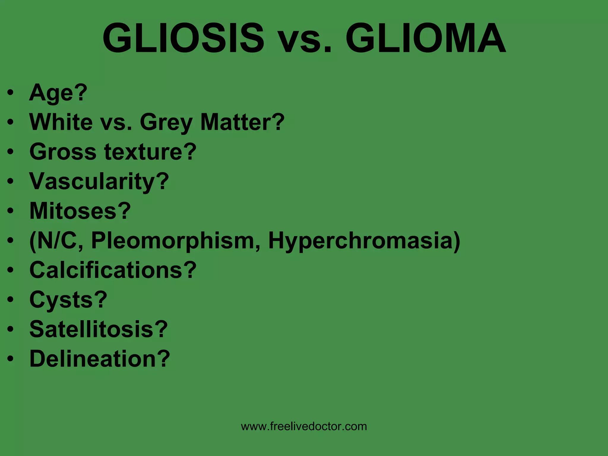
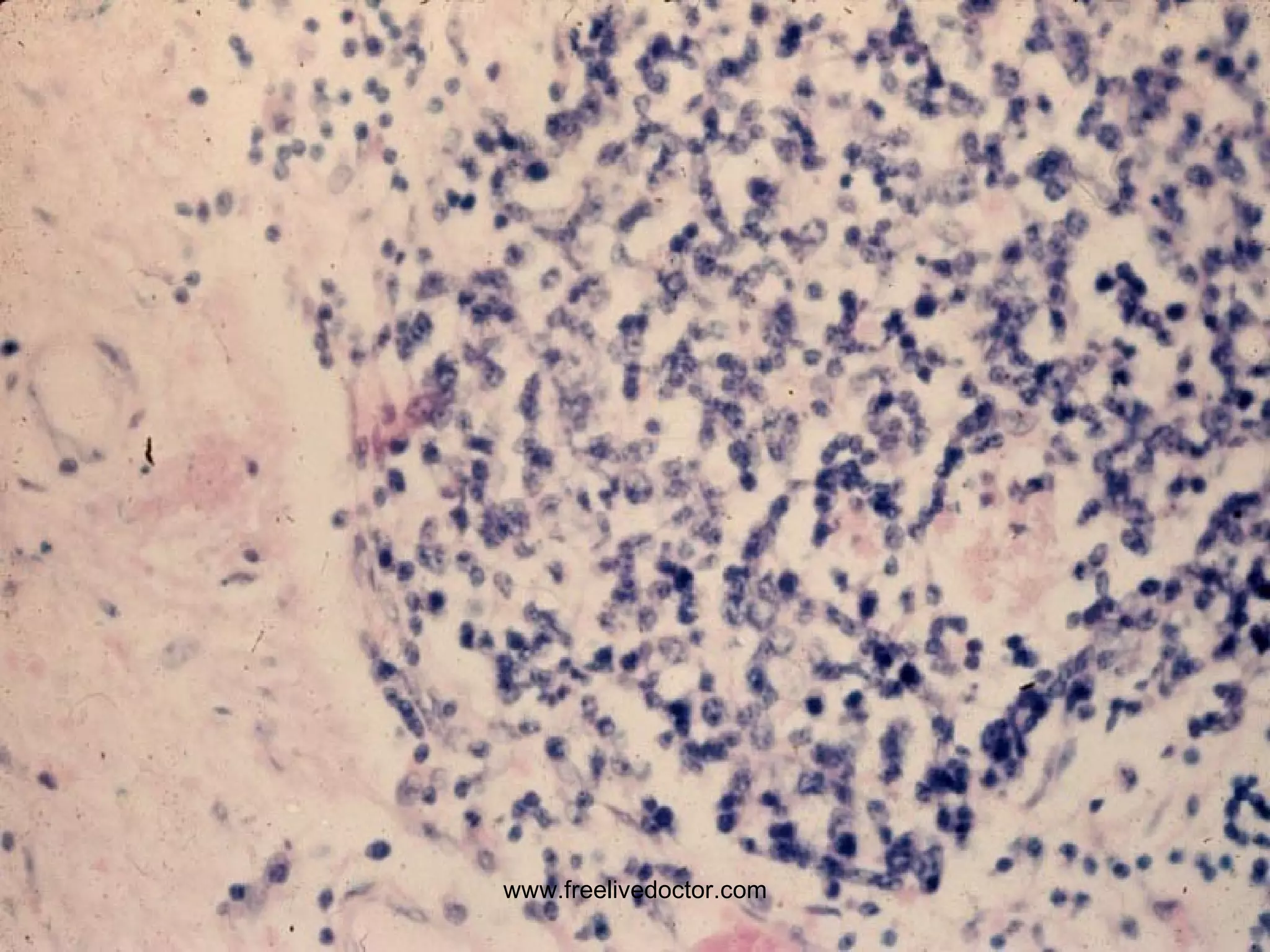
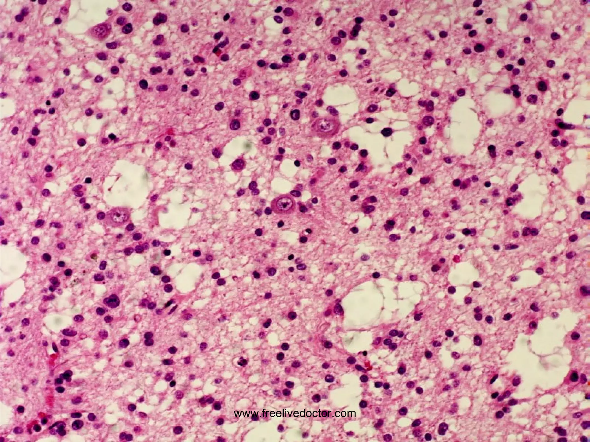
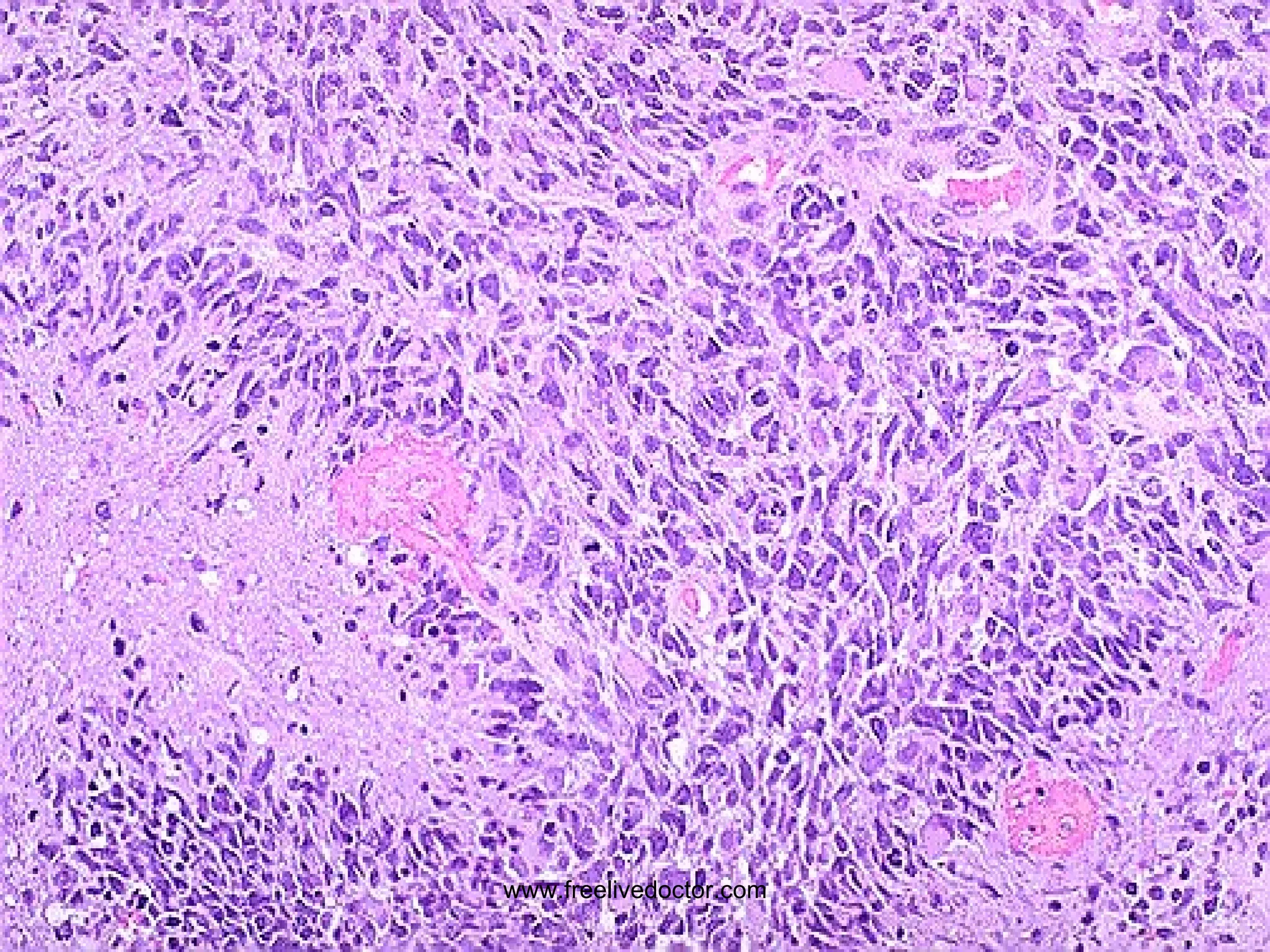
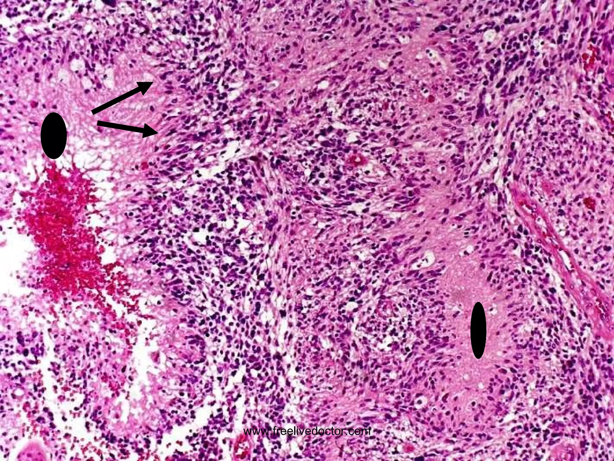
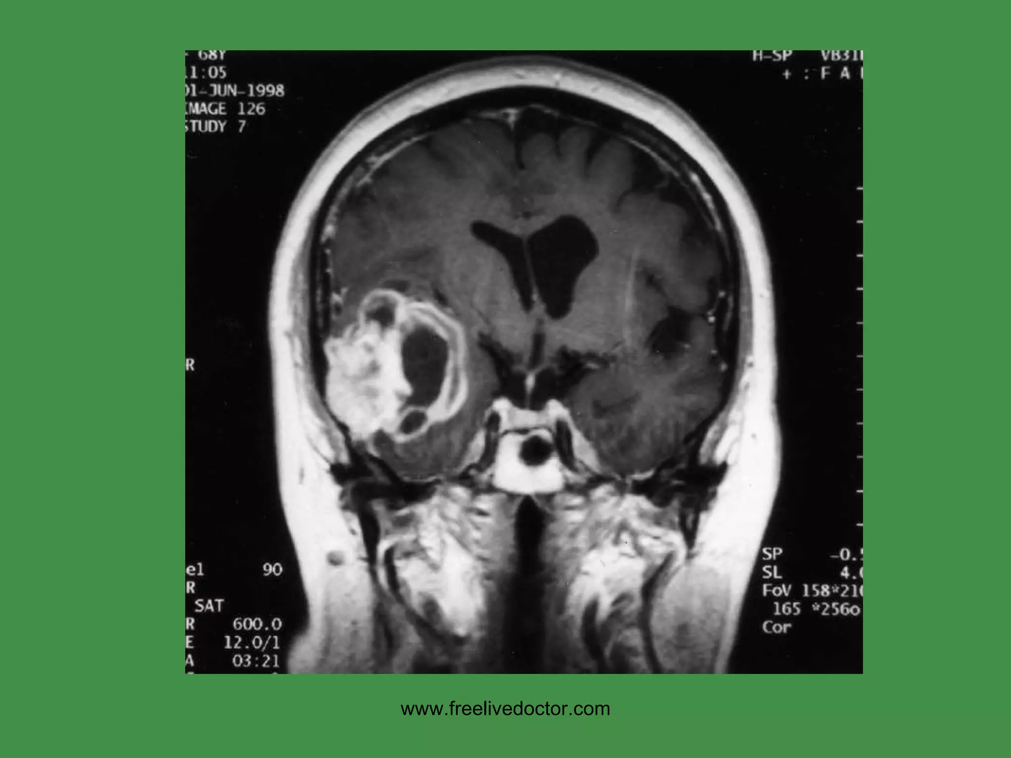
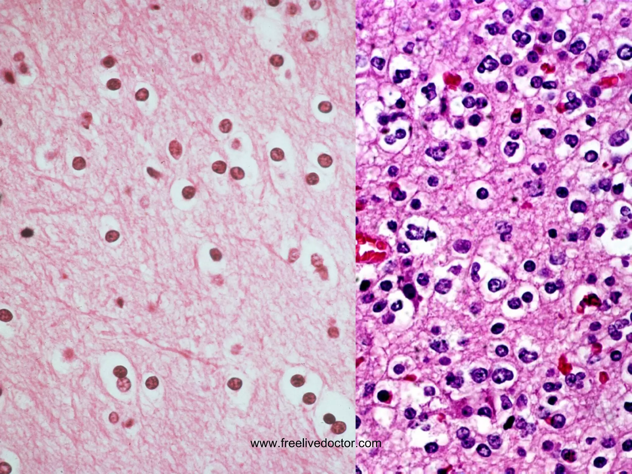
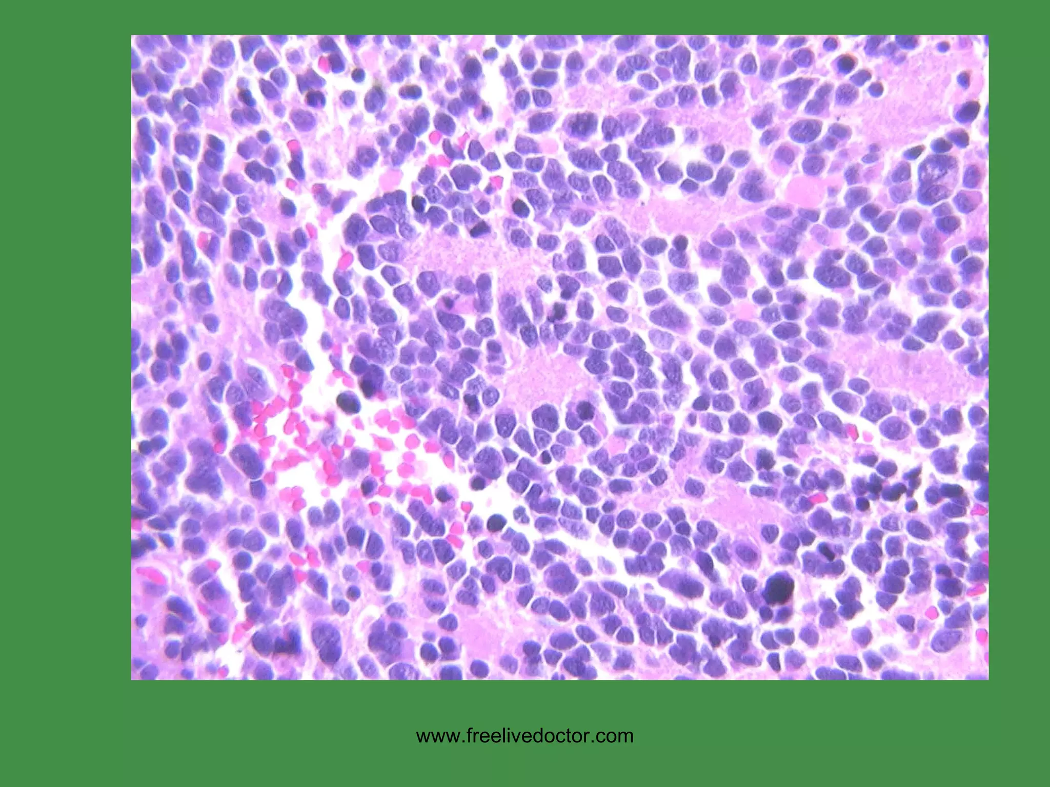
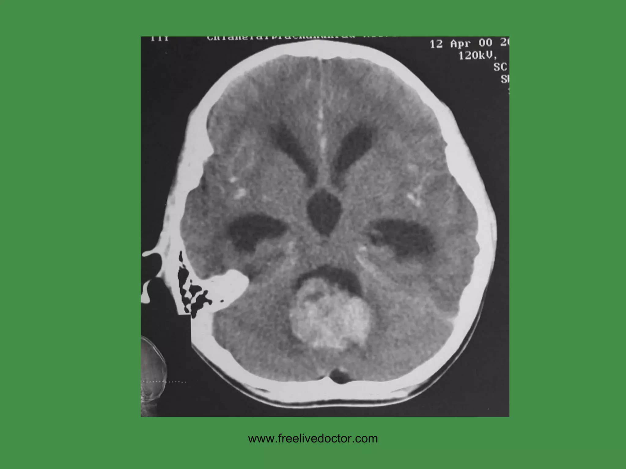
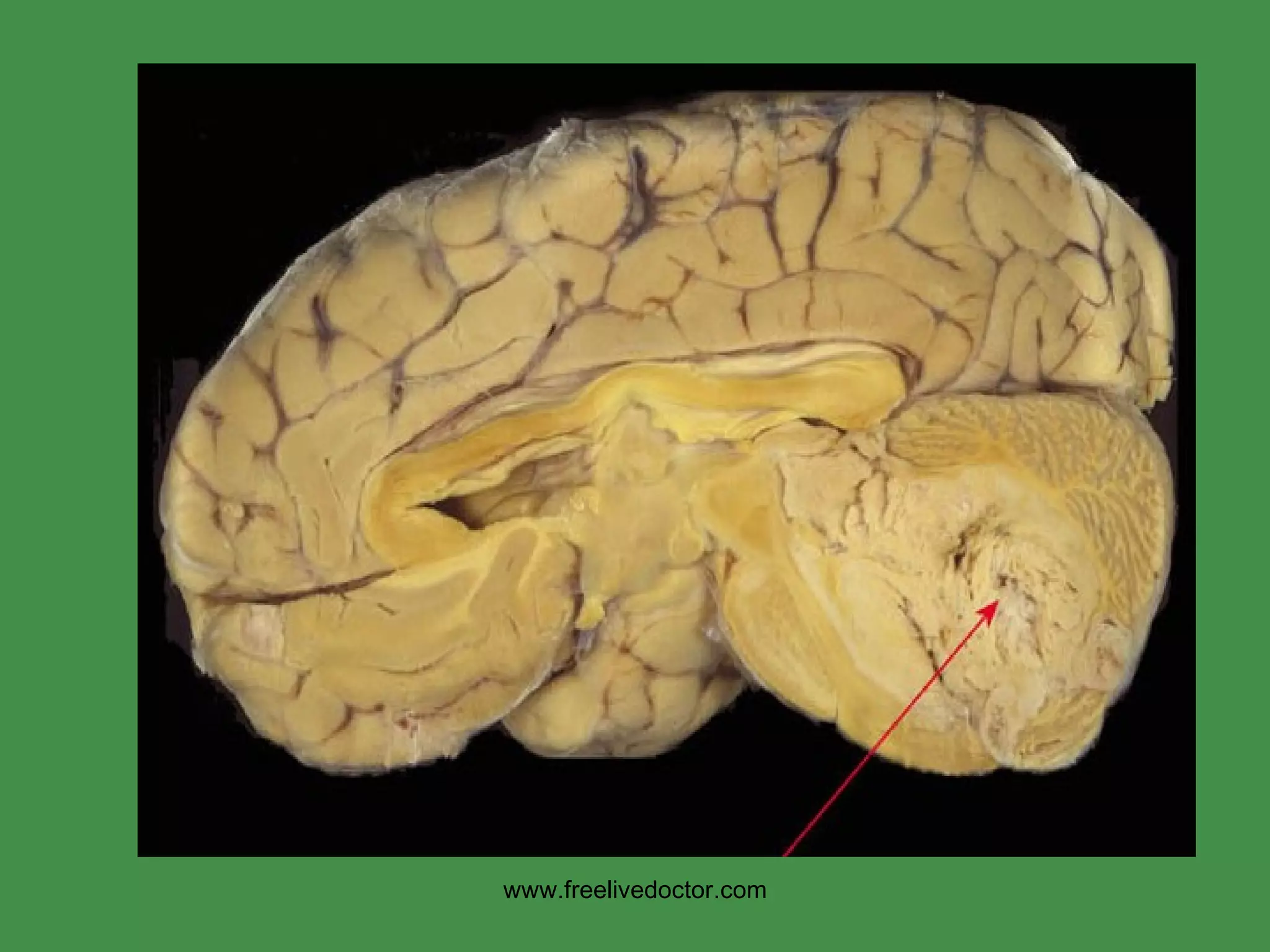
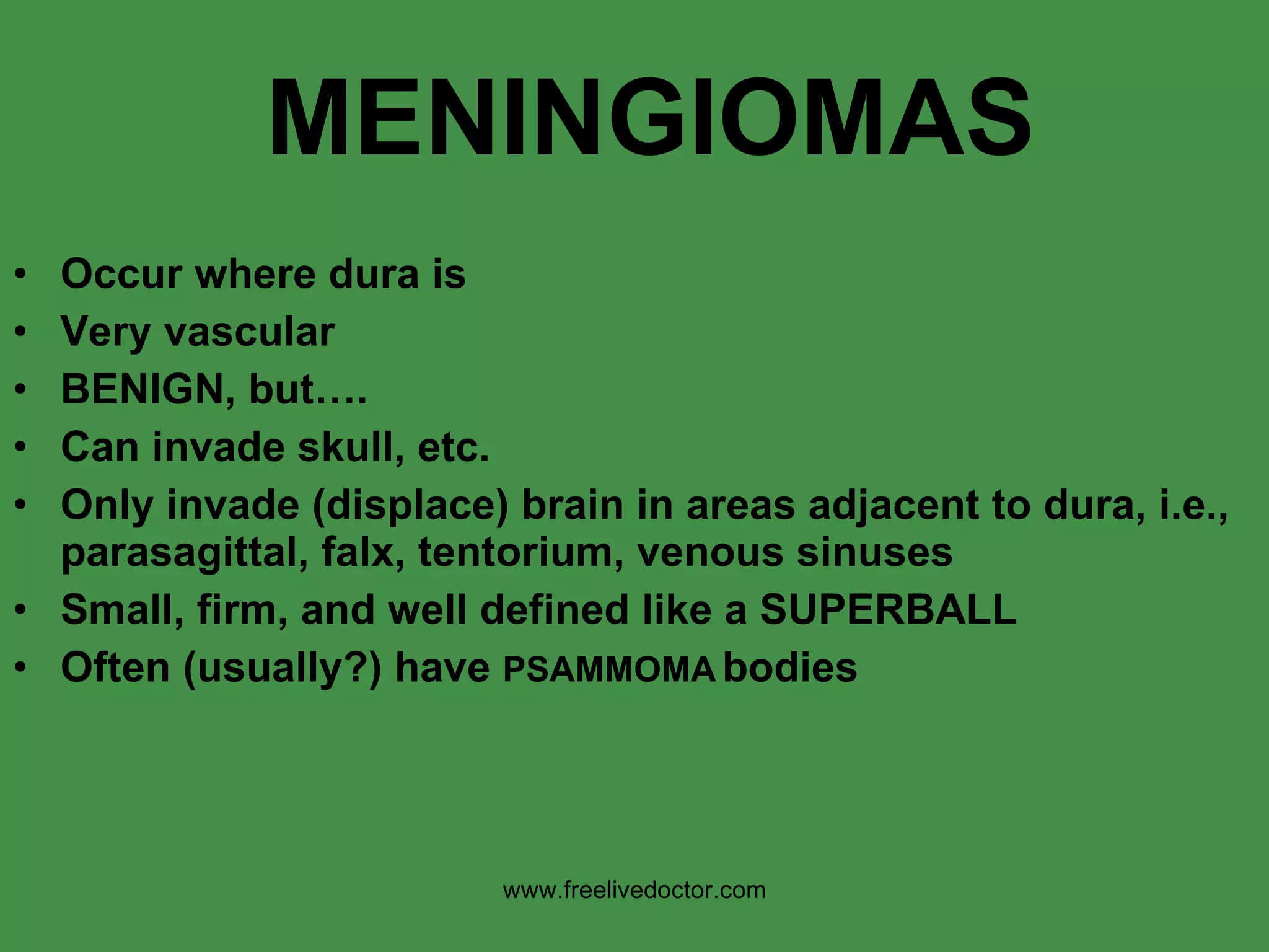
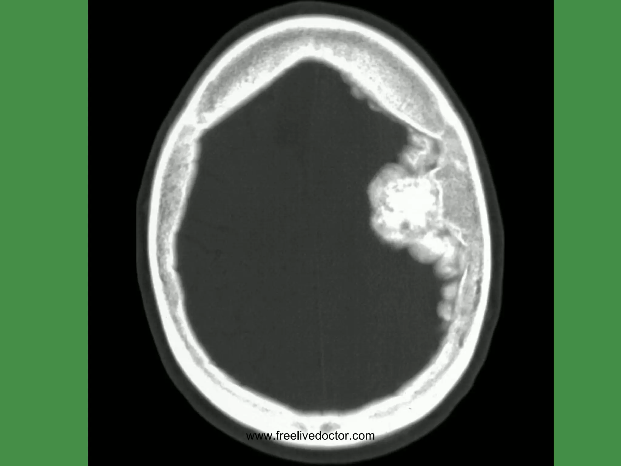
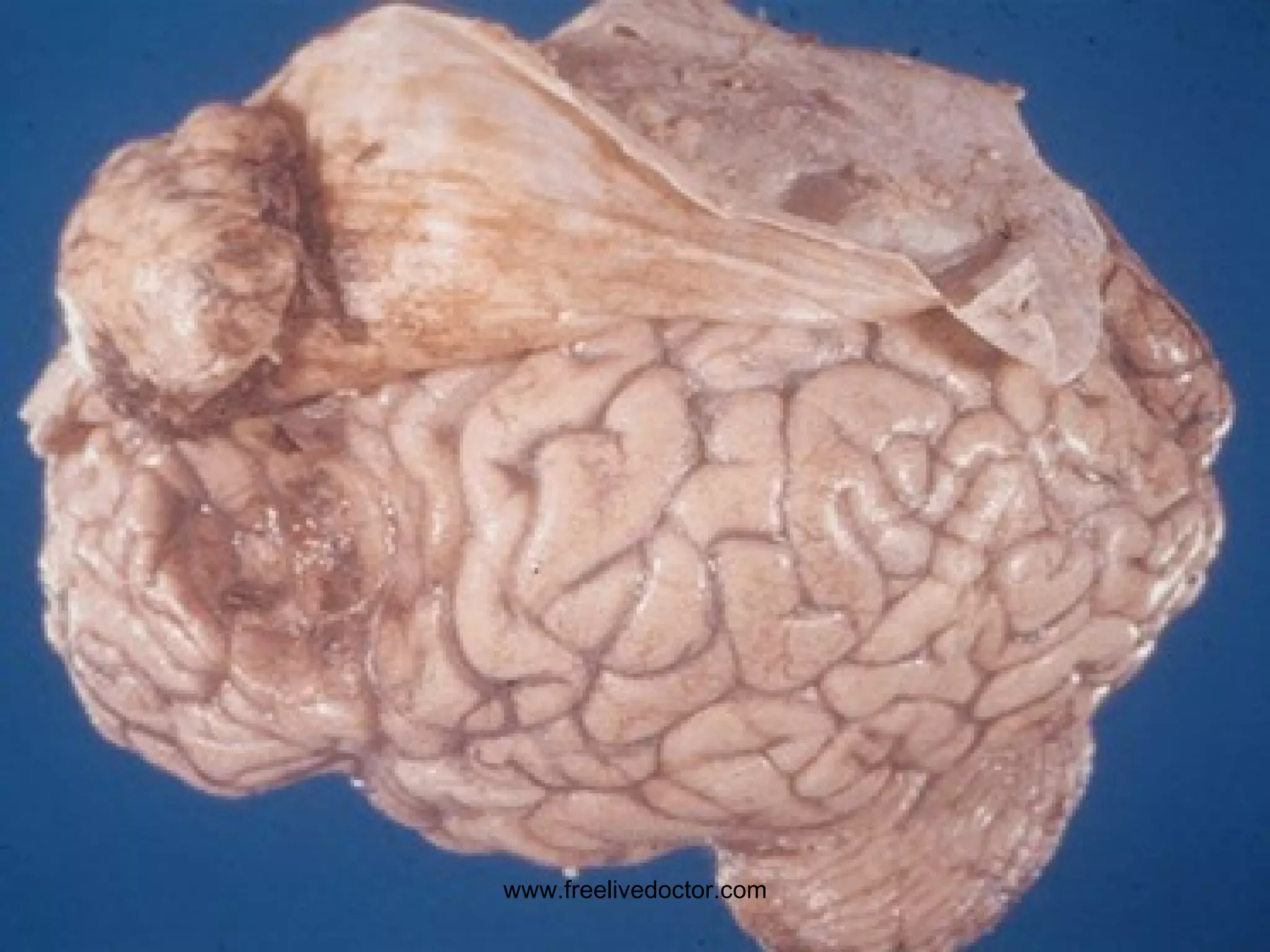
CNS www.freelivedoctor.com
CNS Normal Neurons Glia Astrocytes Oligodendrocytes Ependymal Cells Microglia www.freelivedoctor.com
Classical Disease Patterns Degenerative Inflammatory Neoplastic www.freelivedoctor.com
Classical  CNS  Disease Patterns Degenerative Inflammatory Neoplastic Traumatic www.freelivedoctor.com
www.freelivedoctor.com
CELLULAR REACTIONS Neurons Acute (RED neuron, karyolysis) Subacute, chronic, cell loss, gliosis Axonal Inclusions (lipid, prot., carb., viruses) Glia, “gliosis” Swelling Fibers Inclusions www.freelivedoctor.com
ACUTE NEURONAL INJURY “ RED ” NEURONS www.freelivedoctor.com
www.freelivedoctor.com
CEREBRAL EDEMA (normal weight 1200-1300 grams) Vasogenic (disrupted BBB) Intravascular   INTER-cellular Cytotoxic     INTRA-cellular www.freelivedoctor.com
CEREBRAL EDEMA Subfalcine (SUPRA-tentorial) Cingulate (TENTORIAL) Cerebellar tonsilar (SUB-tentorial, or INFRA-tentorial) www.freelivedoctor.com
www.freelivedoctor.com
DDX: EVERYTHING SYMPTOMS HEADACHE HALLUCINATIONS COMA DEATH CEREBRAL EDEMA www.freelivedoctor.com
HYDROCEPHALUS www.freelivedoctor.com
HYDROCEPHALUS Impaired RESORPTION Increased PRODUCTION OBSTRUCTION COMMUNICATING (entire) NON-COMMUNICATING (part) HIGH Pressure NORMAL Pressure www.freelivedoctor.com
www.freelivedoctor.com
CNS MALFORMATIONS Neural Tube Anencephaly, Encephalocele, Spina Bifida Forebrain Polymicrogyria, Holoprosencephaly, Agenesis of Corpus Callosum Posterior Fossa (Infratentorial) Arnold Chiari (infratentorial herniation, SMALL PF), Dandy-Walker (cerebellar cyst, LARGE PF) Syringomyelia/Hydromyelia www.freelivedoctor.com
SPINA BIFIDA www.freelivedoctor.com
POLYMICROGYRIA www.freelivedoctor.com
HOLOPROSENCEPHALY www.freelivedoctor.com
www.freelivedoctor.com
SYRINGOMYELIA (note “SYRINX”) www.freelivedoctor.com
PERINATAL Brain Injuries Intraparenchymal Hemorrhage Intraventricular hemorrhage (premies) Periventricular “leukomalacia” (i.e., infarcts) Cerebral “Palsy” refers to nonprogressive diffuse cerebral pathology apparent at childbirth www.freelivedoctor.com
CNS TRAUMA Skull Fractures Parenchymal Injuries Traumatic Vascular Injury Sequelae Spinal Cord Trauma www.freelivedoctor.com
BRAIN TRAUMA Contusion (bruise) Laceration (tear) Coup/Contre-Coup Concussion www.freelivedoctor.com
www.freelivedoctor.com
“ HAIRLINE” “ DEPRESSED”,  aka “ DISPLACED” www.freelivedoctor.com
HEMATOMAS/HEMORRHAGE EPIDURAL (fx) SUBDURAL (trauma NO fx) SUBARACHNOID (arterial, no trauma) INTRAPARENCHYMAL (any) INTRAVENTRICULAR (no trauma, rare in adults, common in premies) www.freelivedoctor.com
www.freelivedoctor.com
EPIDURAL HEMATOMA www.freelivedoctor.com
SUBDURAL HEMATOMA www.freelivedoctor.com
SUBARACHNOID www.freelivedoctor.com
INTRAPARENCHYMAL www.freelivedoctor.com
INTRAVENTRICULAR www.freelivedoctor.com
CNS TRAUMA SEQUELAE Hydrocephalus (WHY?) Dementia (Punch Drunk Syndrome) Diffuse Axonal Injury (white matter) www.freelivedoctor.com
www.freelivedoctor.com
SPINAL CORD TRAUMA Parallels BRAIN patterns of injury on a cellular basis Usually secondary to spinal column displacement Level of injury mirrors motor loss: Death   Quadriplegia    Paraplegia www.freelivedoctor.com
www.freelivedoctor.com
Cerebrovascular Diseases  (CVA, “Stroke”) Ischemic ( ↓ blood and 02) Global Focal (regional):  ACUTE: edema    neuronal microvacuolization    pyknosis    karyorrhexis    neutrophils CHRONIC: macrophages    gliosis Hemorrhagic (rupture of artery/aneurysm) www.freelivedoctor.com
www.freelivedoctor.com
THROMBOTIC MCA www.freelivedoctor.com
HEMORRHAGIC ACA www.freelivedoctor.com
EDEMA “ RED” NEURONS POLYs MONO’s (MACs) GLIOSIS Histopathologic progression of CNS infarcts www.freelivedoctor.com
HYPERTENSIVE CVA Intracerebral Basal Ganglia Region (lenticulostriate arteries of internal capsule,  putamen) www.freelivedoctor.com
www.freelivedoctor.com
HYPERTENSIVE CVA www.freelivedoctor.com
LACUNAR INFARCTS www.freelivedoctor.com
“ SLIT” HEMORRHAGE(s) www.freelivedoctor.com
SUBARACHNOID HEMORRHAGE Rupture of large intracerebral arteries which are the primary branches of the anatomical circle (of Willis) Congenital (“berry” aneurysms) Atherosclerotic (atherosclerotic aneurysms, or direct wall rupture) www.freelivedoctor.com
www.freelivedoctor.com
HYPERTENSIVE ENCEPHALOPATHY ACUTE Headaches Confusion Anxiety Convulsions CHRONIC Dementia (MID, Multi-Infarct-Dementia) Gait Disturbances Basal Ganglia symptoms www.freelivedoctor.com
CNS INFECTIONS ACUTE MENINGITIS ACUTE FOCAL SUPPURATIVE  CHRONIC BACTERIAL VIRAL FUNGAL OTHER www.freelivedoctor.com
INFECTIONS Meningitis (generally* bacterial) E. coli, Strep B (neonates) H. influenzae (children) Neisseria meningitidis (adults) Strep. pneumoniae, Listeria (elderly) PMNs in CSF, INCREASED protein, REDUCED glucose Encephalitis (generally viral) Arboviruses, HSV, CMV, V/Z, polio, rabies, HIV Lymphs and macrophages in perivascular “Virchow-Robbins” spaces Meningoencephalitis www.freelivedoctor.com
www.freelivedoctor.com
ACUTE FOCAL SUPPURATIVE CNS INFECTIONS CEREBRAL ABSCESSES Local (mastoiditis, sinusitis) Hematogenous (tooth extraction, sepsis) Staph, Strep Often fibrous capsule, liquid center SUBDURAL EMPYEMA  (IN SINUSITIS) EXTRADURAL ABSCESS  (IN OSTEOMYELITIS) www.freelivedoctor.com
www.freelivedoctor.com
SUBDURAL EMPYEMA www.freelivedoctor.com
CHRONIC BACTERIAL Meningo-encephalits TB, brain and meninges SYPHILIS, gummas in brain LYME DISEASE (Neuro-Borreliosis) www.freelivedoctor.com
TUBERCULOMA www.freelivedoctor.com
VIRAL Meningo-encephalitis ARBO VIRUSES (West Nile, Equines, Venez., many more) HSV1 HSV2 V/Z CMV POLIO RABIES HIV Progressive Multifocal Leukoencephalopathy (JC) Subacute Sclerosing Panencephalitis (Measles) www.freelivedoctor.com
VIRAL ENCEPHALITIS PERIVASCULAR LYMPHOCYTIC “ CUFFING” www.freelivedoctor.com
Bitemporal encephalitis is HSV until proven otherwise! www.freelivedoctor.com
HSV = TEMPORAL lobe(s) www.freelivedoctor.com
www.freelivedoctor.com
PERIVASCULAR GIANT CELLS in  WHITE MATTER in HIV ENCEPHALITIS www.freelivedoctor.com
P ROGRESSIVE  M ULTIFOCAL  L EUKOENCEPHALOPATHY (PML) JC Polyoma virus is the cause Primarilly affects oligodendocytes Ergo, demyelination is the main feature www.freelivedoctor.com
www.freelivedoctor.com
www.freelivedoctor.com Demyelination and gliosis
PML www.freelivedoctor.com
SUBACUTE SCLEROSING PANENCEPHALITIS (SSPE) VERY rare since measles eradicated Thought to be caused by measles virus www.freelivedoctor.com
FUNGAL MENINGO-ENCEPHALITIS CRYPTOCOCCUS CANDIDA ASPERGILLIS MUCOR (Mostly in immunocompromised hosts) www.freelivedoctor.com
www.freelivedoctor.com
CRYPTOCOCCUS MICROABSCESSES www.freelivedoctor.com
OTHERS MALARIA TOXOPLASMOSIS (in HIV) AMEBIASIS TRYPANOSOMES RICKETTSIAE ECHINOCOCCUS www.freelivedoctor.com
CNS  II www.freelivedoctor.com
PRION DISEASES Creutzfeldt-Jakob Disease (CJD) Gerstmann-Straussler-Scheinker syn. (GSS) Fatal familial insomnia Kuru, human variety Scrapie (sheep and goats) Mink transmissible encephalopathy Chronic wasting disease (deer and elk) Bovine Spongiform Encephalopathy (BSE) www.freelivedoctor.com
PRION DISEASES: common features Infectious agents with apparently no DNA DEMENTIA Prion Protein ( PrP ) accumulation “ SPONGIFORM”  changes in neurons and glia TRANSMISSIBLE, FATAL, NO Rx www.freelivedoctor.com
PRION PROTEIN Normally found in humans Exact structure known, 208 amino acids Specific chromosome, #20, specific genes also known Requires a  conformational change  to accumulate and do damage www.freelivedoctor.com
www.freelivedoctor.com
CJD (Creutzfeldt-Jakob) 1 per million incidence, 7 th  decade Sporadic cases,  not  epidemic Transmitted! Familial cases well documented Rapidly progressive dementia Grey Matter Cerebellar ataxia also, usually FATAL, no treatment known, like ALL prion diseases www.freelivedoctor.com
DEMYELINATING DISEASES MS (MULTIPLE SCLEROSIS) MS variants ACUTE DISSEMINATED ENCEPHALOMYELITIS (ADEM) ACUTE NECROTIZING HEMORRHAGIC ENCEPHALOMYELITIS (ANHE) Many, many, many others. Remember: DEMYELINATION is a NON-SPECIFIC reaction to MANY types of CNS injury www.freelivedoctor.com
MS Cause:  ? USA prevalence:  1:1000  F>>M,  Ages: 30’s, 40’s Immune response primarily against CNS myelin (white matter) Regional area of white matter demyelination is called “PLAQUE” Increased CSF gamma globulin, i.e., oligoclonal bands Often presents with VISUAL problems EXACERBATIONS/REMISSIONS www.freelivedoctor.com
www.freelivedoctor.com
PLAQUES, MS www.freelivedoctor.com
www.freelivedoctor.com
CNS DEGENERATIVE DISEASES CORTEX (dementias) BASAL GANGLIA and BRAIN STEM (parkinsonian) SPINOCEREBELLAR (ataxias) MOTOR NEURONS (muscle atrophy) www.freelivedoctor.com
CNS DEGENERATIVE DISEASES CORTEX (dementias) ALZHEIMER DISEASE Frontotemporal Pick Disease (also primarily frontal) Progressive Supranuclear Palsy (PSP) Corticobasal Degeneration (CBD) Vascular Dementias (MID) www.freelivedoctor.com
ALZHEIMER DISEASE Commonest cause of dementias (majority) Sporadic, 5-10% familial CORTICAL (grey matter) ATROPHY NEURITIC PLAQUES  (extraneuronal) NEUROFIBRILLARY TANGLES  (intraneuronal) AMYLOID!!! www.freelivedoctor.com
www.freelivedoctor.com
Neuritic plaques Neuritic plaques, stained with anti- beta amyloid   immunostain www.freelivedoctor.com
www.freelivedoctor.com
OTHER CORTICAL DEMENTIAS (tau gene/protein, tau-opathies) FRONTOTEMPORAL PICK DISEASE (LOBAR ATROPHY) PROGRESSIVE SUPRANUCLEAR PALSY (PSP) CORTICOBASAL DEGENERATION (CBD) VASCULAR DEMENTIA (MID) www.freelivedoctor.com
VASCULAR DEMENTIA Associated with multiple infarcts, hence the name MID (Multiple Infarct Dementia) Lacunar infarcts Cortical microinfarcts Multiple embolic infarcts SECOND commonest form of dementia after Alzheimer www.freelivedoctor.com
www.freelivedoctor.com
CNS DEGENERATIVE DISEASES BASAL GANGLIA and BRAIN STEM Parkinsonism Parkinson Disease Multiple System Atrophy Huntington Disease www.freelivedoctor.com
Parkinsonism Is a clinical “syndrome”, NOT a disease Diminished facial expression  Stooped posture Slowness of voluntary movement “ Festinating” gate (short, fast) Rigidity (cogwheel), intention tremor “ Pillrolling” tremor The above clinical findings involve pathology of the SUBSTANTIA NIGRA, and include: PARKINSON DISEASE MULTIPLE SYSTEM ATROPHY POSTENCEPHALIC PARKINSONISM PSP, CBD (cortical disorders) www.freelivedoctor.com
PARKINSON   DISEASE PALLOR of the SUBSTANTIA NIGRA   (and LOCUS COERULEUS) LEWY BODIES (alpha-synuclein protein) www.freelivedoctor.com
www.freelivedoctor.com
LOCUS COERULEUS in PONS (CERULEUS) www.freelivedoctor.com
www.freelivedoctor.com
PARKINSON   DISEASE Parkinsonism symptoms, i.e., cogwheel rigidity intention tremor Progressive Hallucinations Dementia Symptomatic response to L-DOPA www.freelivedoctor.com
MULTIPLE SYSTEM ATROPHY MSA WIDE SPECTRUM of diseases GLIAL CYTOPLASMIC INCLUSIONS (GCIs) in oligodendrocytes (alpha synuclein) Clinically, parkinsonism symptoms  autonomic dysfunction www.freelivedoctor.com
www.freelivedoctor.com
HUNTINGTON DISEASE Classical familial, genetic disease Progressive motor loss and dementia “ chorea”, i.e. “jerky” movements Progressive, fatal Atrophy of basal ganglia, i.e., corpus striatum Cortical (basal ganglia) atrophy Ventricular enlargement www.freelivedoctor.com
CNS DEGENERATIVE DISEASES SPINOCEREBELLAR DEGENERATIONS (ATAXIAS) Spinocerebellar ataxias Friedrich Ataxia Ataxia-Telangiectasia www.freelivedoctor.com
SPINOCEREBELLAR DEGENERATIONS Cerebellar cortex Spinal cord Peripheral nerves FEATURES: ATAXIA (loss of extremity muscle coordination) SPASTICITY NEUROPATHIES www.freelivedoctor.com
CNS DEGENERATIVE DISEASES MOTOR NEURONS ALS (Amyotrophic Lateral Sclerosis, i.e., Lou Gehrig’s disease) BulboSpinal Atrophy (Kennedy Syndrome) Spinal Muscular Atrophy www.freelivedoctor.com
Amyotrophic Lateral Sclerosis Unknown etiology Progressive muscle atrophy due to motor neuron loss (lower, upper) 5-10% familial Lou Gehrig had it, so does Steven Hawking Hand weakness   diaphragm Anterior horn cells reduced and gliotic www.freelivedoctor.com
ALS, DEMYELINATION IN CORTICOSPINAL TRACTS ALS, pathologic changes in anterior horn cells www.freelivedoctor.com
GENETIC METABOLIC DISEASES NEURONAL STORAGE DISEASES (classical autosomal recessive enzyme deficiencies) “ LEUKO”-DYSTROPHIES (abnormal “myelin” synthesis) MITOCHONDRIAL ENCEPHALOPATHIES (mitochondrial gene mutations) www.freelivedoctor.com
LEUKODYSTROPHIES Krabbe Metachromatic- Adreno- Pelizaeus-Merzbacher Canavan www.freelivedoctor.com
ACQUIRED TOXIC/METABOLIC CNS DISEASES Vitamin B1 deficiency (Wernicke-Korsakoff) Vitamin B12 deficiency (vibratory sense) Diabetes  Increased/Decreased GLUCOSE Hepatic Failure (NH4+) CO (Cortex, hippocampus, Purkinje cells) CH3-OH, Methanol  (Retinal ganglion cells) CH3-CH2-OH (acute/chronic, direct/nutrit’l) Radiation (Brain MOST resistant to Rad. Rx.) Chemo (Methotrexate + Radiation) www.freelivedoctor.com
www.freelivedoctor.com
128 Hz www.freelivedoctor.com
CNS TUMORS GLIOMAS Astrocytes ( I , II,  III,  IV ) Oligodendroglioma Ependymoma NEURONAL  (neuroblastoma) POORLY DIFFERENTIATED  (medulloblastoma) MENINGIOMAS LYMPHOMAS METASTATIC www.freelivedoctor.com
CNS TUMORS SYMPTOMS? Headache Vomiting Mental Changes Motor Problems Seizures Increased Intracranial Pressure ANY  localizing CNS abnormality www.freelivedoctor.com
CNS TUMORS History Physical Neurologic exam LP (including cytology) CT MRI Brain angiography Biopsy www.freelivedoctor.com
CNS TUMORS Benign? Malignant?, Primary vs. met? Location? Age? X-ray Density?  MIR signals? Calcifications? Vascularity? Necrosis?  Liquefaction? Edema? Compression of neighbors? www.freelivedoctor.com
GLIOSIS vs. GLIOMA Age? White vs. Grey Matter? Gross texture? Vascularity? Mitoses?  (N/C, Pleomorphism, Hyperchromasia) Calcifications? Cysts? Satellitosis? Delineation? www.freelivedoctor.com
www.freelivedoctor.com
MENINGIOMAS Occur where dura is Very vascular BENIGN, but…. Can invade skull, etc. Only invade (displace) brain in areas adjacent to dura, i.e., parasagittal, falx, tentorium, venous sinuses Small, firm, and well defined like a SUPERBALL Often (usually?) have  PSAMMOMA   bodies www.freelivedoctor.com
www.freelivedoctor.com
HIV www.freelivedoctor.com
METASTATIC CNS TUMORS LUNG BREAST MELANOMA KIDNEY GI www.freelivedoctor.com
“ PARA”NEOPLASTIC SYNDROMES SMALL CELL, LUNG LYMPHOMAS BREAST CA Purkinje Cell Degeneration Encephalitis, Limbic System Sensory Neuron Degeneration, DRG Eye Movement Disorders www.freelivedoctor.com
FAMILIAL TUMOR SYNDROMES NF1 Neurofibromas Gliomas NF2 Schwannomas Meningiomas Tuberous Sclerosis ,  i.e., CNS and somatic “hamartomas” Von-Hippel-Lindau ,  CNS hemangioblastomas, chiefly cerebellar www.freelivedoctor.com

Classification of diseases of cns

Editor's Notes

  • #6 As amazingly complex as the CNS is, even more amazing is the fact that its components are so remarkably simple----Neuron body, dendrites, axon.
  • #7 Section from spinal cord. Is this grey matter or white matter?
  • #8 Neurons with nucleus, nissl (Franz Nissl) granules, nerve fibers, glia. Glia are many times more common than neurons.
  • #9 Blood vessel with endothelium and/or smooth muscle.
  • #10 Various glia
  • #11 This particular stain shows why the most common glial cell is called an “astro”cyte.
  • #12 Oligodendrocytes, the CNS myelinators. Note the clear space around their nuclei, much in the same way myelin washed out from schwann glial calls as well.
  • #13 Ependymal cells look exactly like ciliated columnar cells and like the ventricular spaced and choroid.
  • #14 Choroid plexus is “papillary” in configuration.
  • #15 Cluster of microglia, the macrophages of the CNS.
  • #18 Neuronal loss and gliosis is a hallmark of more “chronic” CNS injury
  • #19 A comparison of edema “compartments” the ECS (EXTRA Cellular Space) is vasogenic and the ICS (INTRA Cellular Space) is cytotoxic.
  • #21 Flattened gyri often signify edema. Why? Ans: compression against the calvarium
  • #22 1) Falx, 2) Cingulate, and 3) Cereballar tonsillar levels of edema
  • #24 “ Notching” of the cingulate gyrus.
  • #25 Cerebellar tonsillar herniation
  • #28 Basic pathophysiologic concepts about hydrocephalus which is defined as any major deviation from the normal physiology of CSF
  • #29 Hydrocephalus on CT
  • #30 Hydrocephalus on MRI
  • #31 Hydrocephalus also showing cerebral edema, CT or MRI? Ans: CT Why? Ans: Bone is always very dense on CT, and water is always intense on T2 weighted MRI
  • #32 Hydrocephalus, dilated ventricles
  • #33 COMMON CNS malformations
  • #34 Note the neural canal extends to the outside of the body. AFP, the same antigen found in hepatomas, is a good screening test for this.
  • #35 Small gyri
  • #36 Failure of the prosencephalon to develop, and separate, often leads to cyclops.
  • #37 Normal corpus callosum
  • #38 Absent corpus callosum
  • #40 These are the three most common types of perinatal brain injuries
  • #41 Differentiation between CNS trauma is crucial in medicolegal cases.
  • #42 Know the correct definitions
  • #43 Contusion
  • #44 Skull fracture types
  • #47 Epidural
  • #48 Subdural
  • #53 Three common sequelae of CNS trauma
  • #55 You should recall cord injury level versus sensory and motor defects: “C5, still alive”
  • #61 Histopathologic progression of CNS infarcts, parallels the general cellular progression of events in inflammation
  • #69 Classical congenital “berry” aneurysm
  • #70 Extensive basilar subarachnoid hemorrhage
  • #71 Basal ganglia symptoms include tremors (rhythmic, involuntary, oscillatory movements), athetosis (slow, writhing movements of the fingers and hands, and sometimes of the toes), chorea (abrupt movements of the limbs and facial muscles), ballism (violent, flailing movements), and dystonia (a persistent posture of a body part which can result in grotesque movements and distorted positions of the body).
  • #74 Meninges, purulent, at the base of the brain
  • #75 Meninges, purulent, at the top of the brain
  • #76 Meningitis vs. meningoencephalitis
  • #84 Perhaps encephalo-meningitis would be a better term? Why? Ans: viruses primarilly involve CNS parenchyma, rather than meninges
  • #85 Perivascular lymphocytic cuffing is the hallmark viral encephalitis, especially with respect to early, mild, or peripheral considerations.
  • #88 Eosinophilic Negri body of Rabies, also basophilic inclusions of CMV
  • #91 Demyelination is associated with gliosis and edema, therefore bright signals on T2 weighted images
  • #92 Demyelination and gliosis
  • #100 PR -oteinaceous I -nfectious particle = PRI -on
  • #102 “ Replication” is felt to be due to the protein undergoing a conformational change to induce neighboring proteins to become like it.
  • #103 Hence the term “spongiform.”
  • #104 CJ has also been called JC, but the term “JC” makes it confusable with the JC polyoma virus, so CJ-D is probably the politically correct term
  • #105 Demyelination, generically, is a NON-specific pattern of CNS reaction to injury of many types and usually goes hand in hand with edema and gliosis, If it wasn’t for the “edema” associated with demyelination, the “plaques” would not be seen on MRI.
  • #106 The PLAQUE of MS is NOT like a plaque of skin diseases, i.e., it is not a raised lesion, but an area of demyelination.
  • #107 MS gave MRI its first HUGE boom, by virtue of being able to detect these lesions, due to edema!
  • #108 Myelinated white matter stains BLUE, and demyelination is loss of blue. Remember MS is a disease PRIMARILLY of WHITE matter.
  • #109 Plaques grossly
  • #110 Plaques microscopically. Demyelination, edema, gliosis, and, lower right, relative preservation of the actual nerve fibers
  • #111 FOUR classical areas for brain degeneration, a decent anatomic classification.
  • #112 ALZHEIMER disease is many times more common than all the other dementias put together.
  • #114 Normal sulci.
  • #115 Prominent sulci in cortical atrophy. Why are the sulci, NOT the gyri, prominent in atrophy? Ans: cortical LOSS
  • #116 Plaques and tangles and beta-amyloid
  • #117 Plaques and tangles, and central core of AMYLOID
  • #118 Amyloid with congo red stain (LEFT), and Amyloid with congo red stain under polarization (RIGHT.
  • #119 Neurons with tangles displacing nucleus, H & E
  • #120 Neurons with TANGLES, often displacing NUCLEUS.
  • #121 A “tangle” in proximity to a nucleus. A “tangle” is hyperphosphorylation of a neuron microtubule protein called “tau”, causing it to precipitate
  • #122 Tau is a gene, Tau protein is a microtubule protein associated with hyperphosphorylation in tau-opathies. MANY cortical dementias are associated with this.
  • #124 How would one differentiate MID from MS? Ans: MS is purely white matter. In this MRI we see grey matter lesions, so it is more likely MID rather than MS
  • #128 NORMALLY BLACK substantia nigra due to adequate dopamine.
  • #129 PALE substantia nigra in Parkinson’s disease (on LEFT) due to inadequate dopamine.
  • #130 The locus ceruleus is also pale in Parkinson’s disease, which is another pigmented area due to abundant dopamine.
  • #131 Which patient has Parkinson’s disease? Ans: the RIGHT Why? Ans: decreased dopamine
  • #132 Lewy bodies are commonly regarded as diagnostic of Parkinson’s disease also. The main substance of the eosinophilic inclusion is alpha-synuclein .
  • #135 Alpha synuclein stains.
  • #145 Vitamin B1 deficiency (Wernicke-Korsakoff), hemorrhagic mamillary bodies are the most classic finding. B1 deficiency is the culprit here.
  • #146 Posterior column demyelination in B12 deficiency, this is also called SUBACUTE COMBINED DEGENERATION
  • #152 Gliosis vs. Glioma?
  • #153 Glioma, intermediate grade
  • #154 Glioma, high grade, Note NECROSIS. NECROSIS (orangophilia and granularity) is needed for the diagnosis of a HIGH grade glioma.
  • #155 Glioblastoma (multiforme). Why is it called “multiforme”? Note the 1) palisading (two arrows) and 2) necrosis (ovals) which are hallmarks of GBM.
  • #156 Central necrosis is a sign of rapid growth. It outgrows its blood supply.
  • #157 Normal oligodendrocytes on the left.
  • #158 What is this? (Hint: note rosettes) Ans: neuroblastoma
  • #159 Any midline cerebellum tumor in a child is a medulloblastoma till proven otherwise!
  • #160 Midline cerebellum tumor in a kid. What is it? Ans: medulloblastoma
  • #162 Arts of this meningioma are denser than bone.
  • #163 Note cortical compression from this meningioma.
  • #164 Psammoma bodies are diagnostic of meningiomas in brain tumors! What other kinds of tumors have psammoma bodies? Ans: papillary carcinomas, classically in thyroid
  • #165 Toxoplasmosis and lymphomas and encephalitis are very common in AIDS. Might you cal the MRI lesion a “toxoplasmoma”? Ans: Sure
  • #166 A solitary brain mass is statistically just as likely to be metastatic than primary