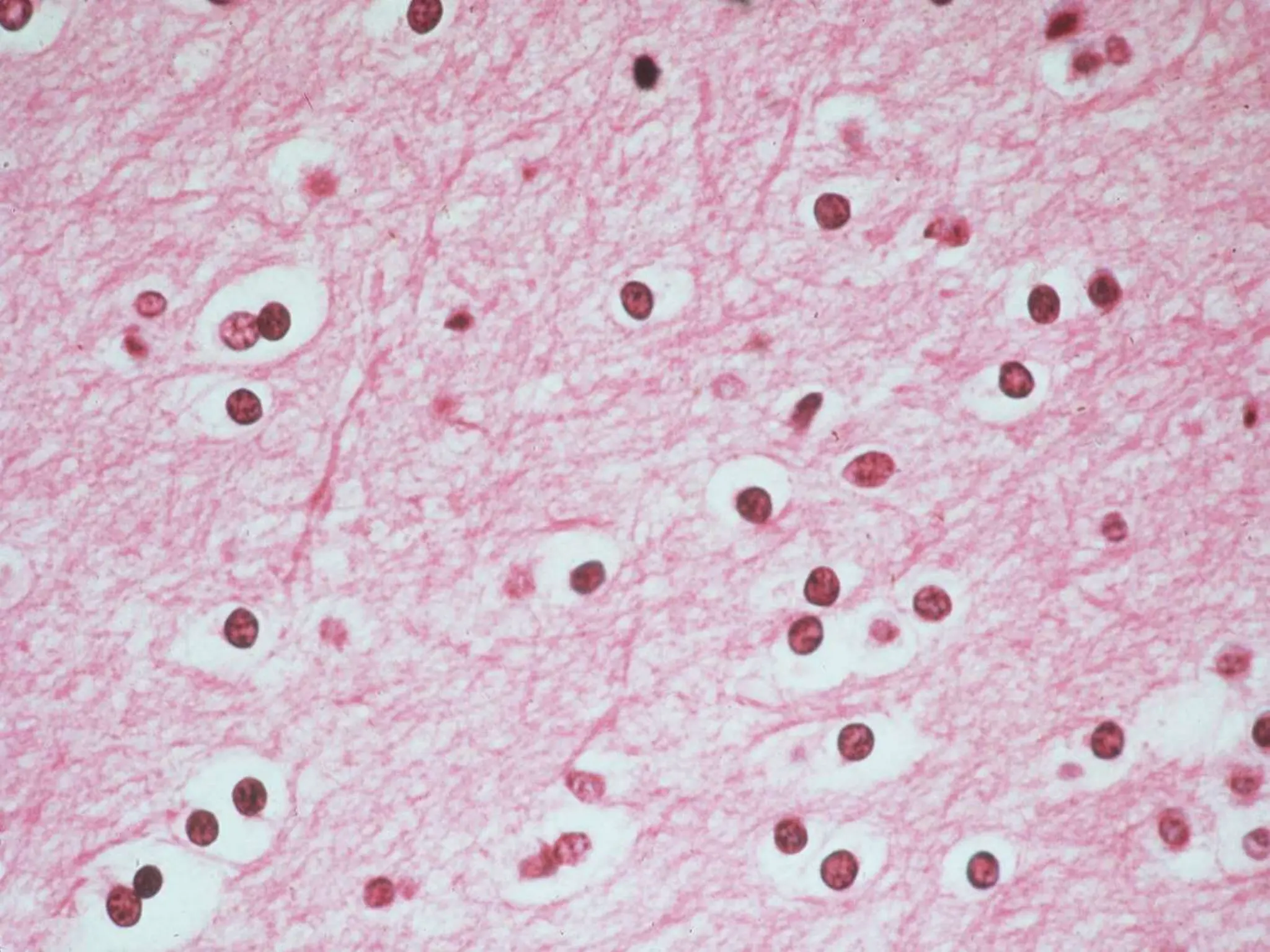
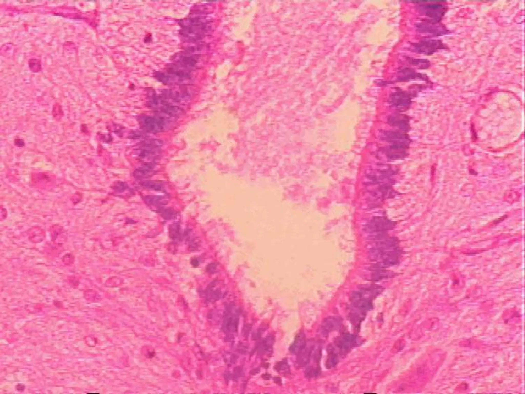
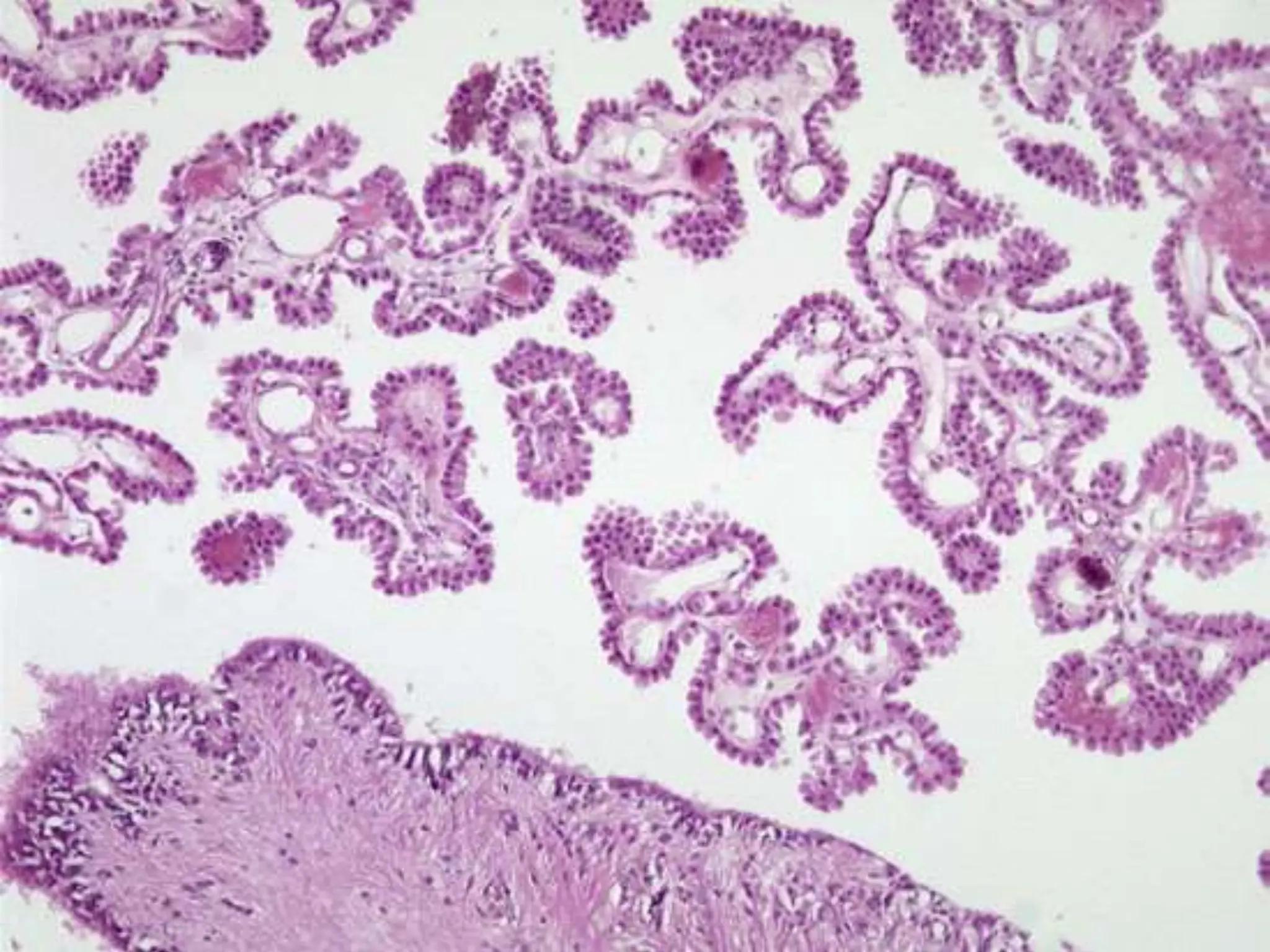
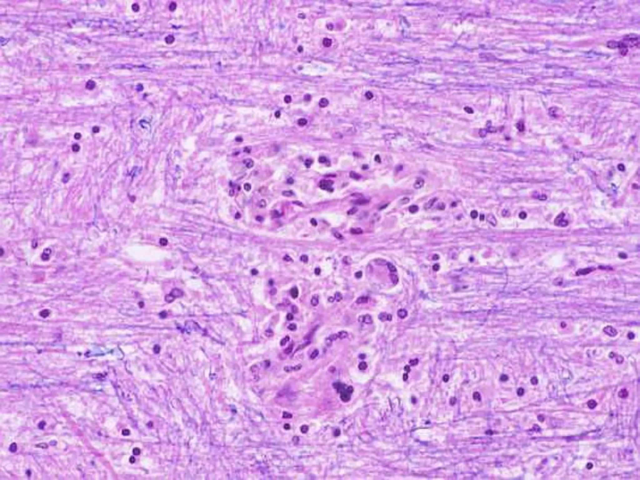
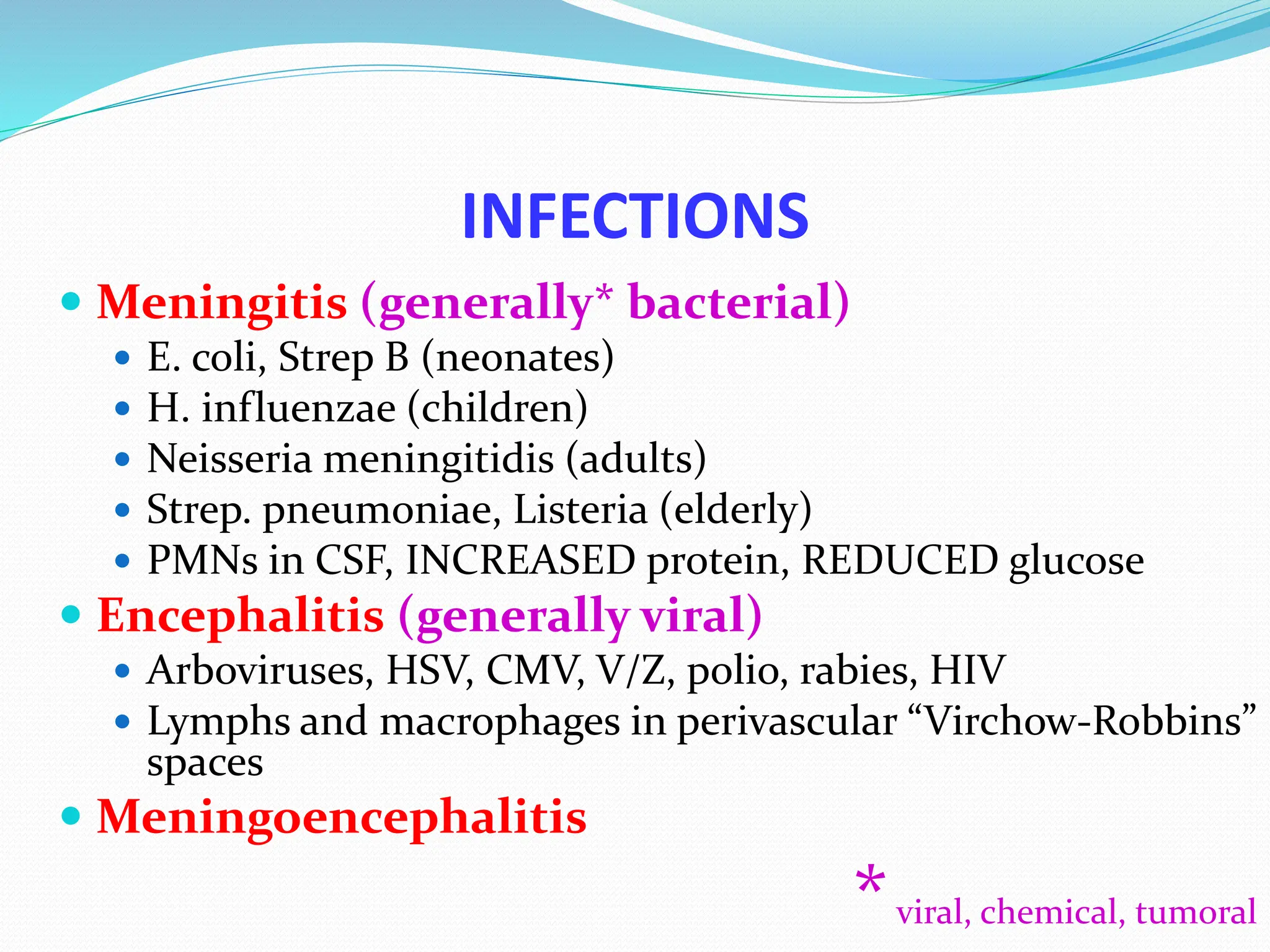
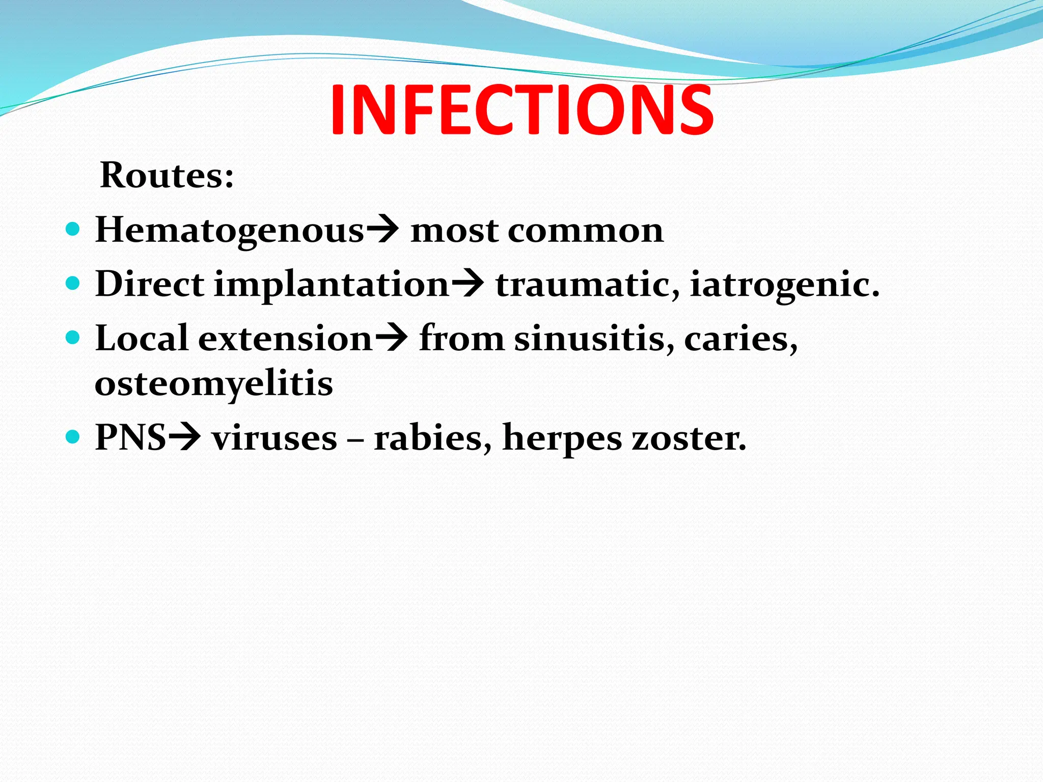
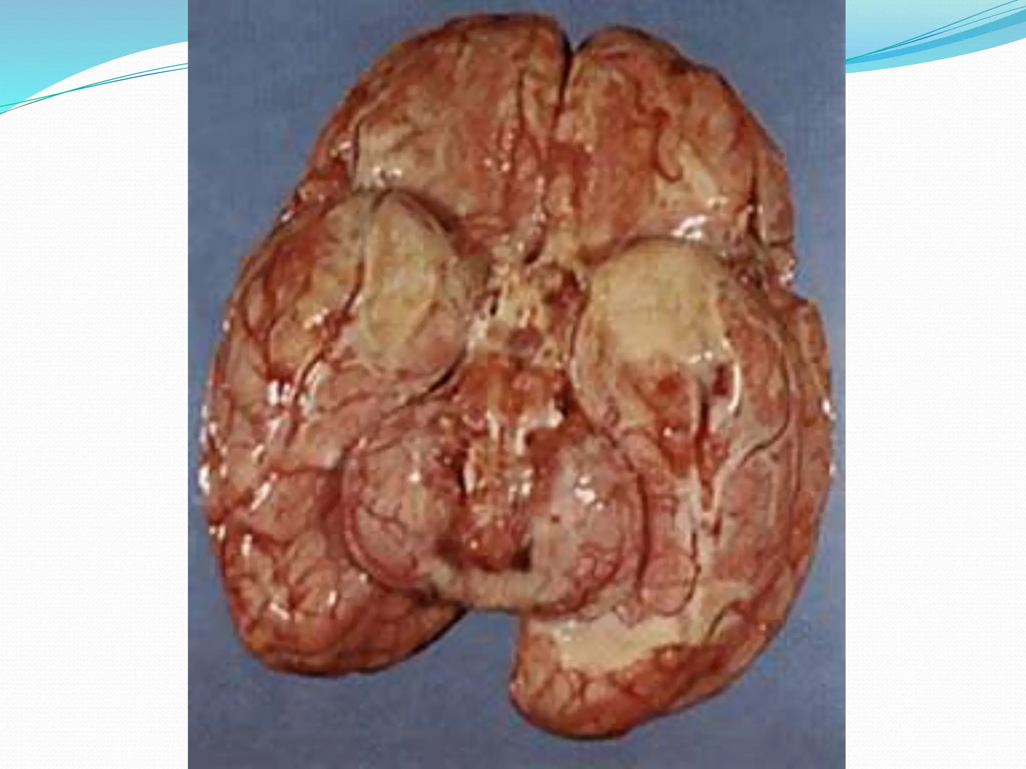
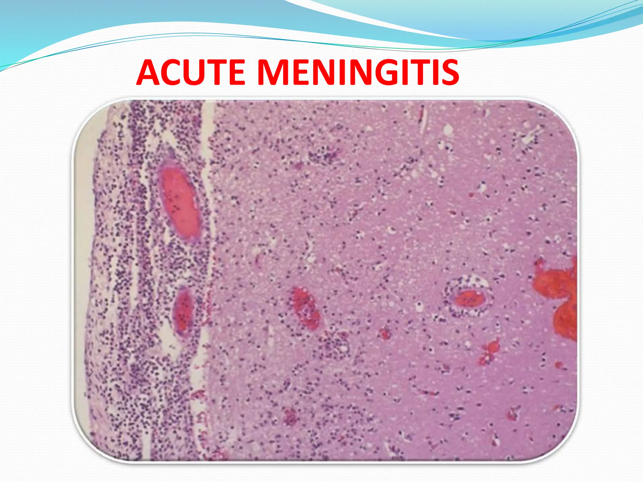
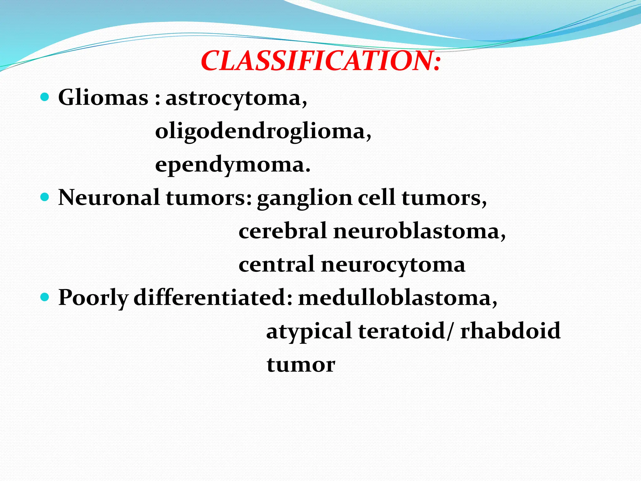
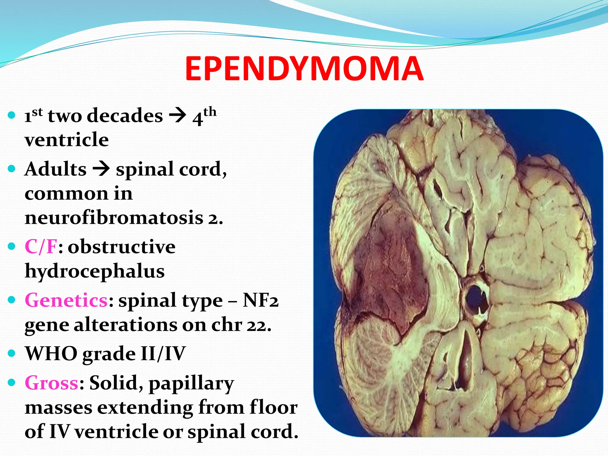
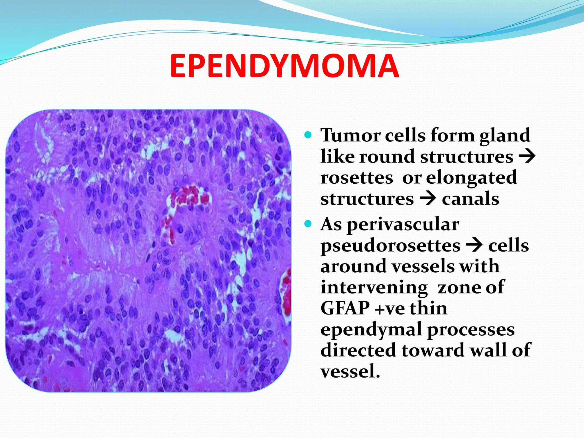
Dr. P.Sravani., M.D.,
Assistant professor.
Govt. medical college
Anantapur
CENTRAL NERVOUS SYSTEM
 Normal cells:
1. neurons
2. glia:
a) derived from neuroectoderm
macroglia: astrocytes,
oligodendrocytes,
ependymocytes.
b) from bone marrow: microglia
3. cells of meninges, blood cells
INFECTIONS
TYPES OF INFECTIONS
1. Acute meningitis
2. Acute focal suppurative infections
3. Chronic bacterial meningoencephalitis
4. Viral meningoencephalitis
5. Fungal meningoencephalitis.
INFECTIONS
 Meningitis (generally* bacterial)
 E. coli, Strep B (neonates)
 H. influenzae (children)
 Neisseria meningitidis (adults)
 Strep. pneumoniae, Listeria (elderly)
 PMNs in CSF, INCREASED protein, REDUCED glucose
 Encephalitis (generally viral)
 Arboviruses, HSV, CMV, V/Z, polio, rabies, HIV
 Lymphs and macrophages in perivascular “Virchow-Robbins”
spaces
 Meningoencephalitis
*viral, chemical, tumoral
INFECTIONS
Routes:
 Hematogenous most common
 Direct implantation traumatic, iatrogenic.
 Local extension from sinusitis, caries,
osteomyelitis
 PNS viruses – rabies, herpes zoster.
ACUTE MENINGITIS
 Meningitis: Inflammation of leptomeninges &
CSF within subarachnoid space
 Meningoencephalitis: Inflammation of meninges
and brain parenchyma.
Types:
 Acute pyogenic ( bacterial),
 Acute aseptic (viral).
CLINICAL FEATURES
 Meningeal irritation & neurologic impairment
 Headache, photophobia, irritability, clouding of
consciousness, neck stiffness
 CSF: purulent, high pressure, high neutrophils,
high protein, markedly low glucose, bacteria +.
 Waterhouse Friderichsen syndrome: meningitis
 adrenal hemorrhagic infarcts.
ACUTE MENINGITIS
Acute aseptic (viral) meningitis
 ECHO, Coxsackie, non-paralytic poliomyelitis
 Less fulminant clinical course
 C/F: Meningeal irritation, fever, altered
consciousness
 CSF: lymphocytic pleocytosis, moderately high
protein, normal glucose.
Acute focal suppurative infections
 Brain abscess
 Subdural empyema (IN SINUSITIS)
 Extradural abscess (IN OSTEOMYELITIS)
Predisposing factors:
 Direct implantation during skull fractures
 Local extention of infection –Otitis media, Sinusitis,
mastoiditis
 Acute bacterial endocarditis
 Cyanotic congenital heart diseases
 Chronic pulmonary sepsis as in bronchiectasis
 C/F: Signs of raised ICP & progressive focal deficits.
 CSF: High pressure, high WBC, high protein, normal
sugar.
BRAIN ABSCESS
Gross: discrete, liquefactive necrosis,
fibrous capsule, edema.
MICROSCOPY
 Exuberant granulation tissue with
neovascularization around necrosis  edema,
 Fibroblasts of vessels  collagen of capsule.
CT BRAIN ABSCESS
SUBDURAL EMPYEMA
 Chronic bacterial meningoencephalitis
A) TB
B) neurosyphilis
C) neuroborreliosis (Lyme Disease)
A) TB:
 C/F: headache, vomitings, confusion
 CSF: moderate pleocytosis mononuclear cells or
mixture of polymorphonuclear and mononuclear
cells, high protein, low or normal glucose.
 Gross: gelatinous or fibrinous exudate in S.A. space
Diffuse meningoencephalitis:
 Discrete, white granules over leptomeninges
 Mixture of lymphocytes, plasma cells & macrophages.
 Granulomas, caseous necrosis, giant cells.
 Obliterative endarteritis.
 Acid fast stains – bacilli.
Tuberculoma:
 Well circumscribed intra parenchymal masses, may
cause mass effect
 Central core of caseous necrosis, surrounded by TB
granulomatous reaction
TUBERCULOMA
Viral meningoencephalitis:
 Viruses:HSV 1 &2, VZV, CMV, Polio, Rabies, HIV.
Fungal:
 Candida albicans, mucor, aspergillus
fumigatus, cryptococcus neoformans.
VIRAL
ENCEPHALITIS
PERIVASCULAR
LYMPHOCYTIC
“CUFFING”
Meningitis Acute
bacterial
Viral Fungal Tuberculous
Csf pressure
90-180mm of
water
Increased Normal-
mild
increased
Increased Increased
Leucocytes
0-5cells
1000-
10,000
5-300 40-400 100-600
Proteins/mg/
dl
(15-45)
100-500 <100 50-300 50-300
Glucose
50-80 mg/dl
<40 normal decreased <45
CNS TUMORS
 Features:
1. Difficult to distinguish benign from malignant,
eg. Benign astrocytoma infiltrates large region of
brain.
2. Difficult to resect without neurological
impairment.
3. Lethality based on site. Eg, meningioma 
compresses medulla  cardiorespiratory arrest.
4. Different pattern of spread. Metastasis outside
brain is rare, spread through CSF common 
seedling along brain & spinal cord.
CLASSIFICATION:
 Gliomas : astrocytoma,
oligodendroglioma,
ependymoma.
 Neuronal tumors: ganglion cell tumors,
cerebral neuroblastoma,
central neurocytoma
 Poorly differentiated: medulloblastoma,
atypical teratoid/ rhabdoid
tumor
 Other parenchymal tumors:
primary CNS lymphoma,
germ cell tumors,
pineal parenchymal tumors
 Meningiomas
 Metastatic tumors
 Peripheral nerve sheath tumors:
schwannoma,
neurofibroma,
MPNST.
CNS TUMORS
SYMPTOMS?
 Headache
 Vomiting
 Mental Changes
 Motor Problems
 Seizures
 Increased Intracranial Pressure
ANY localizing CNS abnormality
ASTROCYTOMAS
 80% of adult primary brain tumors.
 Site: cerebral hemisphere, also in cerebellum,
brain stem, spinal cord.
 Age: 4th to 6th decade.
 C/F: seizures, headache, focal neurologic deficits.
 WHO Grading: grade expressed as “I - IV”.
grade I/IV : pilocytic astrocytoma
II/IV: diffuse fibrillary astrocytoma (well
differentiated)
III/IV: anaplastic astrocytoma
IV/IV: glioblastoma
Genetics:
 Low grade: inactivation of p53, over expression of
PDGF-A & its receptor.
 High grade: additional mutations of tumor
suppressor genes like RB gene.
Pilocytic astrocytoma (WHO I)
 Site: cerebellum, also in 3rd ventricle, optic nerves,
rarely cerebrum.
 Age: Children & young adults,
 p53 mutations rare.
 Benign.
 Slow growth
 Gross:
 Cystic, with a mural nodule in the wall of cyst.
MICROSCOPY
 Bipolar cells, long, thin
hair like cytoplasmic
processes.
 Rosenthal fibers-
granular, brightly
eosinophilic, elongated
cytoplasmic masses
 Microcysts +
DIFFUSE FIBRILLARY ASTROCYTOMA
 WHO grade II/IV
 Gross:
 Poorly defined, gray, infiltrative tumor that
expands and distorts the invaded brain.
 Size : few cms to huge replacing hemisphere.
firm or soft, gelatinous, cystic degeneration
Microscopy:
 Well differentiated
 Tumor cells infiltrate adjacent brain for some
distance
 Mild to moderate increase in number of glial cell
nuclei.
 Variable nuclear pleomorphism
 Intervening feltwork of fine, GFAP positive
astrocytic cell processes  fibrillary background.
ASTROCYTOMA
Anaplastic astrocytoma:
 WHO grade III/IV
 More densely cellular
 More nuclear pleomorphism
 Mitotic figures +
Glioblastoma:
 Was called as glioblastoma multiforme (GBM)
 2 types:
 Primary: de novo, elderly, poor prognosis
 2nd ary : from low grade astrocytoma, young,
better prognosis.
GLIOBLASTOMA
Gross:
 Variegated  firm &
white; soft & yellow
(necrosis); cystic
degeneration,
hemorrhages
 Apparently well
demarcated
 always infiltrates
adjacent brain
Microscopy:
 Anaplastic
astrocytoma + necrosis
& endothelial cell
proliferation.
 Necrosis:
serpentine/
geographic necrosis.
Geographic Necrosis
 Highly malignant
tumor cells crowd
along edge of necrosis
 ‘pseudopalisading’.
Glomeruloid body
 Endothelial hyperplasia:
Tufts of piled up
vascular cells  bulge
into lumen  form a ball
like structure
‘glomeruloid body’.
OLIGODENDROGLIOMA
 Age: 4th & 5th decades
 Site: cerebral hemisphere – white matter
 C/F: neurologic complaints lasting for years.
 Genetics: LOH for chromosomes 1p & 19q.
 Prognosis better than astrocytomas
 WHO Grade II/IV
Gross:
 Well circumscribed, gelatinous, grey masses
 Cysts, hemorrhages & gritty
OLIGODENDROGLIOMA-10X
 Delicate plexiform
network of
anastomosing
capillaries  ‘chicken
wire’ or ‘crow feet’
pattern.
Oligodendroglioma-40x
 Sheets of regular cells
with uniform spherical
nuclei with fine
granular chromatin,
clear halo around
nucleus  ‘fried egg
appearance’.
EPENDYMOMA
 1st two decades  4th
ventricle
 Adults  spinal cord,
common in
neurofibromatosis 2.
 C/F: obstructive
hydrocephalus
 Genetics: spinal type – NF2
gene alterations on chr 22.
 WHO grade II/IV
 Gross: Solid, papillary
masses extending from floor
of IV ventricle or spinal cord.
EPENDYMOMA
 Tumor cells form gland
like round structures 
rosettes or elongated
structures  canals
 As perivascular
pseudorosettes  cells
around vessels with
intervening zone of
GFAP +ve thin
ependymal processes
directed toward wall of
vessel.
MEDULLOBLASTOMA
 Neuroectodermal origin
 Poorly differentiated or embryonal.
 Composed of primitive, undifferentiated cells.
 Children Exclusively in cerebellum
 Adults  cerebral hemisphere
 C/F: obstructive hydrocephaluslethargy,
headache, morning emesis.
 Genetics: isochromosome 17q i(17q).
MEDULLOBLASTOMA
 Gross:
 Well circumscribed,
gray, friable
MICROSCOPY
 Extremely cellular
 Sheets of small
cells, little
cytoplasm,
hyperchromatic
nuclei which are
round or angulated
 Arranged as closely
packed sheets
 Abundant mitoses,
 Highly malignant tumor
 SRBCT.
 Infiltrate as linear cells into cerebellar cortex
penetrate pia  seed into SAS  CSF metastasis
as nodular masses upto cauda equina  “drop
metastasis”.
MENINGIOMA
 Benign tumors, arise from meningothelial cells of
arachnoid.
 Age: adults, sex- F:M::3:2
 Has progesterone receptors – rapid growth in
pregnancy
 Site: external surface of brain, also in ventricles.
Common in parasagittal convexity, wing of
sphenoid, olfactory groove, sella turcica
 Attached to dura.
 Solitary, multiple in NF2.
 Genetics: deletion of chr 22q12 which has NF2
gene.
Gross-meningioma
 round, encapsulated,
bosselated or polypoid
mass- meningioma
Histologic patterns:
 Syncytial or meningotheliomatous:
whorled clusters of cells without visible cell
membranes, tight groups & nests
 Fibroblastic:
elongated cells, dense collagen between cells
 Transitional:
syncytial and fibroblastic
MENINGIOMA
transitional type
PSAMMOMATOUS
MENINGIOMA
Numerous
psammoma
bodies, due to
calcification of
syncytial nests of
meningothelial
cells.
 Microcystic: loose spongy
 Secretory: intracytoplasmic droplets, PAS +ve.
 Atypical meningioma (WHO grade II/IV):
 lower grade meningiomas by the presence of
either a mitotic index of four or more mitoses per
10 high power fields or at least three atypical
features (increased cellularity, small cells with a
high nuclear-to-cytoplasmic ratio, prominent
nucleoli, patternless growth, or necrosis).
 Clear cell & chordoid variants
 Anaplastic (malignant) meningioma (WHO grade
III/IV):
 Papillary: pleomorphic cells around fibrovascular
core
 Rhabdoid: sheets of tumor cells with hyaline
eosinophilic cytoplasm
METASTATIC TUMORS
 Primaries from lung, breast, skin, kidney & GIT.
 Choriocarcinoma, a rare tumor, commonly
metastasizes
Gross
 Sharply demarcated masses at junction of grey &
white matters
 Well defined boundary between tumor & adjacent
brain
 Meningeal carcinomatosis:
tumor nodules stud over brain, spinal cord –
primaries from lung and breast.
METASTASIS BRAIN
PERIPHERAL NERVE SHEATH TUMORS
 Schwannoma
 Neurofibroma
 MPNST
 Myelination by oligodendrocytes is shifted to
that by schwann cells within several millimeters
of substance of brain, as nerves exit brain & spinal
cord.
 Hence, they can arise within cranial vault.
SCHWANNOMA/NEURILEMMOMA
o Benign, arise from schwann cells
o Sites: cerebellopontine angle  vestibular branch
of VIII nerve  vestibular schwannoma / acoustic
neuroma
o C/F: tinnitus, deafness.
o Sensory nerves: branches of V nerve, dorsal roots.
o Gross: well circumscribed, encapsulated masses,
attached to nerve, but can be separated from it .
o Firm, grey
o Cystic change may be +.
o Soft, tan colored, “fish fleshy”
SCHWANNOMA
Microscopy:
 Mixture of 2 growth patterns of elongated cells
with regular, oval nuclei & cytoplasmic processes.
 Antoni A pattern:
 moderate to high cellularity ,
 cells arranged as fascicles,
 little stromal matrix.
 nuclear free zones of cytoplasmic processes that
lie between nuclear palisading  ‘verocay bodies’
 Antoni B pattern: hypocellular, loose meshwork
of cells with microcysts, myxoid areas
ANTONI A WITH
VEROCAY BODIES ANTONI B
NEUROFIBROMA
 Skin: cutaneous NF
 Peripheral nerve: solitary NF
 Arise sporadically or with NF1 plexiform NF
 Cutaneous – nodules , in dermis & fat,
as well demarcated, but unencapsulated masses
of spindle cells, highly collagenised stroma
 Plexiform: large nerve trunk, multiple, nerve
irregularly expanded.
 lesions cannot be separated from nerve,
 Microscopy: loose myxoid background, low
cellularity.
 Schwann cells - Elongated nuclei, extension of pink
cytoplasm
 Large, multipolar fibroblasts
 Inflammatory cells.
 Collagen bundles in the background of myxoid
areas look as shredded carrot
 Malignant transformation high for plexiform NF as
MPNST
MPNST (MALIGNANT SCHWANNOMA)
 High grade sarcomas, NF1 gene, p53 & p16
mutations.
 Gross: poorly defined, invades parent nerve,
necrosis
 Microscopy : schwann cells – elongated nucleus,
prominent bipolar cytoplasmic processes.
 As fascicles, bundles, storiform, herring bone,
geographic necrosis.
FAMILIAL TUMOR SYNDROMES
 Neurofibromatosis Type 1
 This autosomal dominant disorder, one of the
more common genetic disorders, having a
frequency of 1 in 3000, is characterized by
neurofibromas (plexiform and solitary), gliomas
of the optic nerve, pigmented nodules of the iris
(Lisch nodules), and cutaneous hyperpigmented
macules (café au lait spots).
 Neurofibromatosis Type 2 (NF2)
 This is an autosomal dominant disorder resulting
in a range of tumors, most commonly bilateral
eighth-nerve schwannomas and multiple
meningiomas. Gliomas, typically ependymomas
of the spinal cord
 Tuberous Sclerosis Complex
 Tuberous sclerosis is an autosomal dominant syndrome,
hamartomas and benign neoplasms involving the brain and
other tissues.
 Hamartomas within the CNS take the form of cortical tubers
and subependymal nodules;
 subependymal giant-cell astrocytomas are low grade
neoplasms that appear to develop from the hamartomatous
nodules in the same location.
 renal angiomyolipomas, retinal glial hamartomas,
pulmonary lymphangioleiomyomatosis and cardiac
rhabdomyomas.
 Cysts may be found at various sites, including the liver,
kidneys, and pancreas.
 Cutaneous lesions include angiofibromas, localized
leathery thickenings (shagreen patches), hypopigmented
areas (ash-leaf patches), and subungual fibromas.
THANK
YOU
THANK YOU

CENTRAL NERVOUS SYSTEM PATHOGENESIS AND PATHOLOGY

  • 1.
    Dr. P.Sravani., M.D., Assistantprofessor. Govt. medical college Anantapur
  • 2.
    CENTRAL NERVOUS SYSTEM Normal cells: 1. neurons 2. glia: a) derived from neuroectoderm macroglia: astrocytes, oligodendrocytes, ependymocytes. b) from bone marrow: microglia 3. cells of meninges, blood cells
  • 9.
  • 10.
    TYPES OF INFECTIONS 1.Acute meningitis 2. Acute focal suppurative infections 3. Chronic bacterial meningoencephalitis 4. Viral meningoencephalitis 5. Fungal meningoencephalitis.
  • 11.
    INFECTIONS  Meningitis (generally*bacterial)  E. coli, Strep B (neonates)  H. influenzae (children)  Neisseria meningitidis (adults)  Strep. pneumoniae, Listeria (elderly)  PMNs in CSF, INCREASED protein, REDUCED glucose  Encephalitis (generally viral)  Arboviruses, HSV, CMV, V/Z, polio, rabies, HIV  Lymphs and macrophages in perivascular “Virchow-Robbins” spaces  Meningoencephalitis *viral, chemical, tumoral
  • 12.
    INFECTIONS Routes:  Hematogenous mostcommon  Direct implantation traumatic, iatrogenic.  Local extension from sinusitis, caries, osteomyelitis  PNS viruses – rabies, herpes zoster.
  • 13.
    ACUTE MENINGITIS  Meningitis:Inflammation of leptomeninges & CSF within subarachnoid space  Meningoencephalitis: Inflammation of meninges and brain parenchyma. Types:  Acute pyogenic ( bacterial),  Acute aseptic (viral).
  • 14.
    CLINICAL FEATURES  Meningealirritation & neurologic impairment  Headache, photophobia, irritability, clouding of consciousness, neck stiffness  CSF: purulent, high pressure, high neutrophils, high protein, markedly low glucose, bacteria +.  Waterhouse Friderichsen syndrome: meningitis  adrenal hemorrhagic infarcts.
  • 16.
  • 17.
    Acute aseptic (viral)meningitis  ECHO, Coxsackie, non-paralytic poliomyelitis  Less fulminant clinical course  C/F: Meningeal irritation, fever, altered consciousness  CSF: lymphocytic pleocytosis, moderately high protein, normal glucose. Acute focal suppurative infections  Brain abscess  Subdural empyema (IN SINUSITIS)  Extradural abscess (IN OSTEOMYELITIS)
  • 18.
    Predisposing factors:  Directimplantation during skull fractures  Local extention of infection –Otitis media, Sinusitis, mastoiditis  Acute bacterial endocarditis  Cyanotic congenital heart diseases  Chronic pulmonary sepsis as in bronchiectasis  C/F: Signs of raised ICP & progressive focal deficits.  CSF: High pressure, high WBC, high protein, normal sugar. BRAIN ABSCESS
  • 19.
    Gross: discrete, liquefactivenecrosis, fibrous capsule, edema.
  • 21.
    MICROSCOPY  Exuberant granulationtissue with neovascularization around necrosis  edema,  Fibroblasts of vessels  collagen of capsule.
  • 22.
  • 23.
  • 24.
     Chronic bacterialmeningoencephalitis A) TB B) neurosyphilis C) neuroborreliosis (Lyme Disease) A) TB:  C/F: headache, vomitings, confusion  CSF: moderate pleocytosis mononuclear cells or mixture of polymorphonuclear and mononuclear cells, high protein, low or normal glucose.
  • 25.
     Gross: gelatinousor fibrinous exudate in S.A. space Diffuse meningoencephalitis:  Discrete, white granules over leptomeninges  Mixture of lymphocytes, plasma cells & macrophages.  Granulomas, caseous necrosis, giant cells.  Obliterative endarteritis.  Acid fast stains – bacilli. Tuberculoma:  Well circumscribed intra parenchymal masses, may cause mass effect  Central core of caseous necrosis, surrounded by TB granulomatous reaction
  • 26.
  • 27.
    Viral meningoencephalitis:  Viruses:HSV1 &2, VZV, CMV, Polio, Rabies, HIV. Fungal:  Candida albicans, mucor, aspergillus fumigatus, cryptococcus neoformans.
  • 28.
  • 30.
    Meningitis Acute bacterial Viral FungalTuberculous Csf pressure 90-180mm of water Increased Normal- mild increased Increased Increased Leucocytes 0-5cells 1000- 10,000 5-300 40-400 100-600 Proteins/mg/ dl (15-45) 100-500 <100 50-300 50-300 Glucose 50-80 mg/dl <40 normal decreased <45
  • 31.
    CNS TUMORS  Features: 1.Difficult to distinguish benign from malignant, eg. Benign astrocytoma infiltrates large region of brain. 2. Difficult to resect without neurological impairment. 3. Lethality based on site. Eg, meningioma  compresses medulla  cardiorespiratory arrest. 4. Different pattern of spread. Metastasis outside brain is rare, spread through CSF common  seedling along brain & spinal cord.
  • 32.
    CLASSIFICATION:  Gliomas :astrocytoma, oligodendroglioma, ependymoma.  Neuronal tumors: ganglion cell tumors, cerebral neuroblastoma, central neurocytoma  Poorly differentiated: medulloblastoma, atypical teratoid/ rhabdoid tumor
  • 33.
     Other parenchymaltumors: primary CNS lymphoma, germ cell tumors, pineal parenchymal tumors  Meningiomas  Metastatic tumors  Peripheral nerve sheath tumors: schwannoma, neurofibroma, MPNST.
  • 34.
    CNS TUMORS SYMPTOMS?  Headache Vomiting  Mental Changes  Motor Problems  Seizures  Increased Intracranial Pressure ANY localizing CNS abnormality
  • 35.
    ASTROCYTOMAS  80% ofadult primary brain tumors.  Site: cerebral hemisphere, also in cerebellum, brain stem, spinal cord.  Age: 4th to 6th decade.  C/F: seizures, headache, focal neurologic deficits.  WHO Grading: grade expressed as “I - IV”. grade I/IV : pilocytic astrocytoma II/IV: diffuse fibrillary astrocytoma (well differentiated) III/IV: anaplastic astrocytoma IV/IV: glioblastoma
  • 36.
    Genetics:  Low grade:inactivation of p53, over expression of PDGF-A & its receptor.  High grade: additional mutations of tumor suppressor genes like RB gene. Pilocytic astrocytoma (WHO I)  Site: cerebellum, also in 3rd ventricle, optic nerves, rarely cerebrum.  Age: Children & young adults,  p53 mutations rare.  Benign.
  • 37.
     Slow growth Gross:  Cystic, with a mural nodule in the wall of cyst.
  • 38.
    MICROSCOPY  Bipolar cells,long, thin hair like cytoplasmic processes.  Rosenthal fibers- granular, brightly eosinophilic, elongated cytoplasmic masses  Microcysts +
  • 39.
    DIFFUSE FIBRILLARY ASTROCYTOMA WHO grade II/IV  Gross:  Poorly defined, gray, infiltrative tumor that expands and distorts the invaded brain.  Size : few cms to huge replacing hemisphere.
  • 40.
    firm or soft,gelatinous, cystic degeneration
  • 41.
    Microscopy:  Well differentiated Tumor cells infiltrate adjacent brain for some distance  Mild to moderate increase in number of glial cell nuclei.  Variable nuclear pleomorphism  Intervening feltwork of fine, GFAP positive astrocytic cell processes  fibrillary background.
  • 42.
  • 43.
    Anaplastic astrocytoma:  WHOgrade III/IV  More densely cellular  More nuclear pleomorphism  Mitotic figures + Glioblastoma:  Was called as glioblastoma multiforme (GBM)  2 types:  Primary: de novo, elderly, poor prognosis  2nd ary : from low grade astrocytoma, young, better prognosis.
  • 44.
    GLIOBLASTOMA Gross:  Variegated firm & white; soft & yellow (necrosis); cystic degeneration, hemorrhages  Apparently well demarcated  always infiltrates adjacent brain
  • 45.
    Microscopy:  Anaplastic astrocytoma +necrosis & endothelial cell proliferation.  Necrosis: serpentine/ geographic necrosis.
  • 46.
    Geographic Necrosis  Highlymalignant tumor cells crowd along edge of necrosis  ‘pseudopalisading’.
  • 47.
    Glomeruloid body  Endothelialhyperplasia: Tufts of piled up vascular cells  bulge into lumen  form a ball like structure ‘glomeruloid body’.
  • 48.
    OLIGODENDROGLIOMA  Age: 4th& 5th decades  Site: cerebral hemisphere – white matter  C/F: neurologic complaints lasting for years.  Genetics: LOH for chromosomes 1p & 19q.  Prognosis better than astrocytomas  WHO Grade II/IV Gross:  Well circumscribed, gelatinous, grey masses  Cysts, hemorrhages & gritty
  • 49.
    OLIGODENDROGLIOMA-10X  Delicate plexiform networkof anastomosing capillaries  ‘chicken wire’ or ‘crow feet’ pattern.
  • 50.
    Oligodendroglioma-40x  Sheets ofregular cells with uniform spherical nuclei with fine granular chromatin, clear halo around nucleus  ‘fried egg appearance’.
  • 52.
    EPENDYMOMA  1st twodecades  4th ventricle  Adults  spinal cord, common in neurofibromatosis 2.  C/F: obstructive hydrocephalus  Genetics: spinal type – NF2 gene alterations on chr 22.  WHO grade II/IV  Gross: Solid, papillary masses extending from floor of IV ventricle or spinal cord.
  • 53.
    EPENDYMOMA  Tumor cellsform gland like round structures  rosettes or elongated structures  canals  As perivascular pseudorosettes  cells around vessels with intervening zone of GFAP +ve thin ependymal processes directed toward wall of vessel.
  • 55.
    MEDULLOBLASTOMA  Neuroectodermal origin Poorly differentiated or embryonal.  Composed of primitive, undifferentiated cells.  Children Exclusively in cerebellum  Adults  cerebral hemisphere  C/F: obstructive hydrocephaluslethargy, headache, morning emesis.  Genetics: isochromosome 17q i(17q).
  • 56.
    MEDULLOBLASTOMA  Gross:  Wellcircumscribed, gray, friable
  • 57.
    MICROSCOPY  Extremely cellular Sheets of small cells, little cytoplasm, hyperchromatic nuclei which are round or angulated  Arranged as closely packed sheets  Abundant mitoses,
  • 58.
     Highly malignanttumor  SRBCT.  Infiltrate as linear cells into cerebellar cortex penetrate pia  seed into SAS  CSF metastasis as nodular masses upto cauda equina  “drop metastasis”.
  • 59.
    MENINGIOMA  Benign tumors,arise from meningothelial cells of arachnoid.  Age: adults, sex- F:M::3:2  Has progesterone receptors – rapid growth in pregnancy  Site: external surface of brain, also in ventricles. Common in parasagittal convexity, wing of sphenoid, olfactory groove, sella turcica  Attached to dura.  Solitary, multiple in NF2.  Genetics: deletion of chr 22q12 which has NF2 gene.
  • 60.
  • 61.
    Histologic patterns:  Syncytialor meningotheliomatous: whorled clusters of cells without visible cell membranes, tight groups & nests  Fibroblastic: elongated cells, dense collagen between cells  Transitional: syncytial and fibroblastic
  • 62.
  • 63.
  • 64.
     Microcystic: loosespongy  Secretory: intracytoplasmic droplets, PAS +ve.  Atypical meningioma (WHO grade II/IV):  lower grade meningiomas by the presence of either a mitotic index of four or more mitoses per 10 high power fields or at least three atypical features (increased cellularity, small cells with a high nuclear-to-cytoplasmic ratio, prominent nucleoli, patternless growth, or necrosis).  Clear cell & chordoid variants
  • 65.
     Anaplastic (malignant)meningioma (WHO grade III/IV):  Papillary: pleomorphic cells around fibrovascular core  Rhabdoid: sheets of tumor cells with hyaline eosinophilic cytoplasm
  • 66.
    METASTATIC TUMORS  Primariesfrom lung, breast, skin, kidney & GIT.  Choriocarcinoma, a rare tumor, commonly metastasizes Gross  Sharply demarcated masses at junction of grey & white matters  Well defined boundary between tumor & adjacent brain  Meningeal carcinomatosis: tumor nodules stud over brain, spinal cord – primaries from lung and breast.
  • 67.
  • 68.
    PERIPHERAL NERVE SHEATHTUMORS  Schwannoma  Neurofibroma  MPNST  Myelination by oligodendrocytes is shifted to that by schwann cells within several millimeters of substance of brain, as nerves exit brain & spinal cord.  Hence, they can arise within cranial vault.
  • 69.
    SCHWANNOMA/NEURILEMMOMA o Benign, arisefrom schwann cells o Sites: cerebellopontine angle  vestibular branch of VIII nerve  vestibular schwannoma / acoustic neuroma o C/F: tinnitus, deafness. o Sensory nerves: branches of V nerve, dorsal roots. o Gross: well circumscribed, encapsulated masses, attached to nerve, but can be separated from it . o Firm, grey o Cystic change may be +. o Soft, tan colored, “fish fleshy”
  • 70.
  • 71.
    Microscopy:  Mixture of2 growth patterns of elongated cells with regular, oval nuclei & cytoplasmic processes.  Antoni A pattern:  moderate to high cellularity ,  cells arranged as fascicles,  little stromal matrix.  nuclear free zones of cytoplasmic processes that lie between nuclear palisading  ‘verocay bodies’  Antoni B pattern: hypocellular, loose meshwork of cells with microcysts, myxoid areas
  • 72.
    ANTONI A WITH VEROCAYBODIES ANTONI B
  • 73.
    NEUROFIBROMA  Skin: cutaneousNF  Peripheral nerve: solitary NF  Arise sporadically or with NF1 plexiform NF  Cutaneous – nodules , in dermis & fat, as well demarcated, but unencapsulated masses of spindle cells, highly collagenised stroma  Plexiform: large nerve trunk, multiple, nerve irregularly expanded.  lesions cannot be separated from nerve,
  • 74.
     Microscopy: loosemyxoid background, low cellularity.  Schwann cells - Elongated nuclei, extension of pink cytoplasm  Large, multipolar fibroblasts  Inflammatory cells.  Collagen bundles in the background of myxoid areas look as shredded carrot  Malignant transformation high for plexiform NF as MPNST
  • 75.
    MPNST (MALIGNANT SCHWANNOMA) High grade sarcomas, NF1 gene, p53 & p16 mutations.  Gross: poorly defined, invades parent nerve, necrosis  Microscopy : schwann cells – elongated nucleus, prominent bipolar cytoplasmic processes.  As fascicles, bundles, storiform, herring bone, geographic necrosis.
  • 76.
    FAMILIAL TUMOR SYNDROMES Neurofibromatosis Type 1  This autosomal dominant disorder, one of the more common genetic disorders, having a frequency of 1 in 3000, is characterized by neurofibromas (plexiform and solitary), gliomas of the optic nerve, pigmented nodules of the iris (Lisch nodules), and cutaneous hyperpigmented macules (café au lait spots).
  • 77.
     Neurofibromatosis Type2 (NF2)  This is an autosomal dominant disorder resulting in a range of tumors, most commonly bilateral eighth-nerve schwannomas and multiple meningiomas. Gliomas, typically ependymomas of the spinal cord
  • 78.
     Tuberous SclerosisComplex  Tuberous sclerosis is an autosomal dominant syndrome, hamartomas and benign neoplasms involving the brain and other tissues.  Hamartomas within the CNS take the form of cortical tubers and subependymal nodules;  subependymal giant-cell astrocytomas are low grade neoplasms that appear to develop from the hamartomatous nodules in the same location.  renal angiomyolipomas, retinal glial hamartomas, pulmonary lymphangioleiomyomatosis and cardiac rhabdomyomas.  Cysts may be found at various sites, including the liver, kidneys, and pancreas.  Cutaneous lesions include angiofibromas, localized leathery thickenings (shagreen patches), hypopigmented areas (ash-leaf patches), and subungual fibromas.
  • 79.