Download to read offline
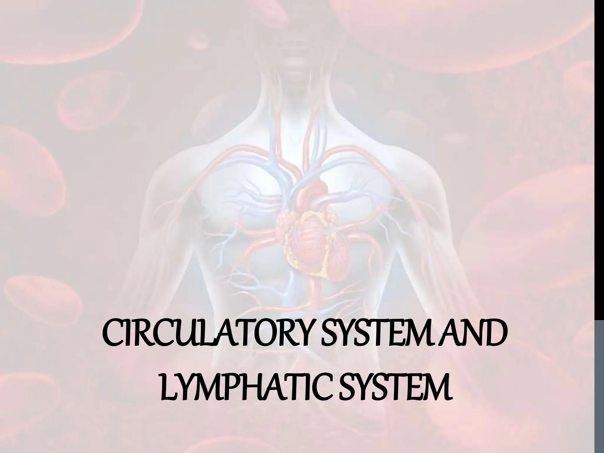
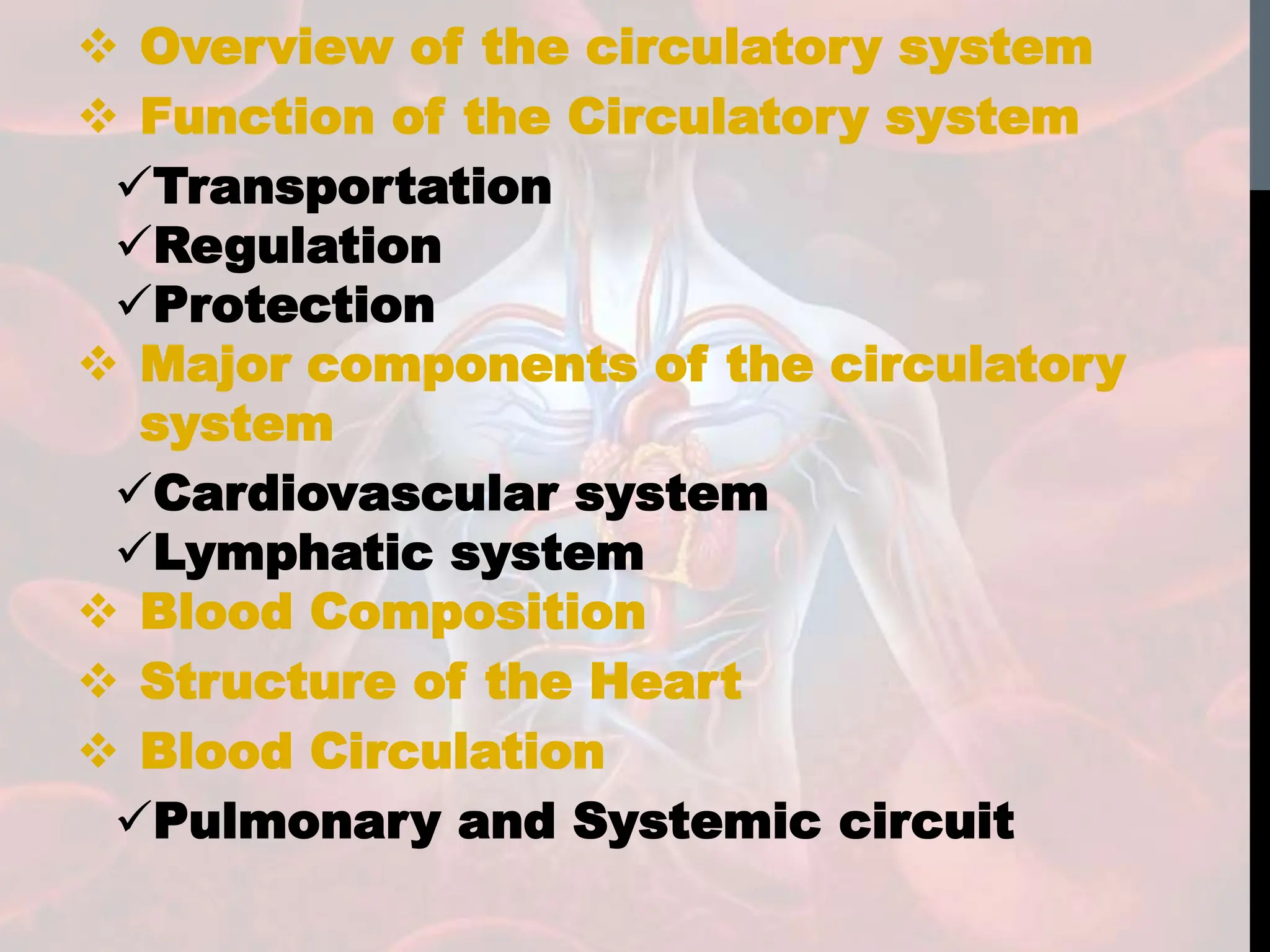
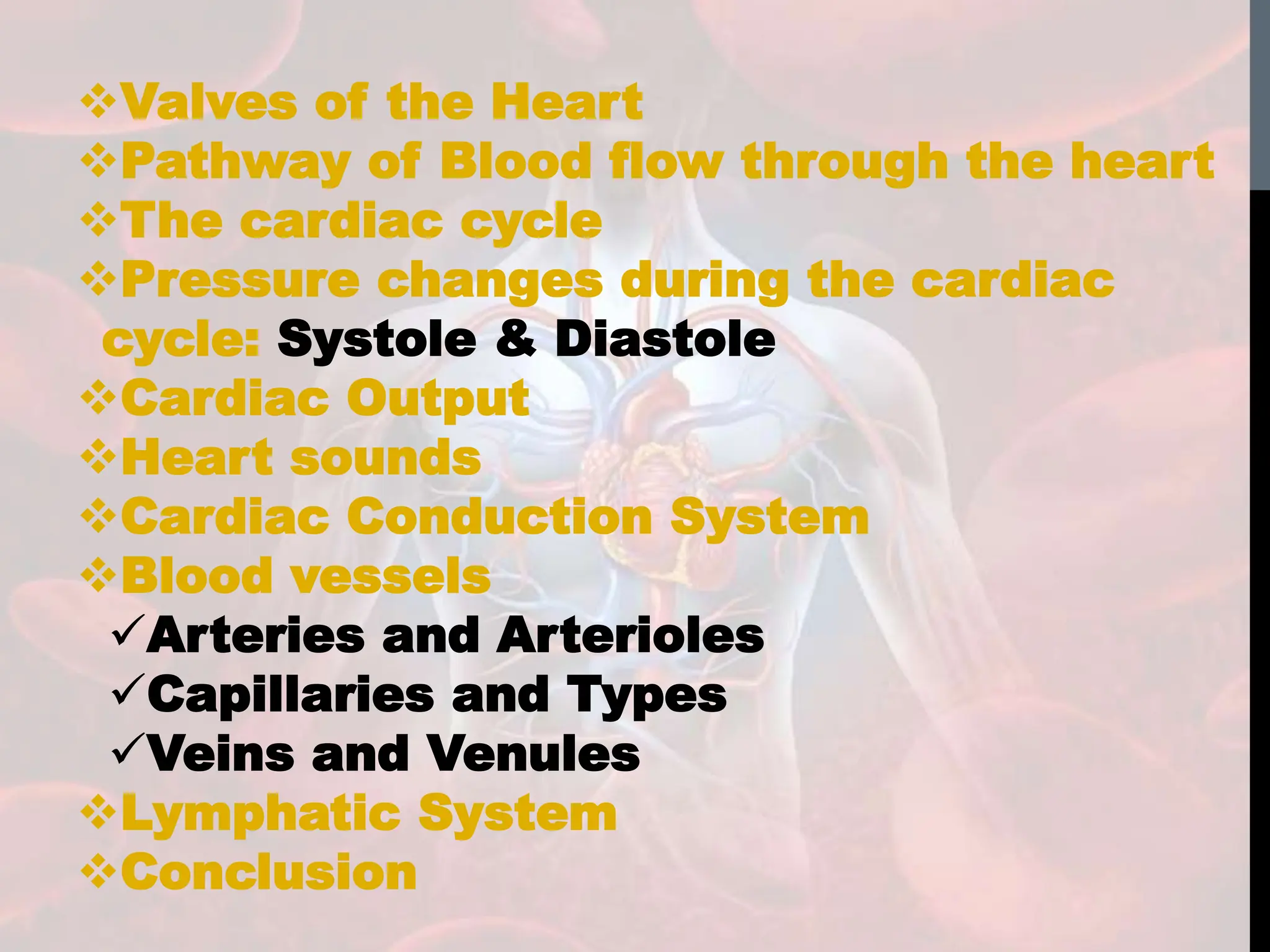
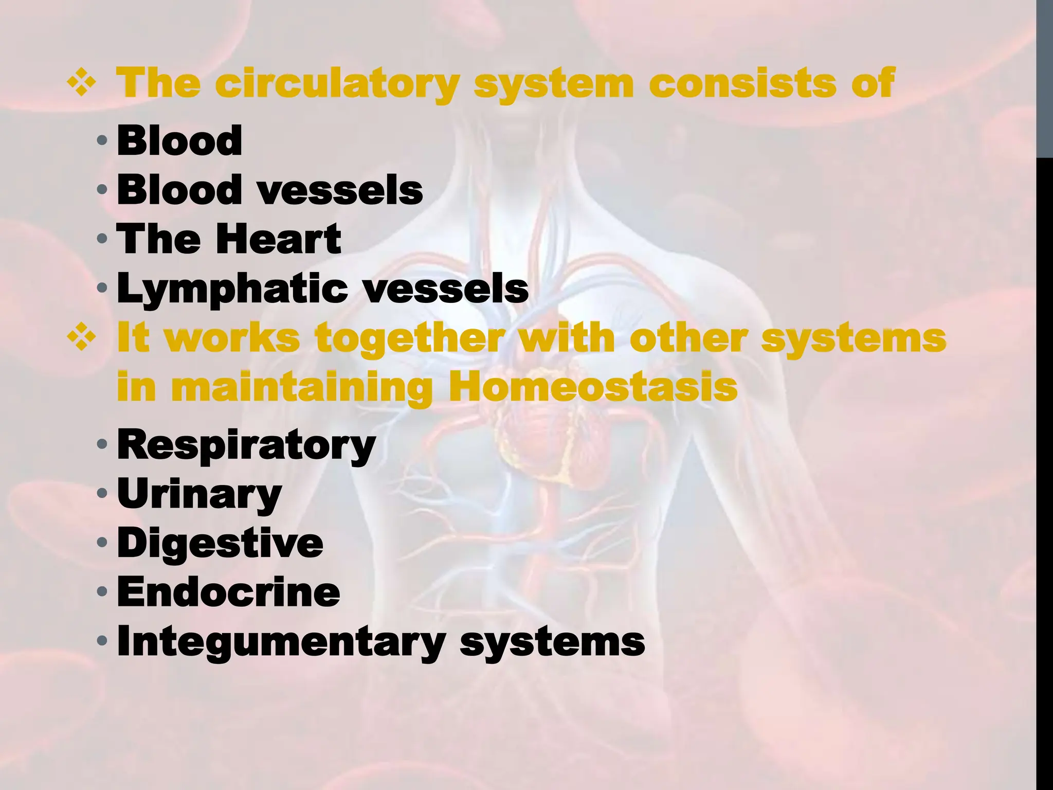

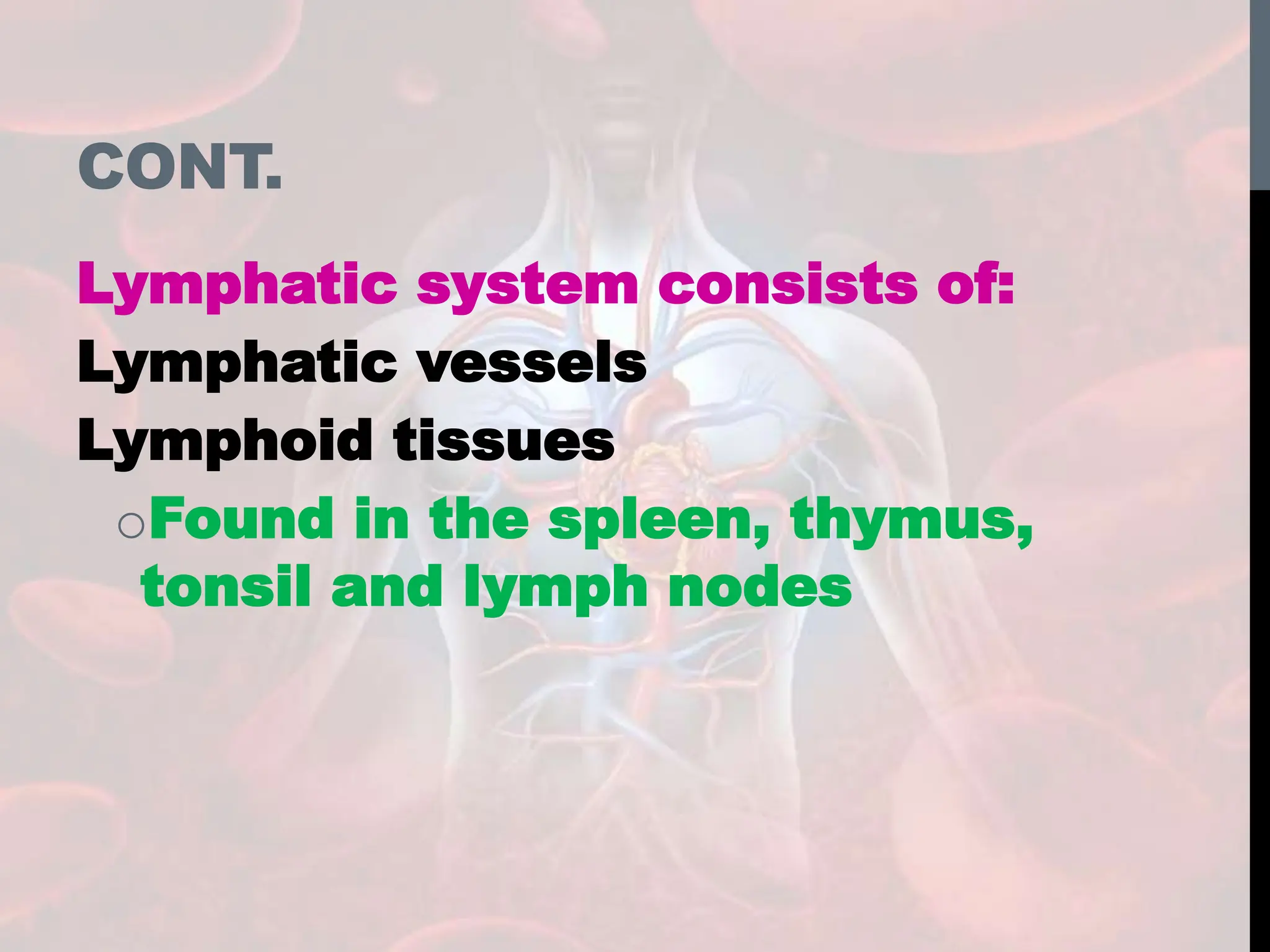
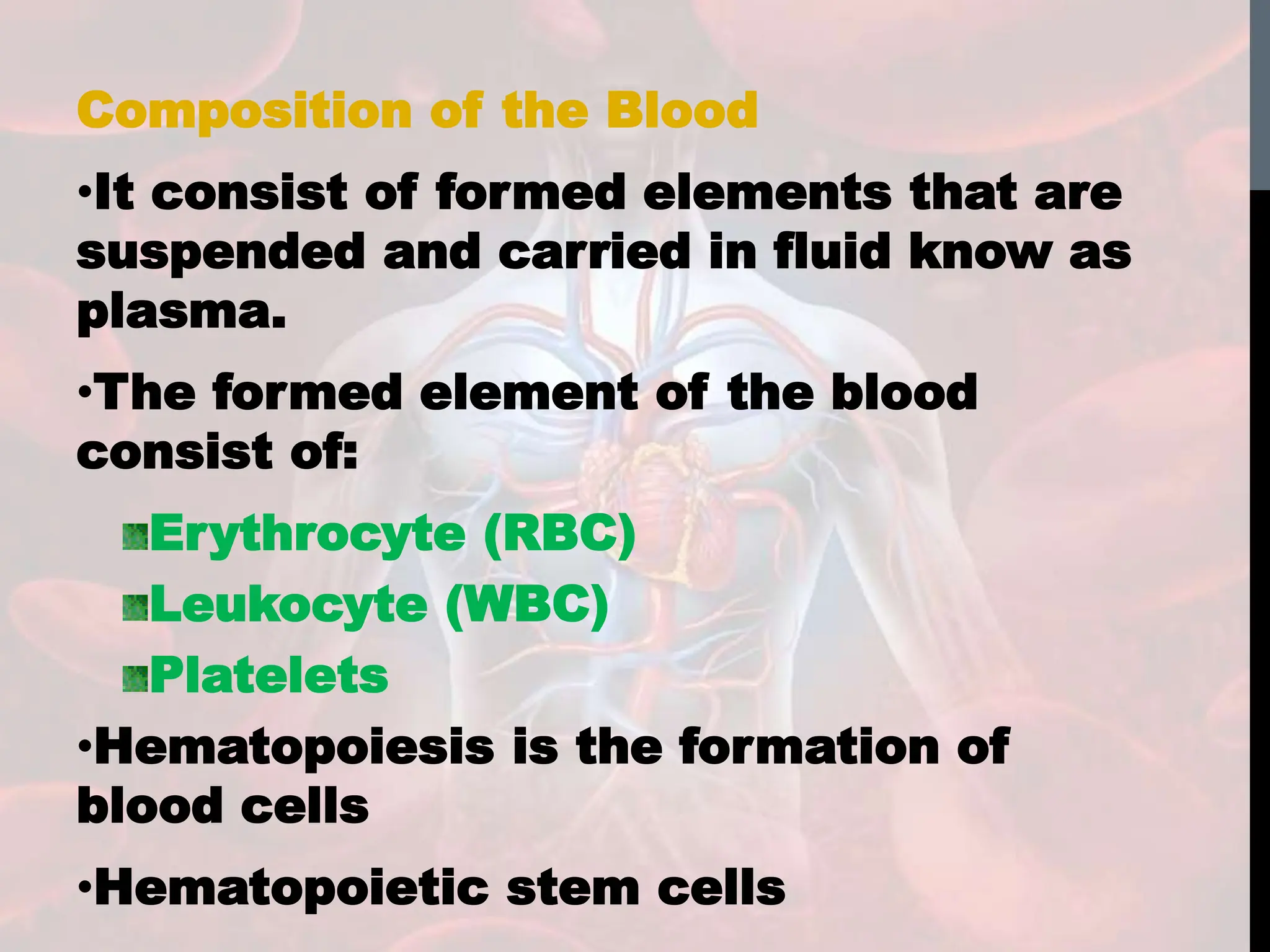
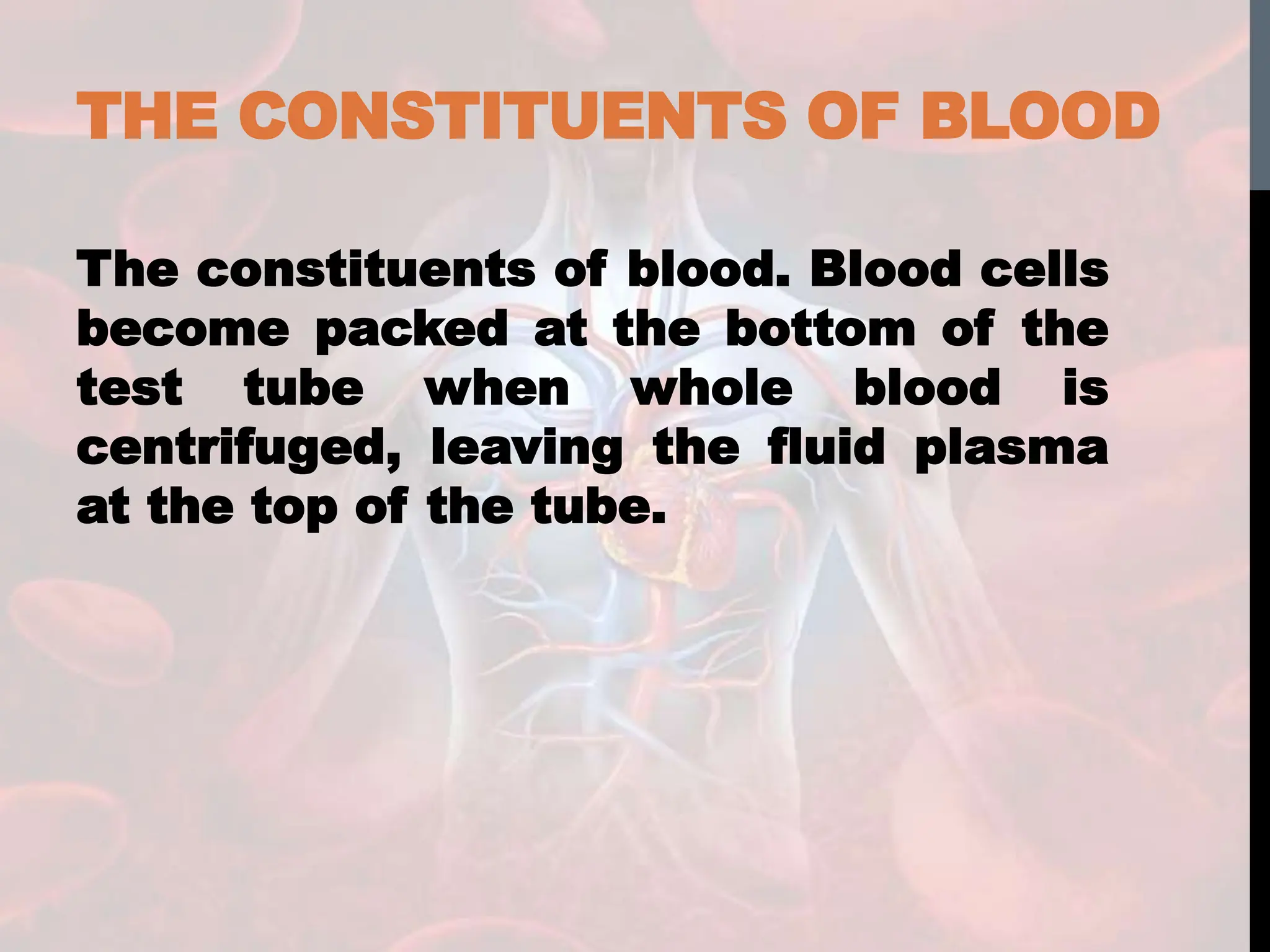
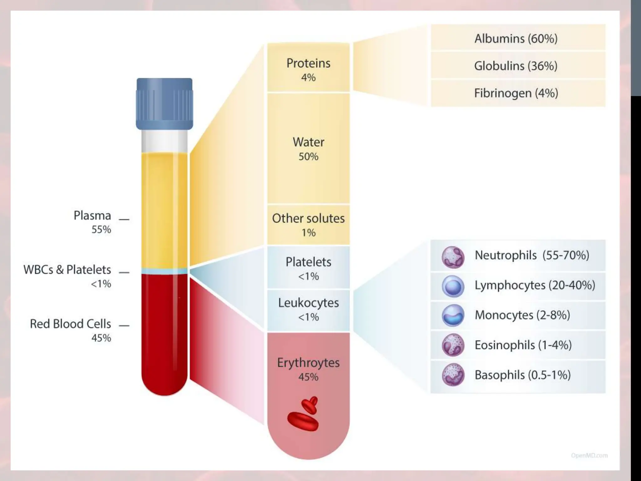
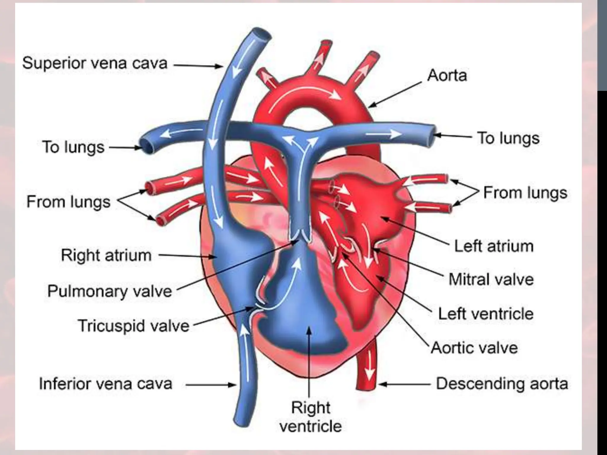
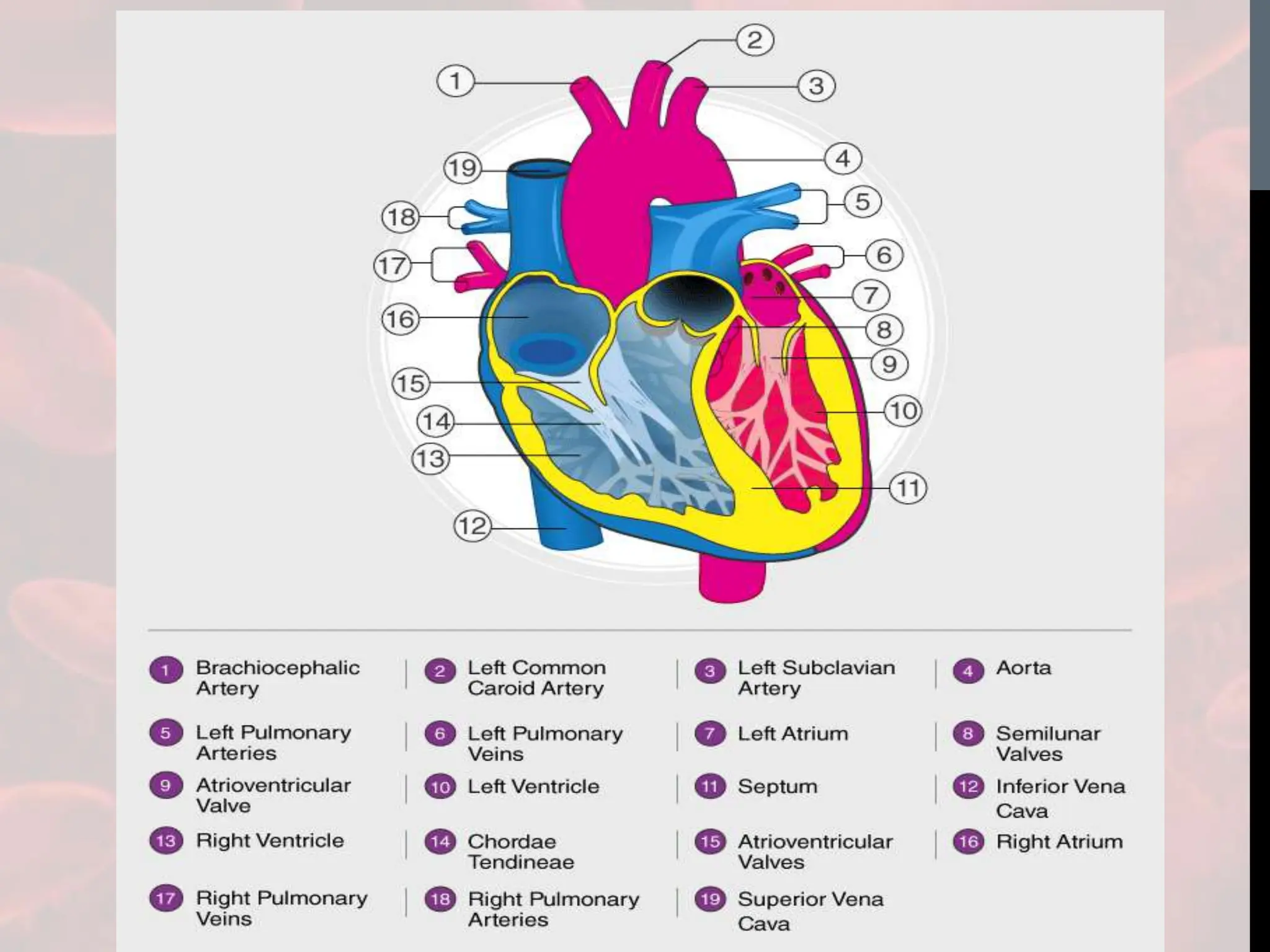

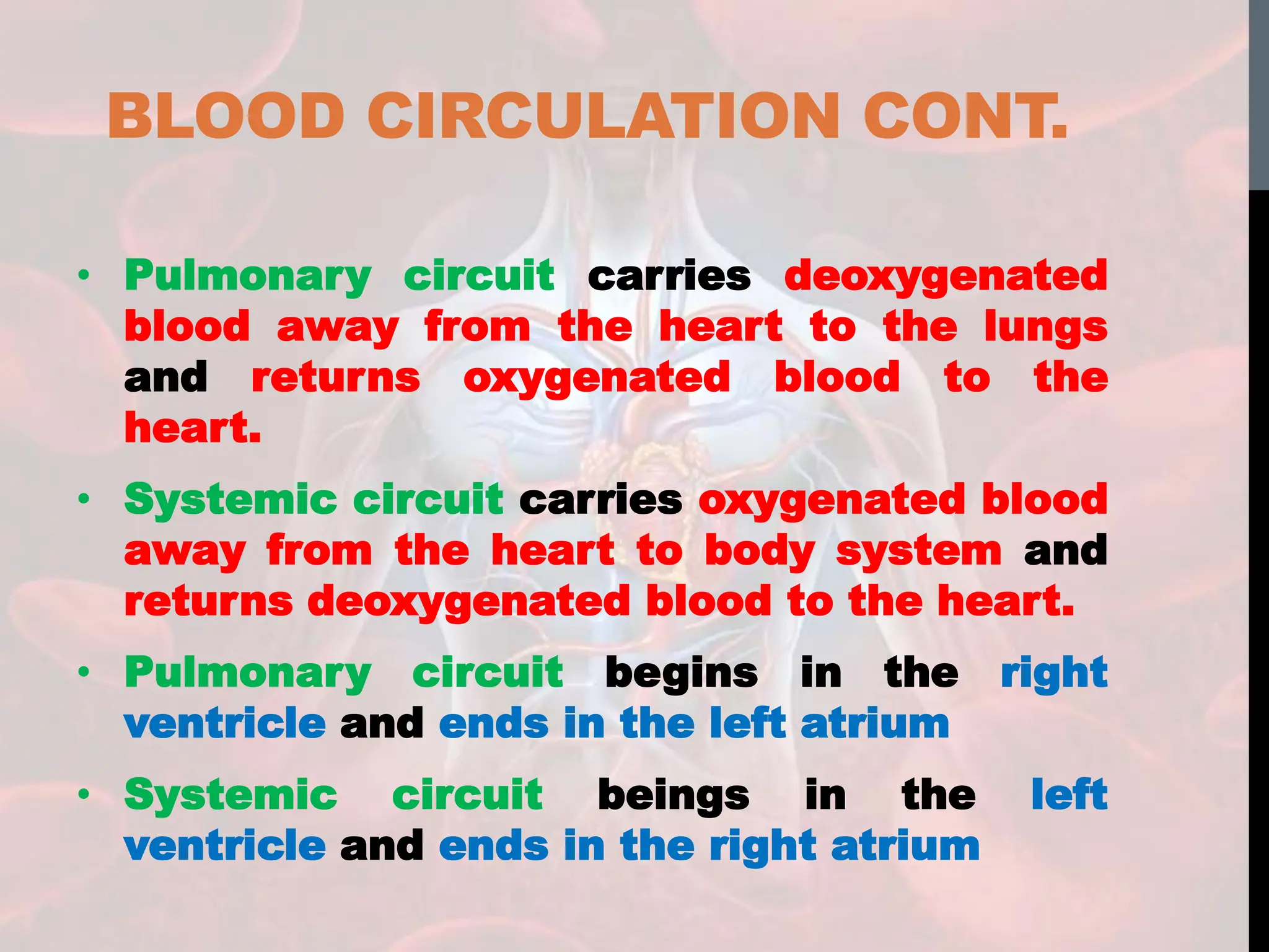
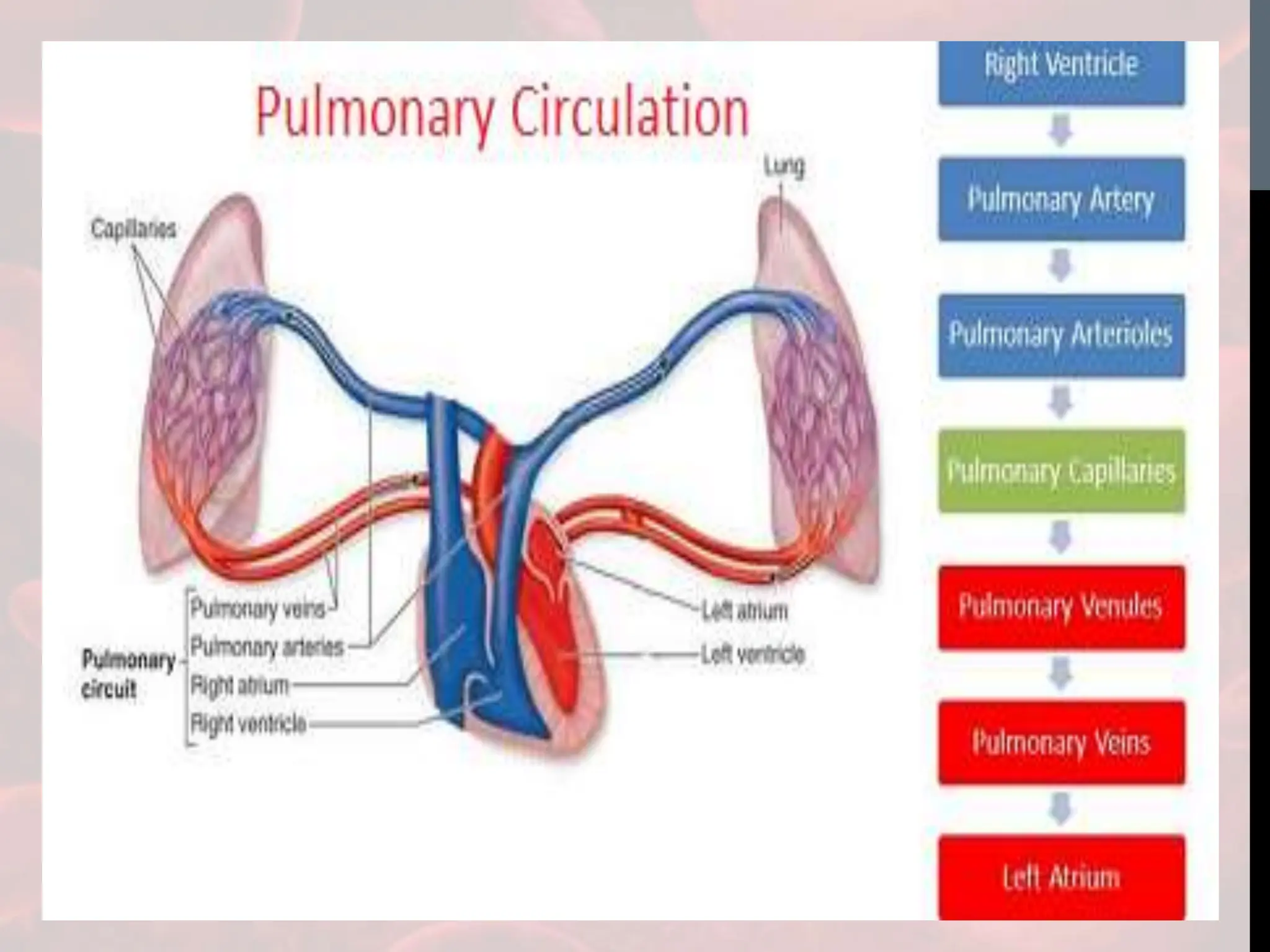
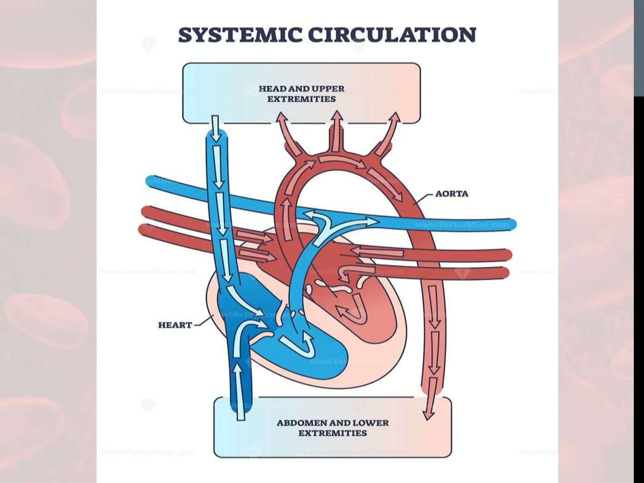


The document provides an extensive overview of the circulatory and lymphatic systems, detailing their major components, functions, and the processes involved in blood circulation and heart function. It discusses the structure of the heart, the cardiac cycle, various blood vessels, and the components of blood, alongside the functions of the lymphatic system in fluid transport and immune defense. Overall, the document emphasizes the interconnectedness of these systems in maintaining homeostasis within the body.


![Circulatory System Physiology [Zoo 403]](https://cdn.slidesharecdn.com/ss_thumbnails/circulatorysystemphysiologygroup-1-190416144432-thumbnail.jpg?width=640&height=640&fit=bounds)