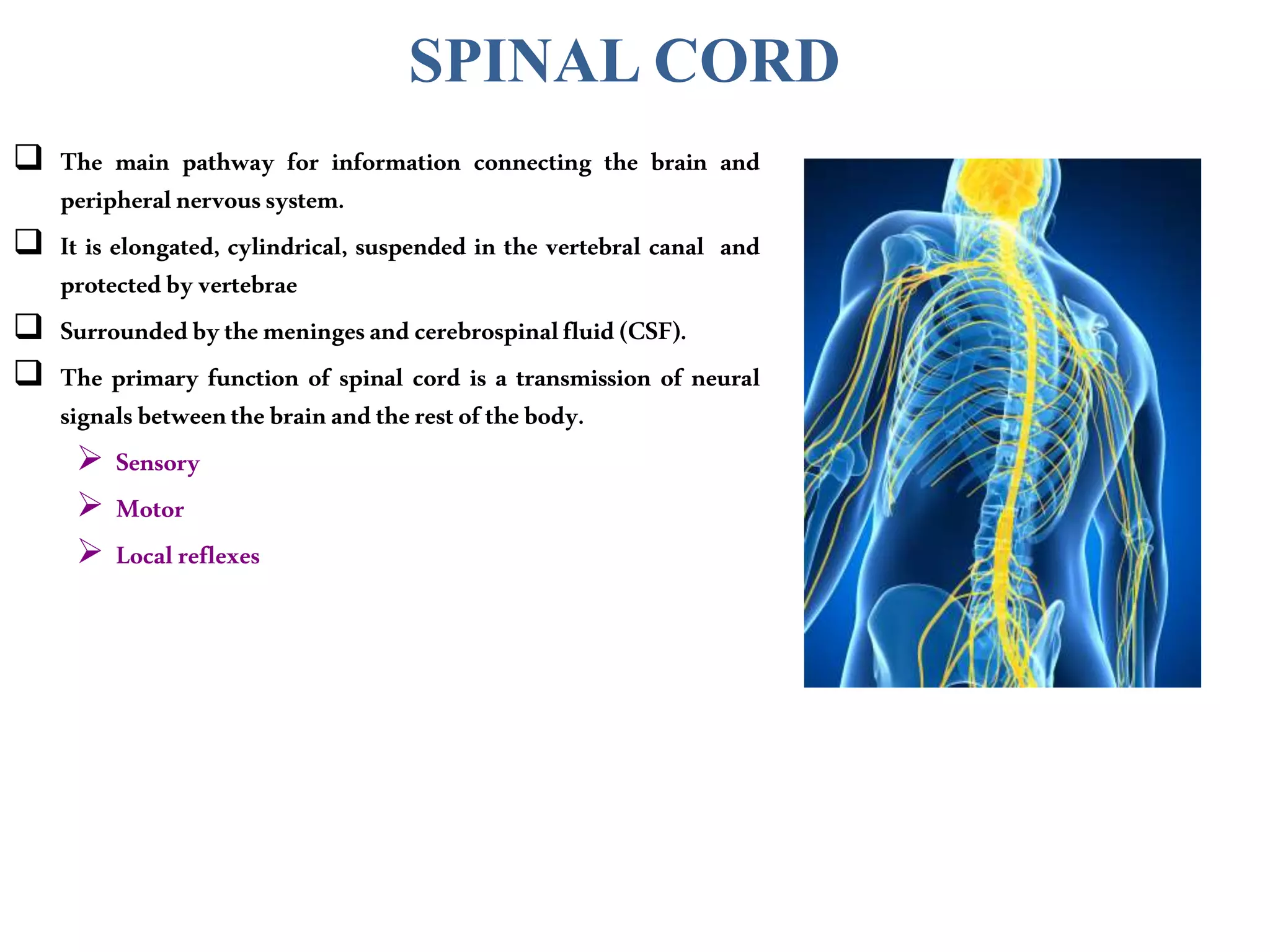
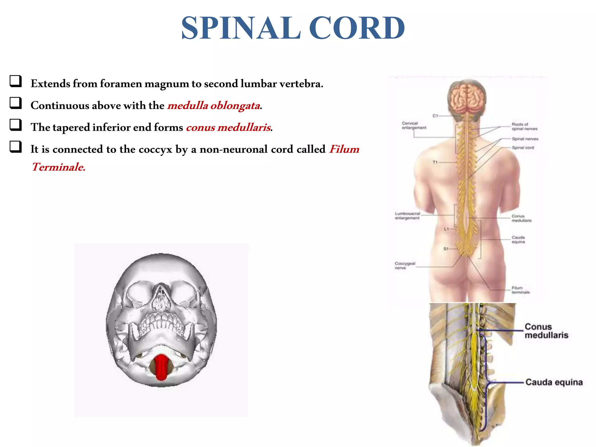
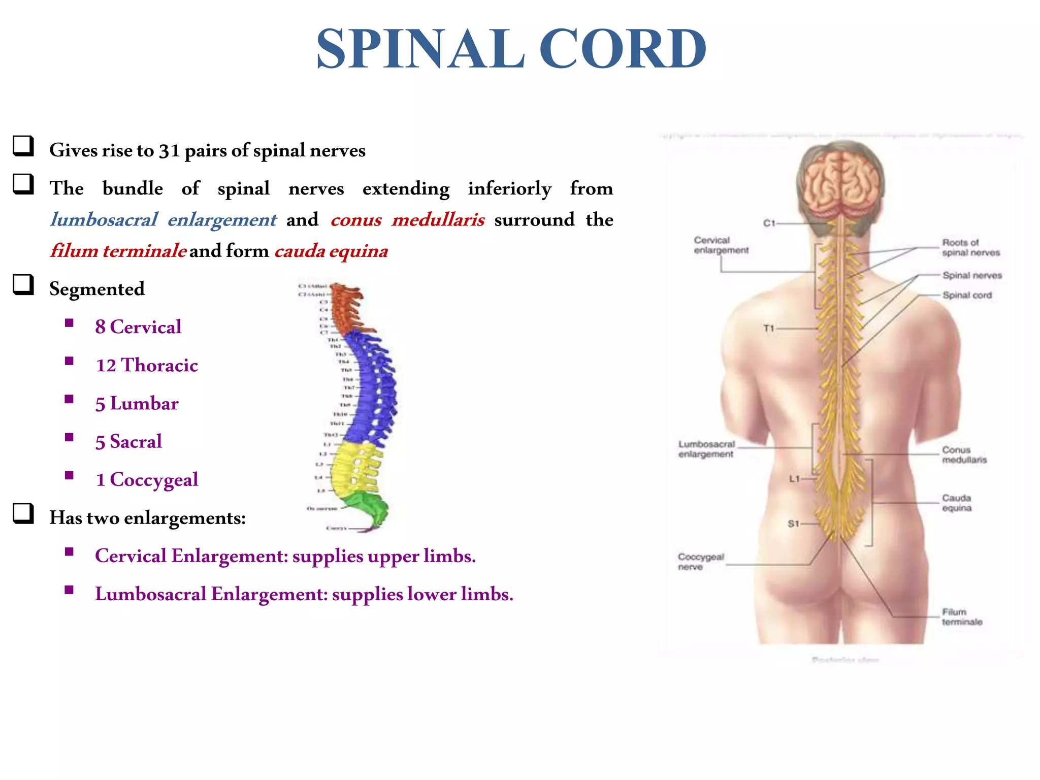
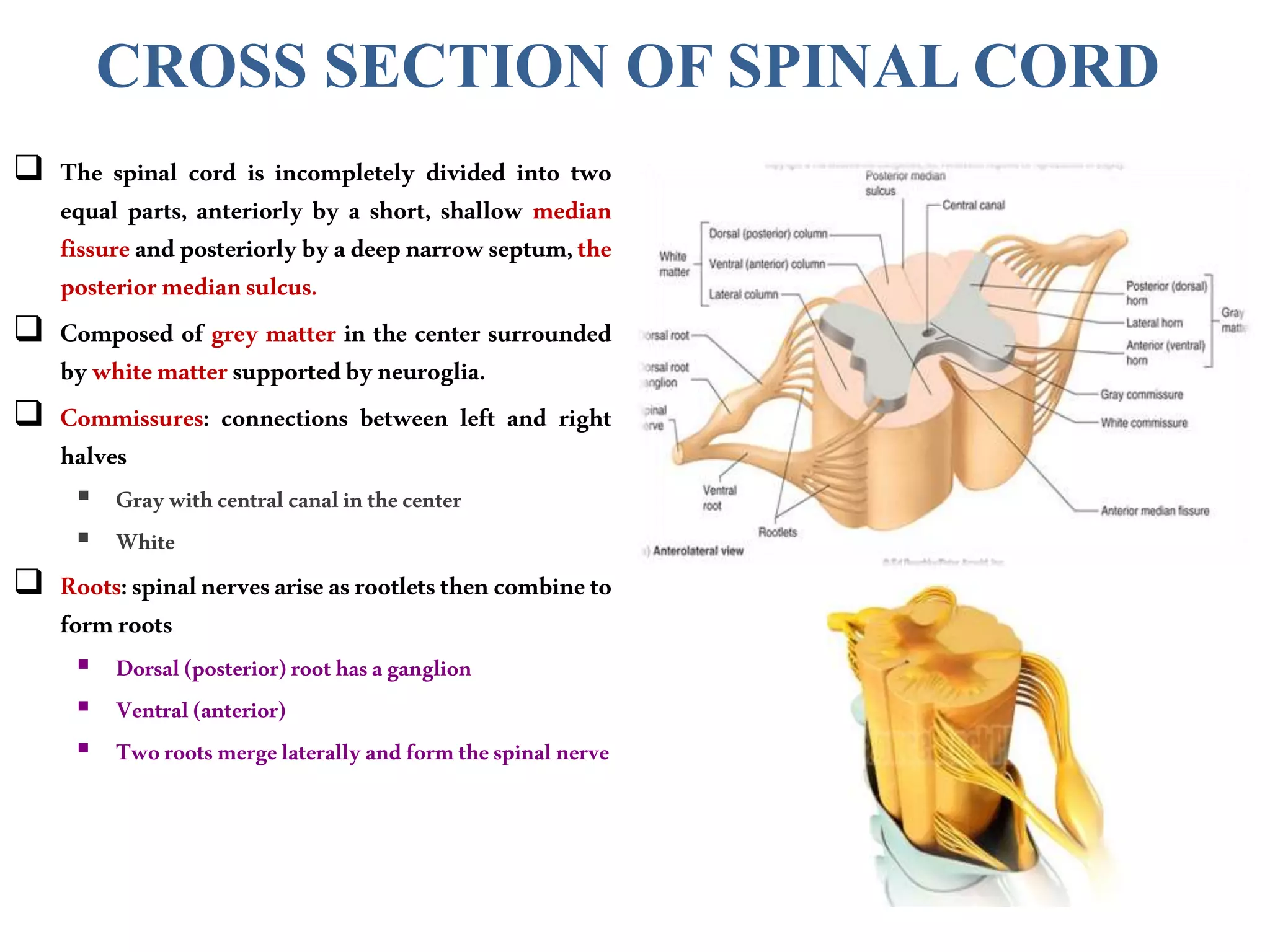
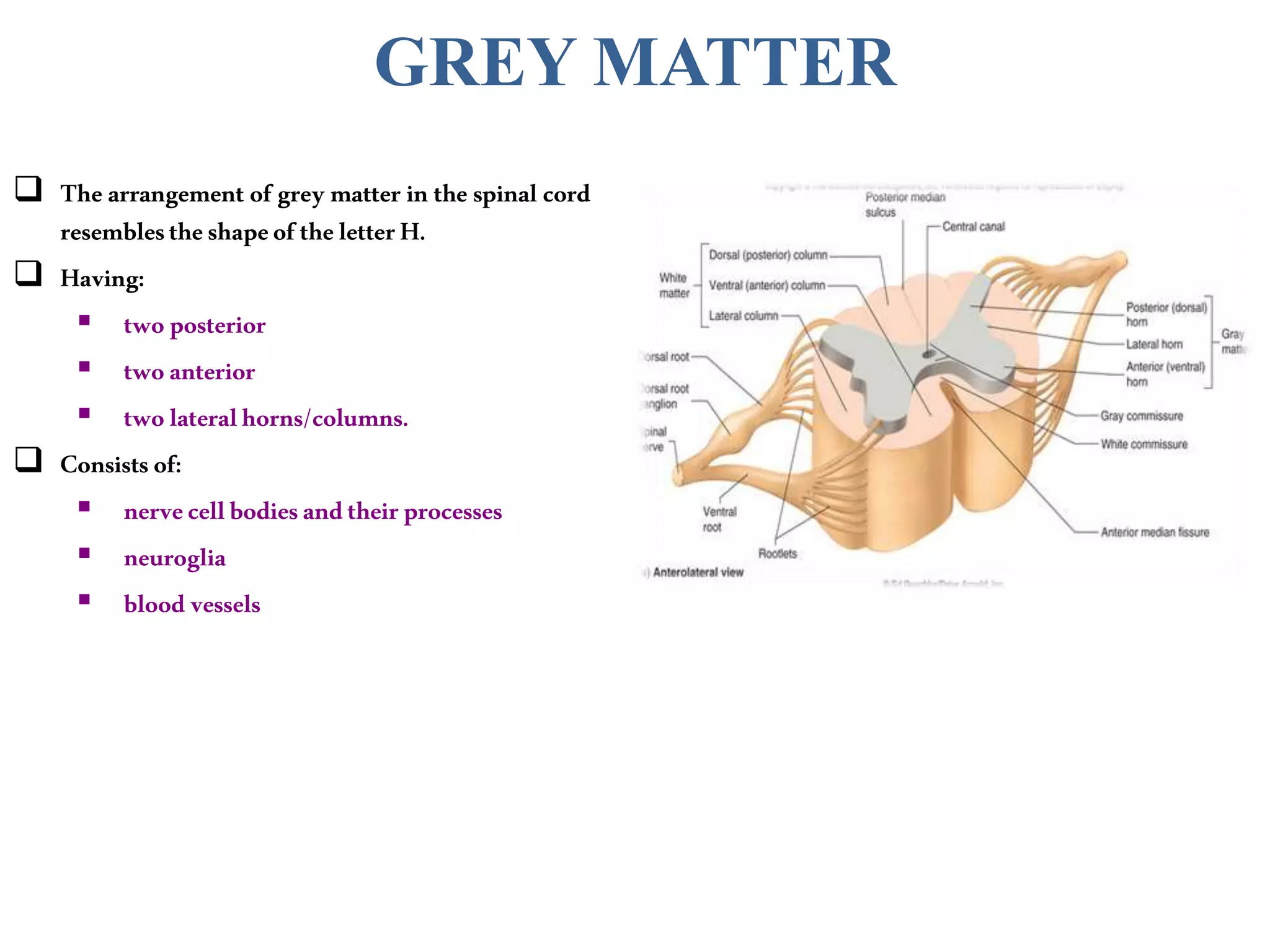
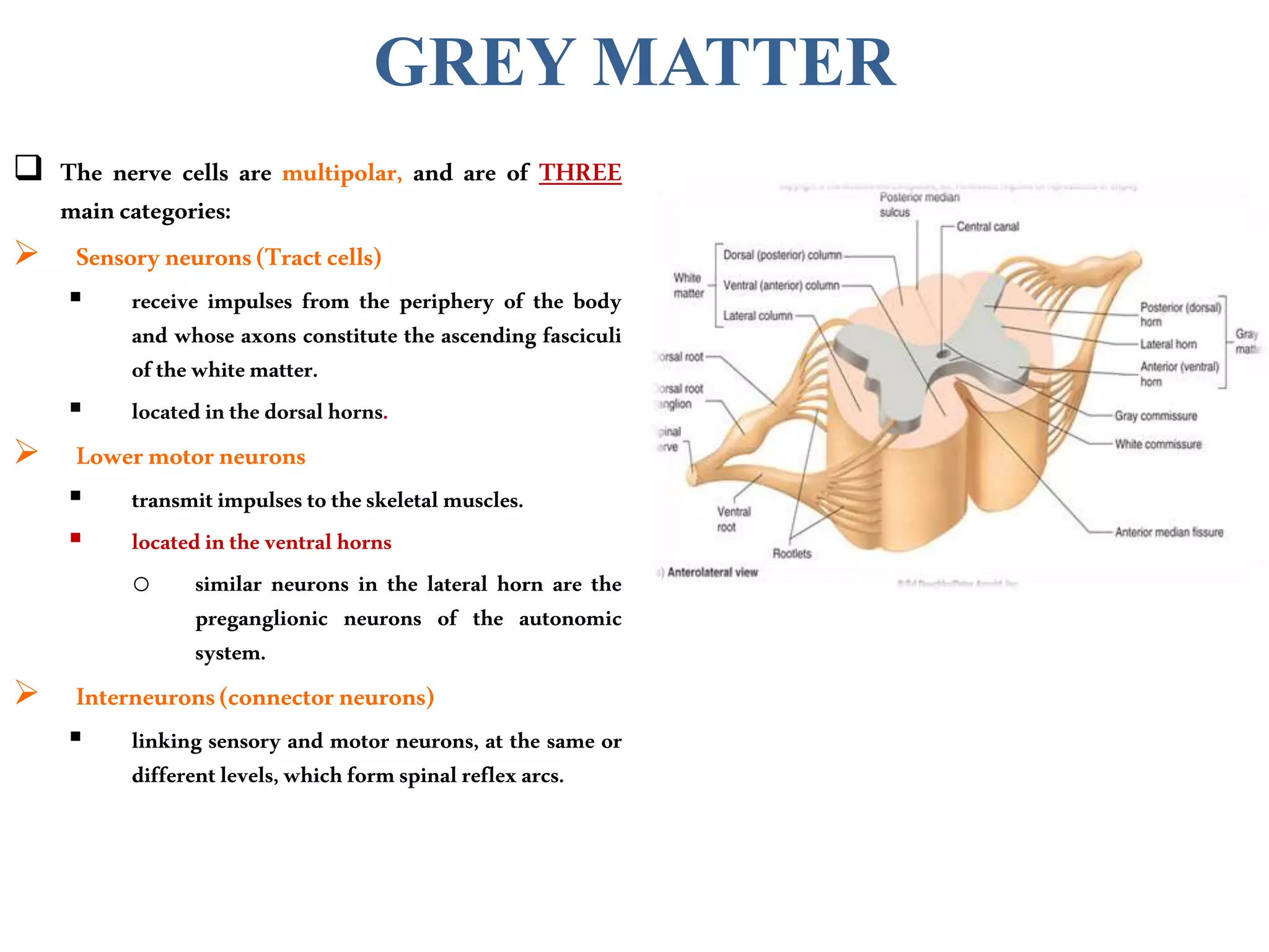
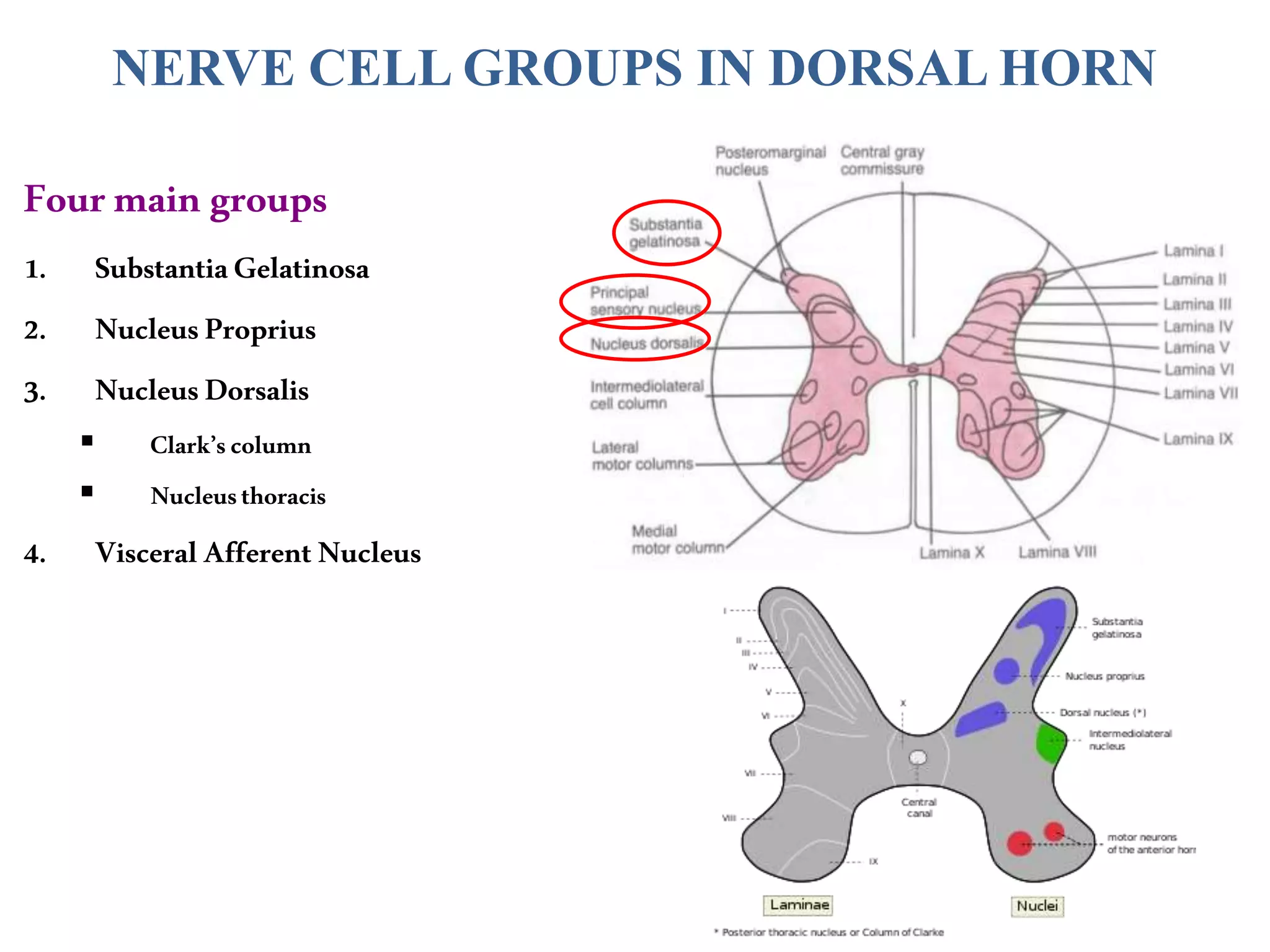
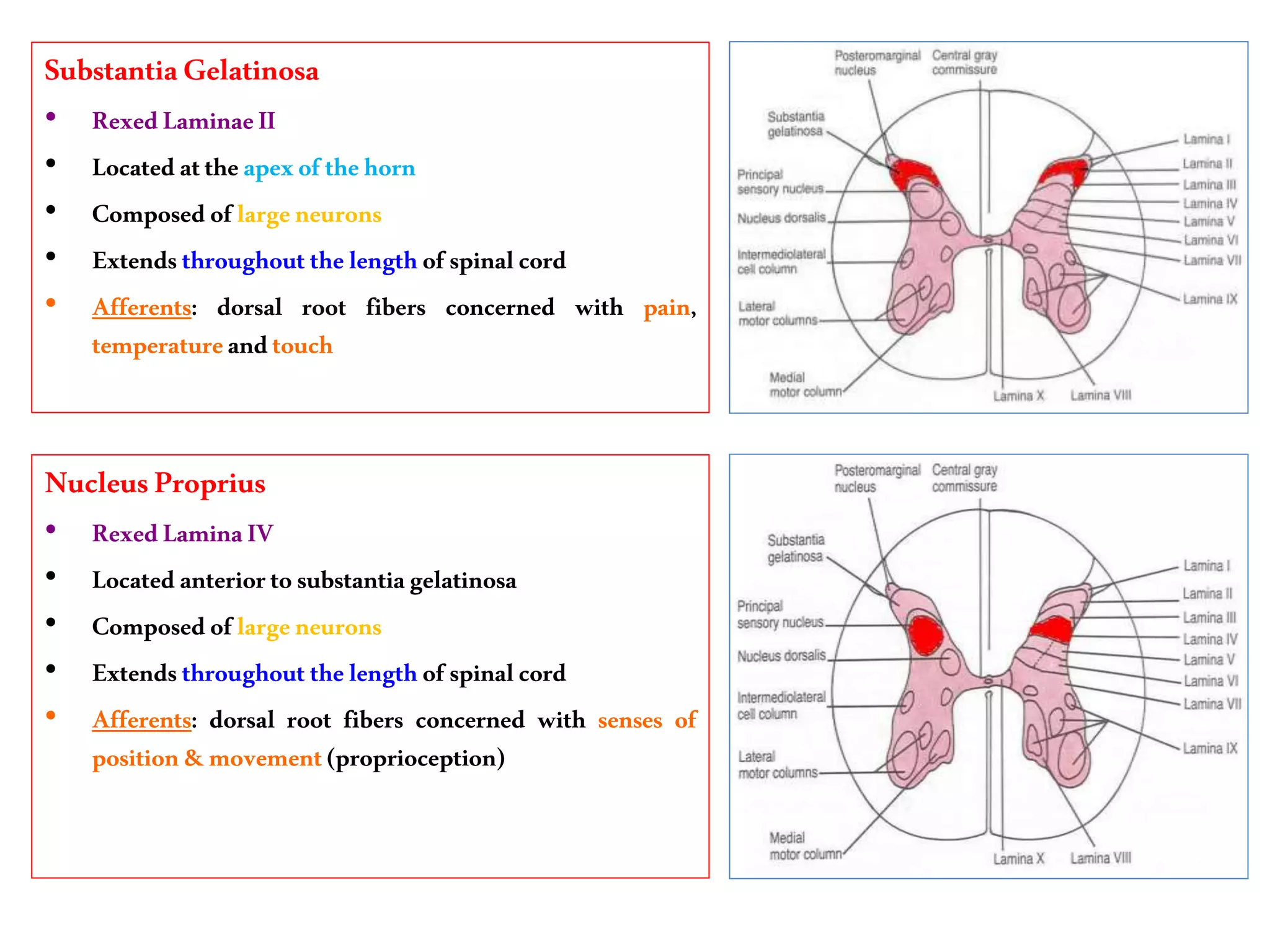
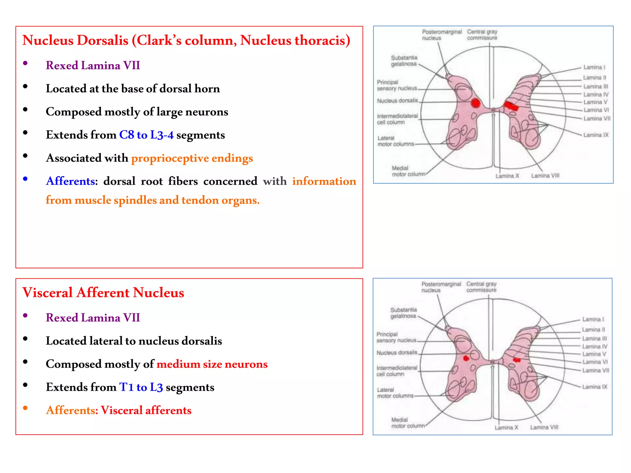
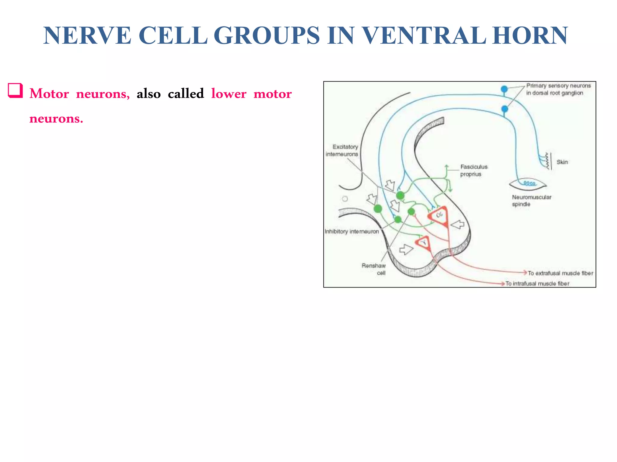
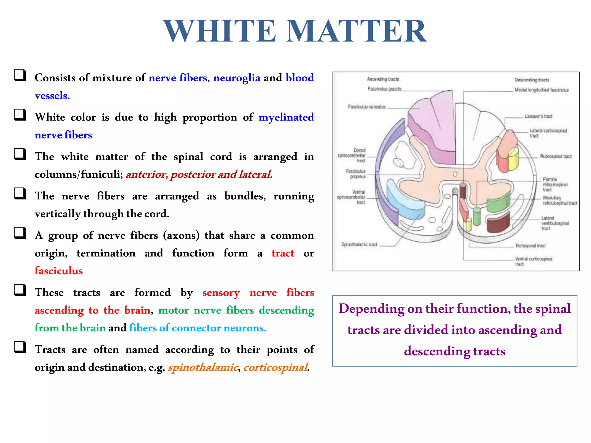
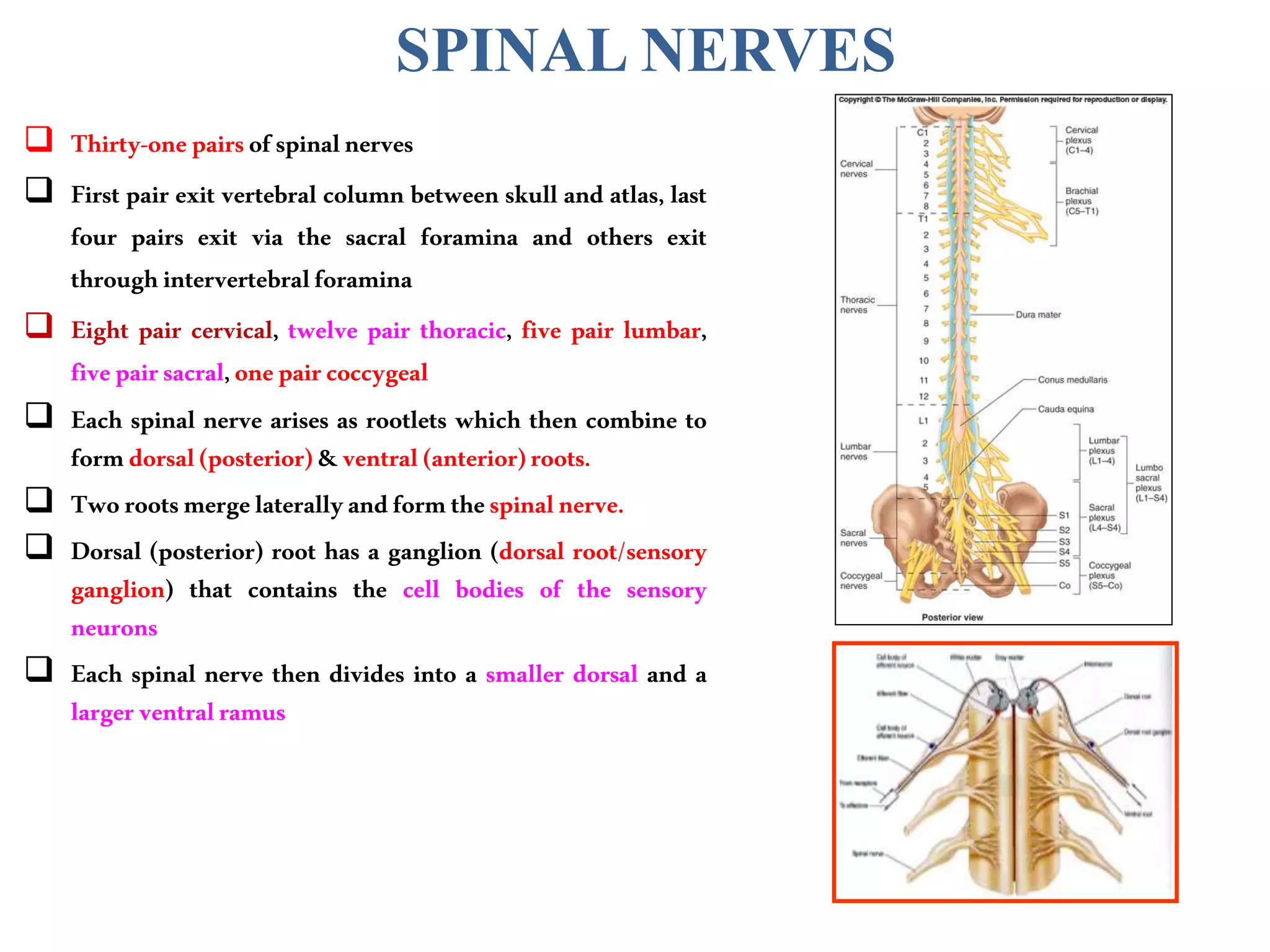
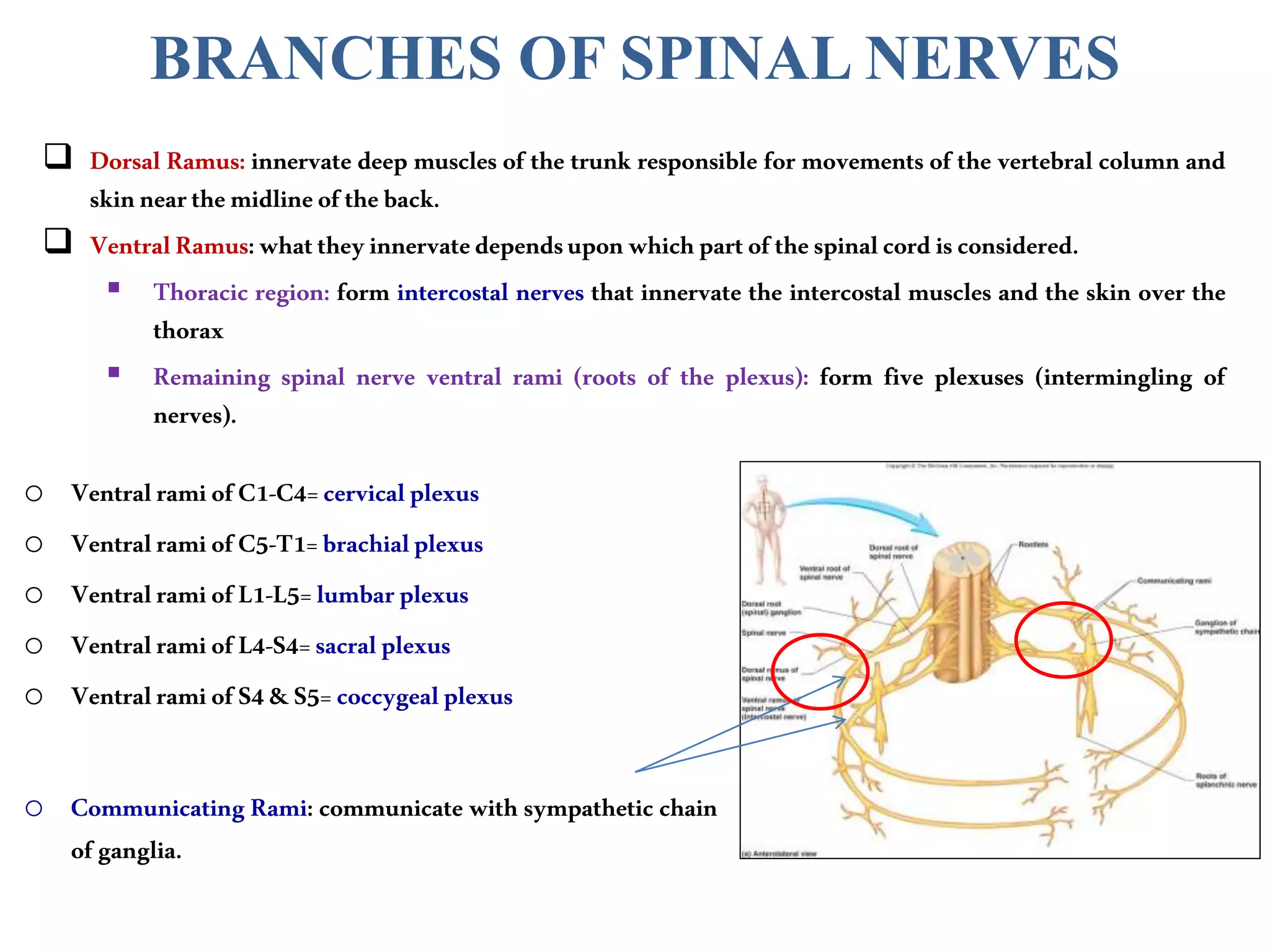
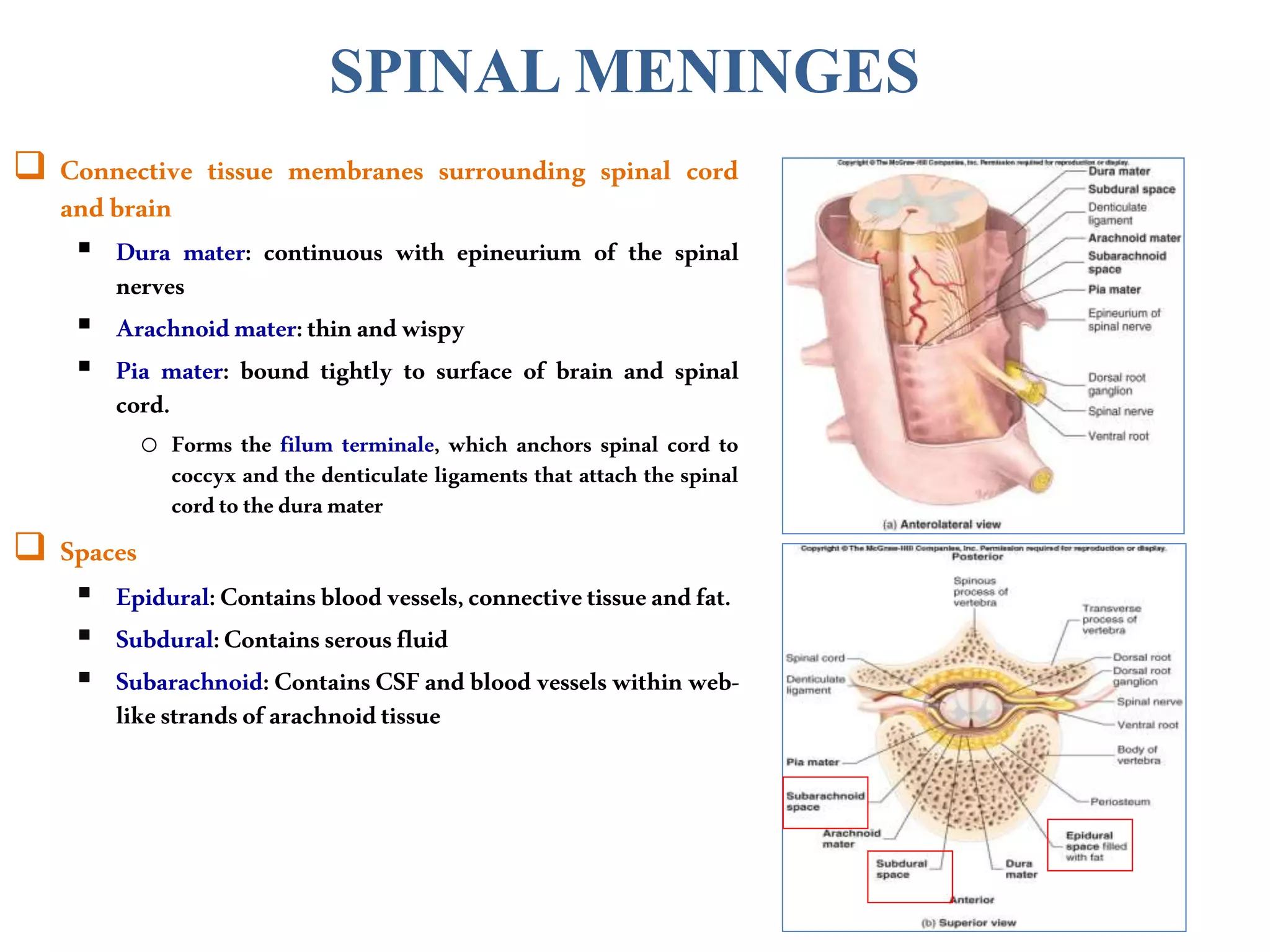
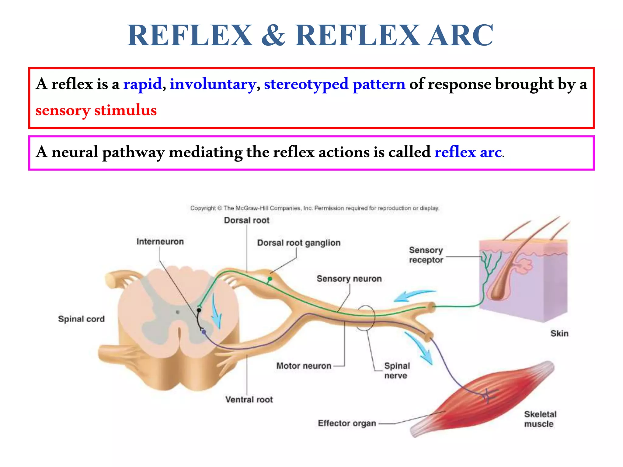
6. FUNCTIONS
The nervous system has 3 functions:
Collection of Sensory Input
Identifies changes occurring
inside and outside the body by
using sensory receptors. These
changes are called stimuli
Integration
Processes, analyses & interprets
these changes and makes
decisions
Motor Output
It then effects a response by
activating muscles or glands
(effectors) via motor output
9. Nucleus
A group of neurons
within the CNS
Ganglion
A group of neurons outside
the CNS
Tract
A group of nerve fibers (axons)
within the CNS
Nerve
A group of nerve fibers
(axons) outside the CNS
10. SPINAL CORD
The main pathway for information connecting the brain and
peripheralnervoussystem.
It is elongated, cylindrical, suspended in the vertebral canal and
protectedbyvertebrae
Surroundedbythemeningesandcerebrospinalfluid(CSF).
The primary function of spinal cord is a transmission of neural
signalsbetweenthebrainandtherestofthebody.
Sensory
Motor
Localreflexes
12. SPINAL CORD
Givesriseto31pairsofspinalnerves
The bundle of spinal nerves extending inferiorly from
lumbosacral enlargement and conus medullaris surround the
filumterminaleandformcaudaequina
Segmented
8Cervical
12Thoracic
5Lumbar
5Sacral
1Coccygeal
Hastwoenlargements:
CervicalEnlargement:suppliesupperlimbs.
LumbosacralEnlargement:supplieslowerlimbs.
13. CROSS SECTION OF SPINAL CORD
The spinal cord is incompletely divided into two
equal parts, anteriorly by a short, shallow median
fissureandposteriorlybyadeepnarrowseptum,the
posteriormediansulcus.
Composed of grey matter in the center surrounded
bywhitemattersupportedbyneuroglia.
Commissures: connections between left and right
halves
Graywithcentral canalin thecenter
White
Roots:spinalnervesariseasrootletsthencombineto
formroots
Dorsal(posterior)root hasaganglion
Ventral (anterior)
Tworootsmergelaterallyand formthespinal nerve
14. The arrangement of grey matter in the spinal cord
resemblestheshapeoftheletterH.
Having:
twoposterior
twoanterior
twolateralhorns/columns.
Consistsof:
nervecellbodiesandtheirprocesses
neuroglia
blood vessels
GREY MATTER
15. The nerve cells are multipolar, and are of THREE
maincategories:
Sensoryneurons(Tractcells)
receive impulses from the periphery of the body
and whose axons constitute the ascending fasciculi
ofthewhitematter.
locatedinthe dorsalhorns.
Lowermotorneurons
transmitimpulses to theskeletal muscles.
locatedinthe ventral horns
o similar neurons in the lateral horn are the
preganglionic neurons of the autonomic
system.
Interneurons(connectorneurons)
linking sensory and motor neurons, at the same or
differentlevels,whichformspinal reflexarcs.
GREY MATTER
16. NEURONALARCHITECTURE OF
SPINAL GREY MATTER
Cells of the same type are clustered into groups, which
occurinlongcolumns
In transverse section, these columns appear as layers,
especiallywithinthedorsalhorn
These layers are called the laminae of Rexed that are
numbered by Roman numerals, starting from the tip of
the dorsal horn and moving ventrally into the ventral
horn.
The Rexed laminae comprise a system of ten layers
of grey matter (I-X), identified in the early 1950s by
Swedishneuroscientist.
17. NERVE CELL GROUPS IN DORSAL HORN
Fourmain groups
1. SubstantiaGelatinosa
2. NucleusProprius
3. NucleusDorsalis
Clark’scolumn
Nucleusthoracis
4. VisceralAfferentNucleus
20. NERVE CELL GROUPS IN VENTRAL HORN
Motor neurons, also called lower motor
neurons.
21. MOTOR NEURONS IN VENTRAL HORN
AreofTwo types
Largemultipolarcells
whose axons pass out in the ventral roots
of spinal nerves as alpha efferents which
innervate extrafusal muscle fibers of
skeletalmuscles.
Lessnumerous smallermultipolar cells
whose axons pass out in the ventral roots
of spinal nerves as gamma efferents
which innervate intrafusal muscle fibers
ofneuromuscularspindles
Bothalphaand gamma motorneuronsare
undertheinfluenceofdescending
pathwaysfrombrain
22. Motor neurons areorganizedin3groups:
Medial:
Present in most segments, innervates
muscles of neck and trunk (including
intercostalandabdominalmuscles)
Central:
Smallest, present in some cervical (phrenic
C3-5, spinal accessory C1-6) and
lumbosacral(L2-S1)segments
Lateral:
Present in cervical and lumbosacral
segments,innervatesmusclesofthelimbs
Neuronssupplyingflexormusclesare
locateddorsaltoneuronsforextensor
muscles
MOTOR NEURONS IN VENTRAL HORN
23. NERVE CELL GROUPS IN LATERAL HORN
SmallColumncomposed of smallneurons
ExtendsfromT1toL2-3segments:
o Giverisetopreganglionicsympatheticfibers
ExtendsfromS2-4 segments:
o Give rise to preganglionic parasympathetic
fibers
24. WHITE MATTER
Consists of mixture of nerve fibers, neuroglia and blood
vessels.
White color is due to high proportion of myelinated
nervefibers
The white matter of the spinal cord is arranged in
columns/funiculi;anterior,posteriorandlateral.
The nerve fibers are arranged as bundles, running
verticallythroughthecord.
A group of nerve fibers (axons) that share a common
origin, termination and function form a tract or
fasciculus
These tracts are formed by sensory nerve fibers
ascending to the brain, motor nerve fibers descending
fromthebrainandfibersofconnectorneurons.
Tracts are often named according to their points of
originanddestination,e.g.spinothalamic,corticospinal.
Dependingontheirfunction,thespinal
tractsaredividedintoascendingand
descendingtracts
25.
26. COMMISSURES OF THE SPINAL CORD
Grey Commissure:
Transverse bridge of grey matter connecting the
anteriorandposteriorgrayhornsoneachside
Is pierced by the central canal that divides it into
anteriorandposteriorparts
White Commissure:
Liesventraltothegraycommissure
Mainlycontainsdecussatingnervefibers
27. CENTRAL CANAL
The cerebrospinal-filled space that runs longitudinally
throughtheentirelengthofthespinalcord.
Linedbyependyma(ciliatedcolumnarepithelium)
Continuouswiththeventricularsystemofthebrain
Superiorlyopensintothe4th ventricle
Inferiorly in the conus medullaris, it expands into the
fusiformterminalventricleand terminatesbelowatthe
rootoffilumterminale
28. REGIONAL DIFFERENCES
Although the general pattern of gray matter is the
same throughout spinal cord, regional differences
areapparentintransversesections
The amount of white matter increases in a caudal-to-
cranial direction because fibers are added to
ascendingtractsandfibersleavedescendingtracts
The gray matter is in increased volume in cervical &
lumbosacral enlargements for innervation of upper
&lowerlimbs
The lateral horn is characteristics of thoracic and
upperlumbarsegments
Cervical
Thoracic
Lumbar
Sacral
29. SPINAL NERVES
Thirty-onepairsofspinalnerves
First pair exit vertebral column between skull and atlas, last
four pairs exit via the sacral foramina and others exit
throughintervertebralforamina
Eight pair cervical, twelve pair thoracic, five pair lumbar,
fivepairsacral,onepaircoccygeal
Each spinal nerve arises as rootlets which then combine to
formdorsal(posterior)&ventral(anterior)roots.
Tworootsmergelaterallyandformthespinalnerve.
Dorsal (posterior) root has a ganglion (dorsal root/sensory
ganglion) that contains the cell bodies of the sensory
neurons
Each spinal nerve then divides into a smaller dorsal and a
largerventralramus
30. BRANCHES OF SPINAL NERVES
Dorsal Ramus: innervate deep muscles of the trunk responsible for movements of the vertebral column and
skinnearthemidlineoftheback.
VentralRamus:whattheyinnervatedependsuponwhichpartofthespinalcordisconsidered.
Thoracic region: form intercostal nerves that innervate the intercostal muscles and the skin over the
thorax
Remaining spinal nerve ventral rami (roots of the plexus): form five plexuses (intermingling of
nerves).
o VentralramiofC1-C4=cervicalplexus
o VentralramiofC5-T1=brachialplexus
o VentralramiofL1-L5=lumbarplexus
o VentralramiofL4-S4=sacralplexus
o VentralramiofS4&S5=coccygealplexus
o Communicating Rami: communicate with sympathetic chain
ofganglia.
31. SPINAL MENINGES
Connective tissue membranes surrounding spinal cord
andbrain
Dura mater: continuous with epineurium of the spinal
nerves
Arachnoidmater:thinandwispy
Pia mater: bound tightly to surface of brain and spinal
cord.
o Forms the filum terminale, which anchors spinal cord to
coccyx and the denticulate ligaments that attach the spinal
cordto theduramater
Spaces
Epidural:Containsbloodvessels,connectivetissueandfat.
Subdural:Containsserousfluid
Subarachnoid: Contains CSFand blood vesselswithin web-
likestrandsofarachnoidtissue
32. REFLEX & REFLEX ARC
Areflexisarapid,involuntary,stereotypedpatternof responsebroughtby a
sensorystimulus
Aneuralpathwaymediatingthereflexactionsiscalledreflexarc.
33. COMPONENTS OF A REFLEX ARC
Actionpotentialsproducedin Sensoryneuron
Sensoryreceptor
Interneuron
Motorneuron
Effector organ which responds
withareflex