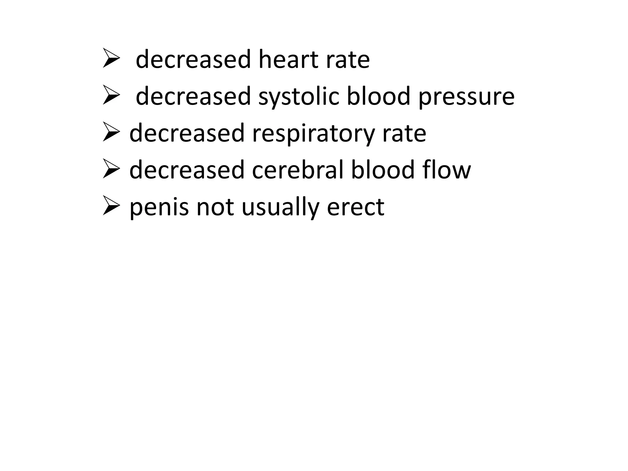
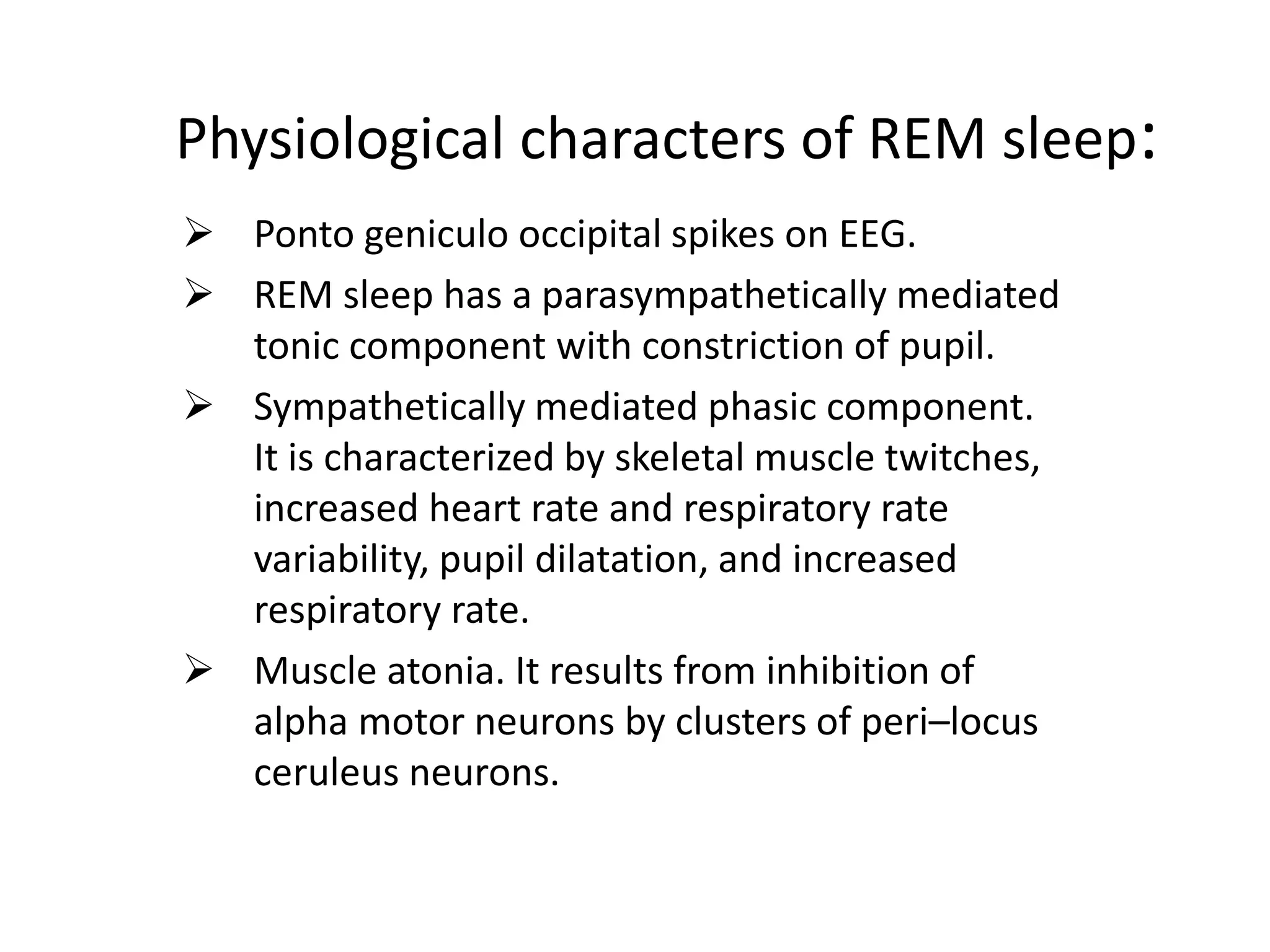
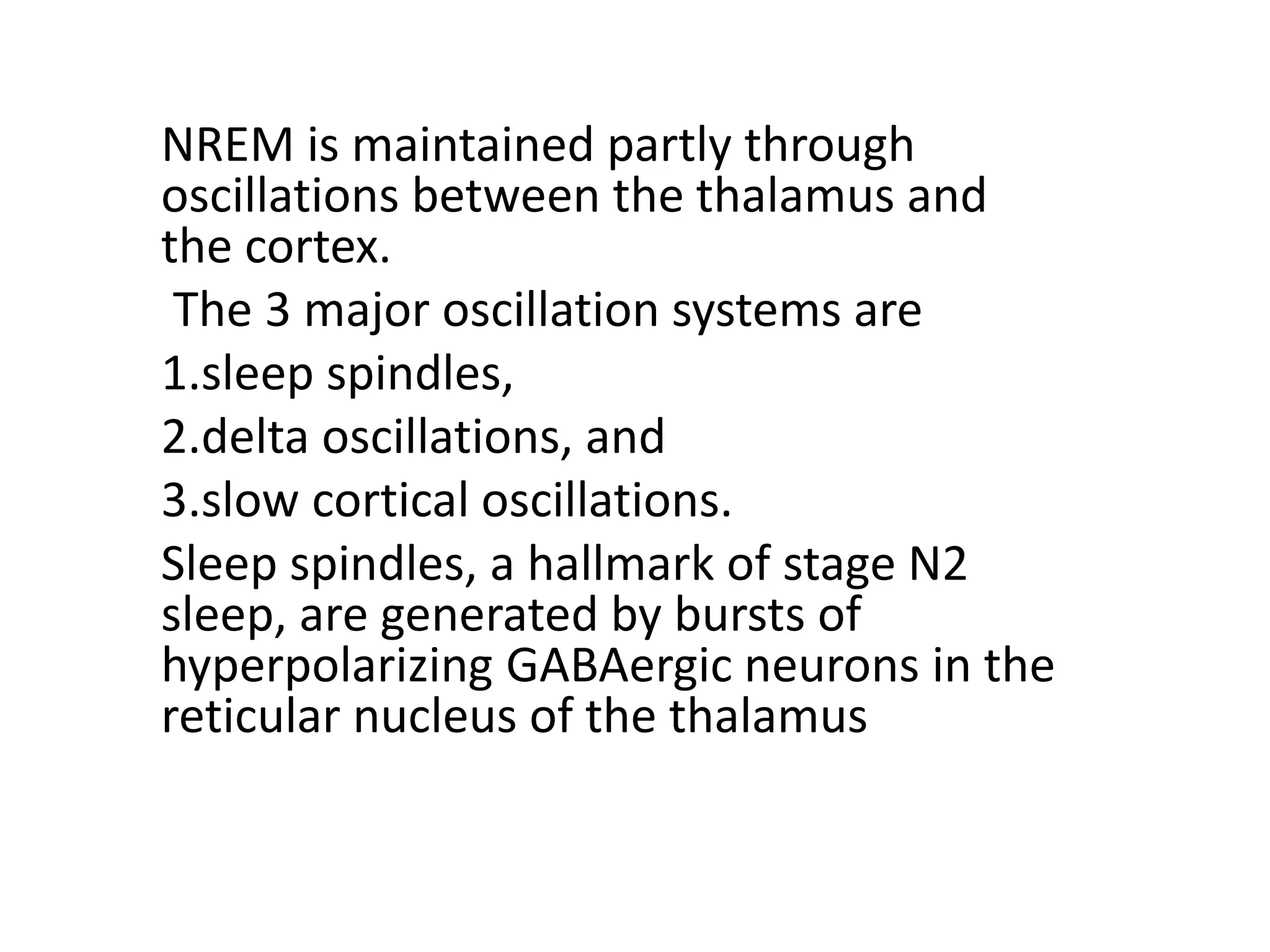
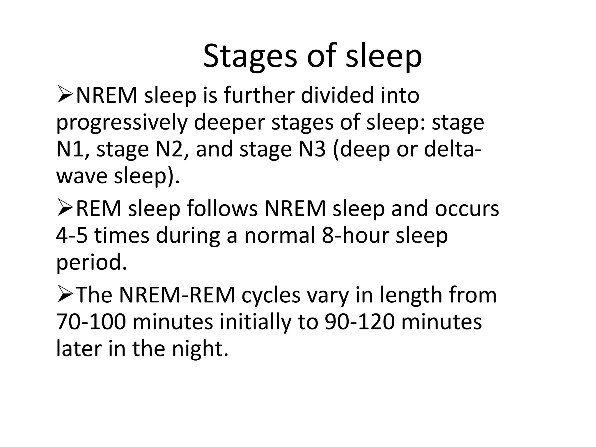
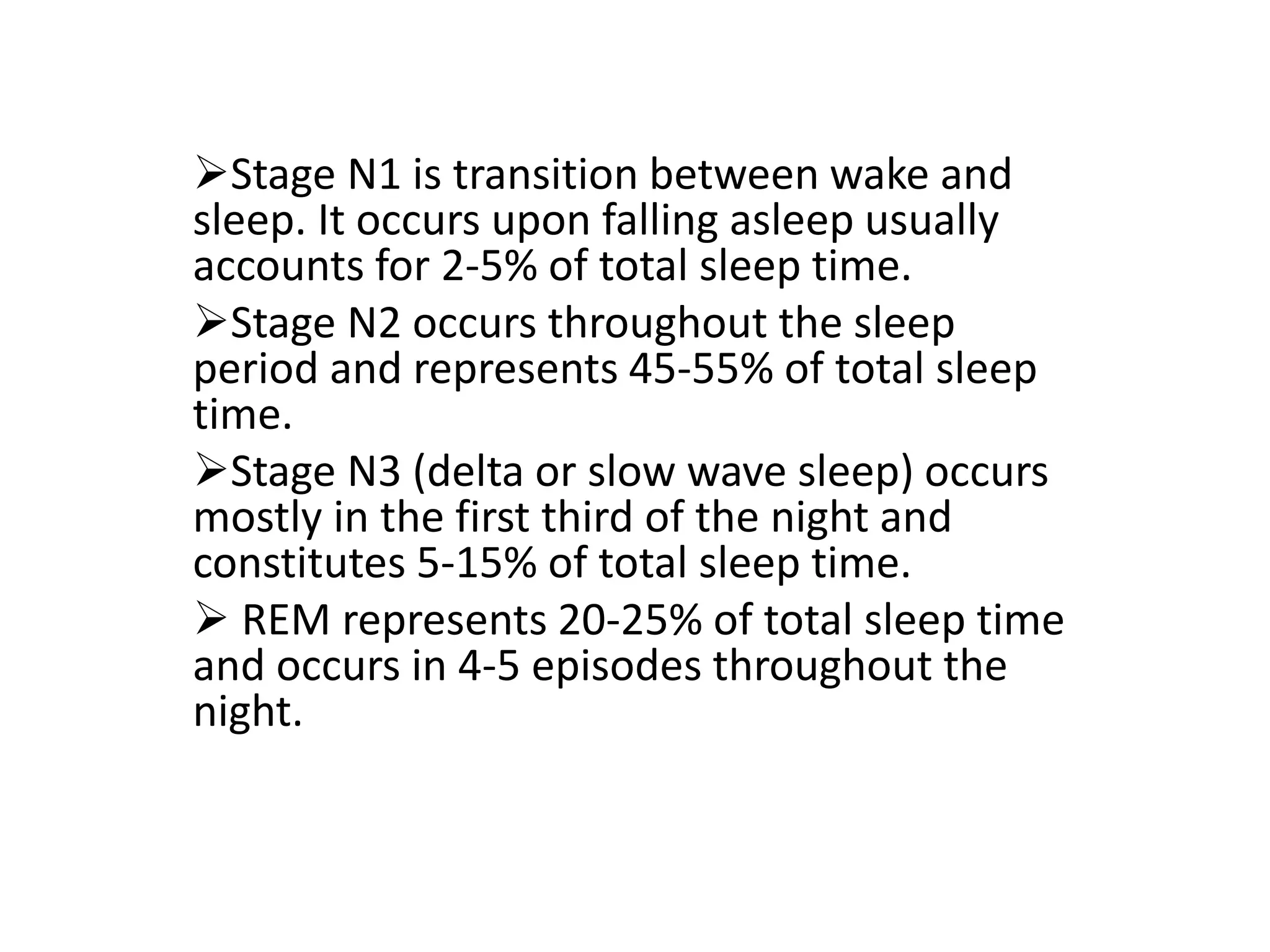
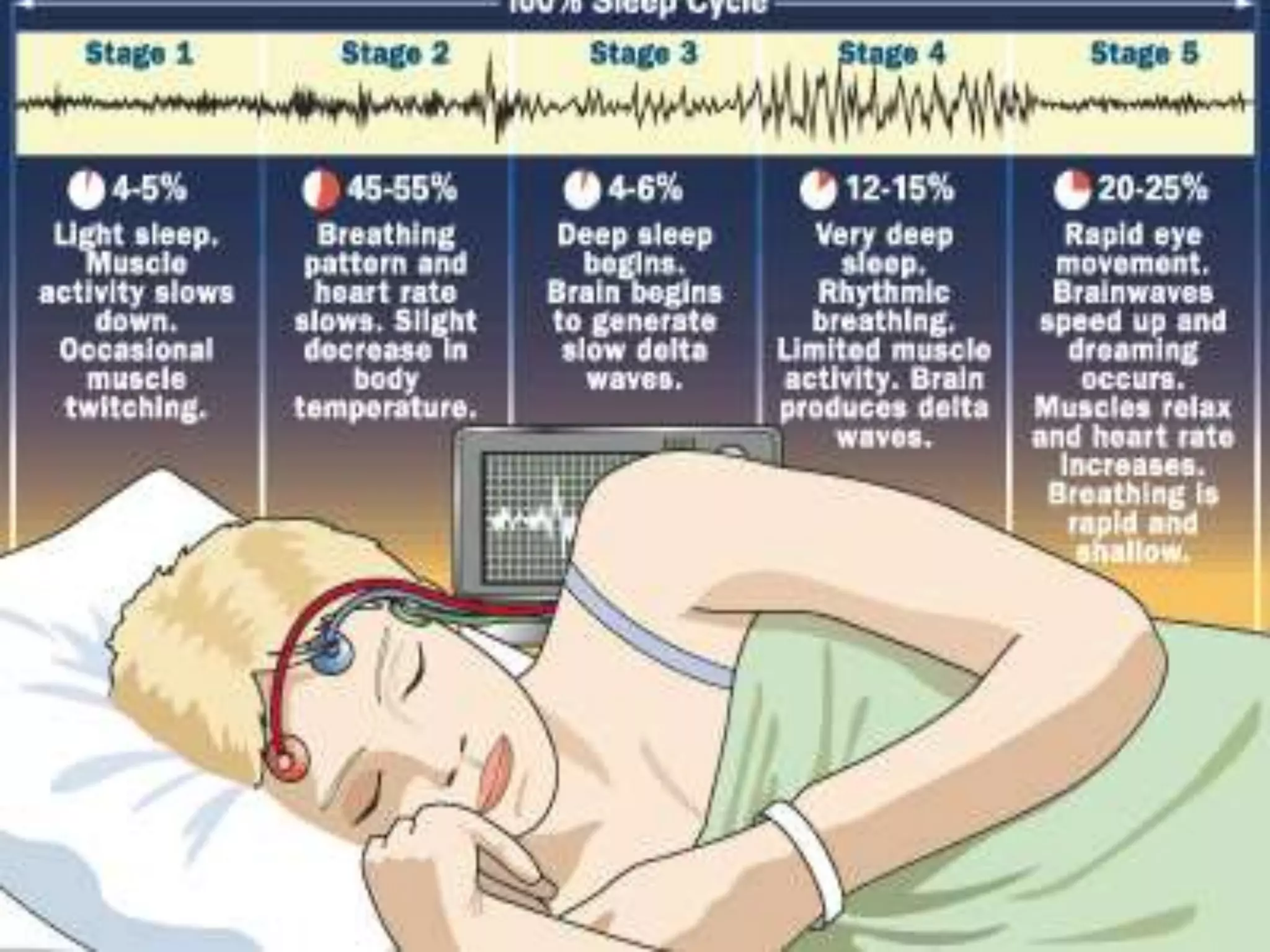
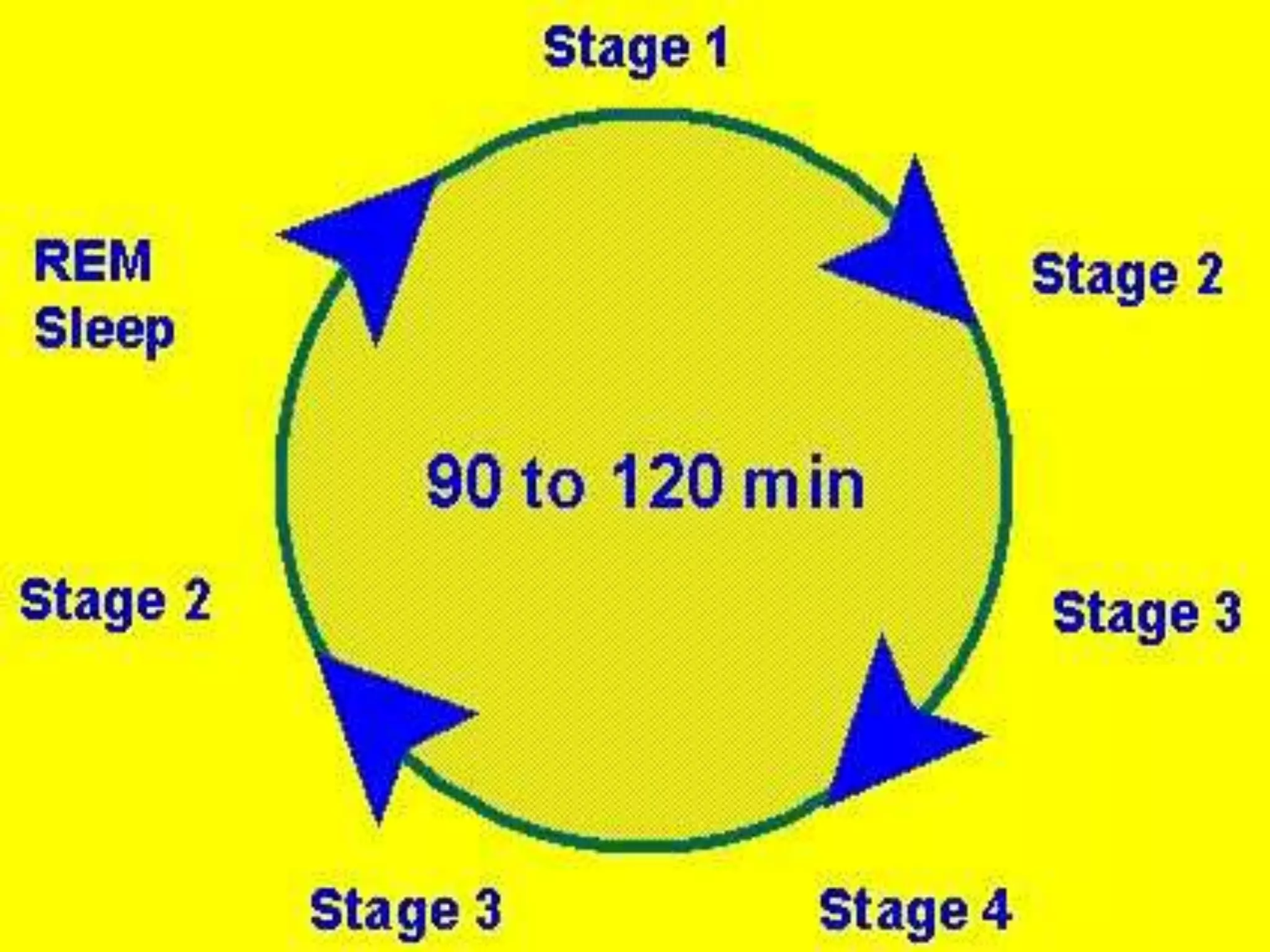
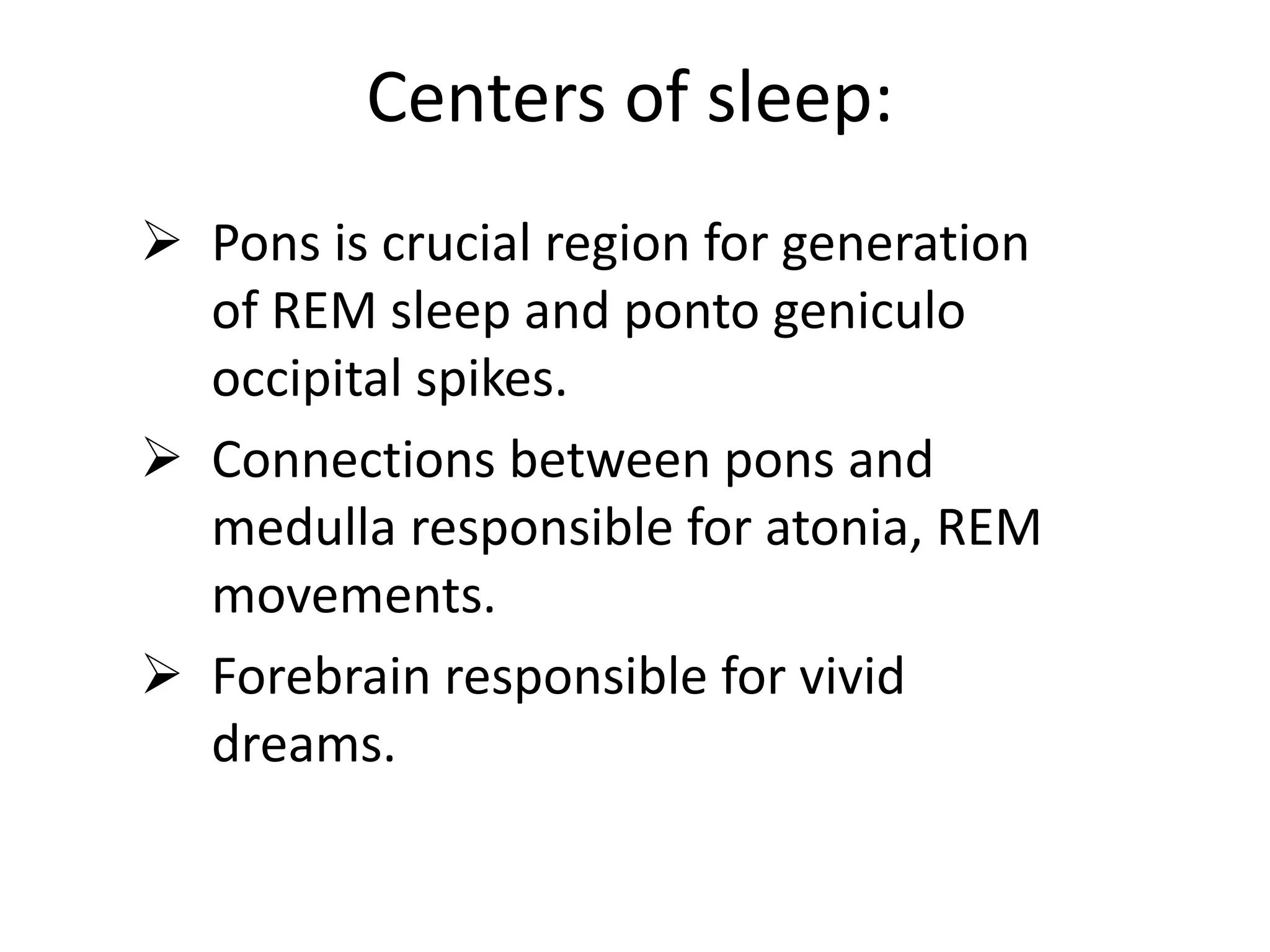
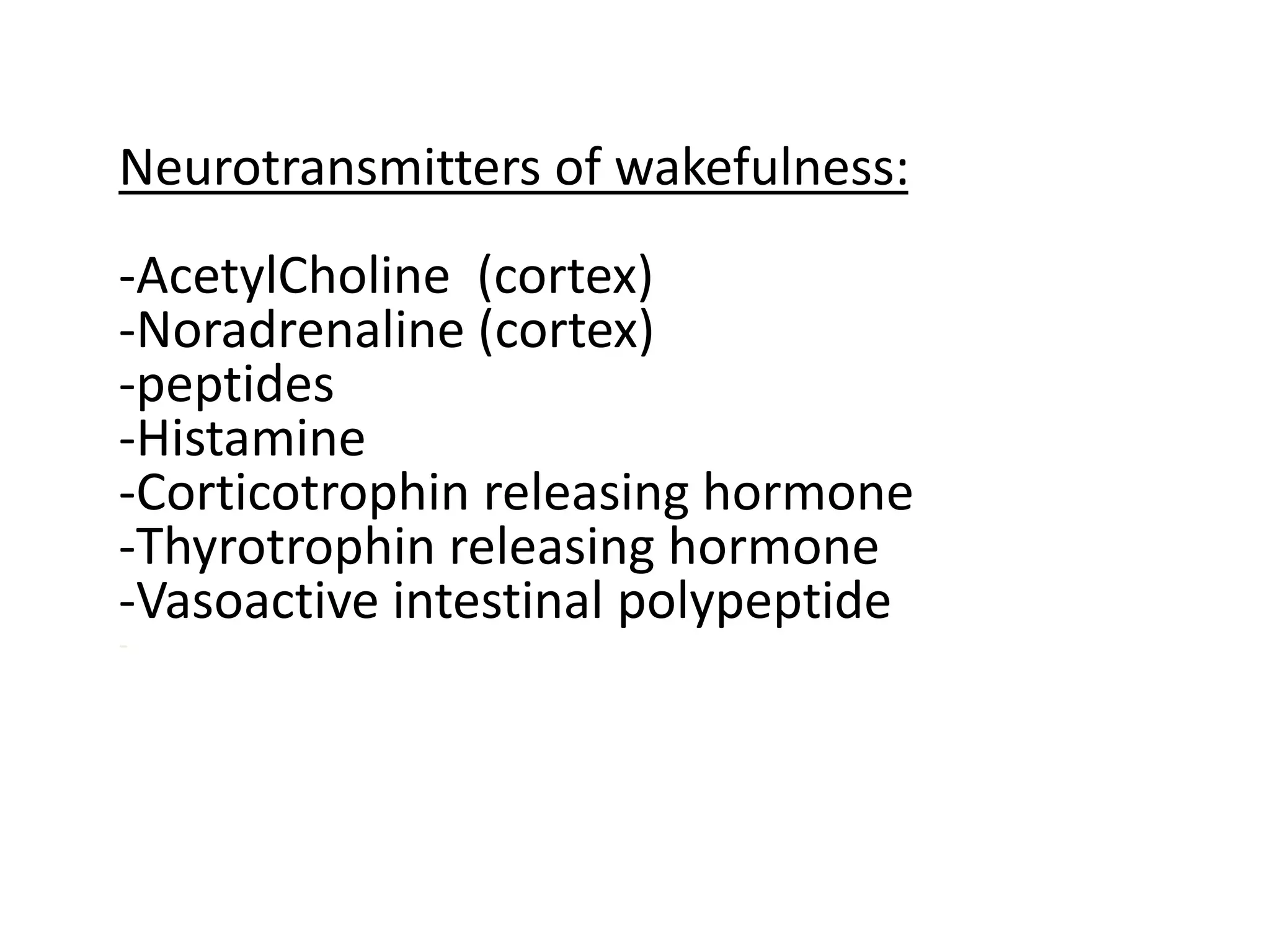
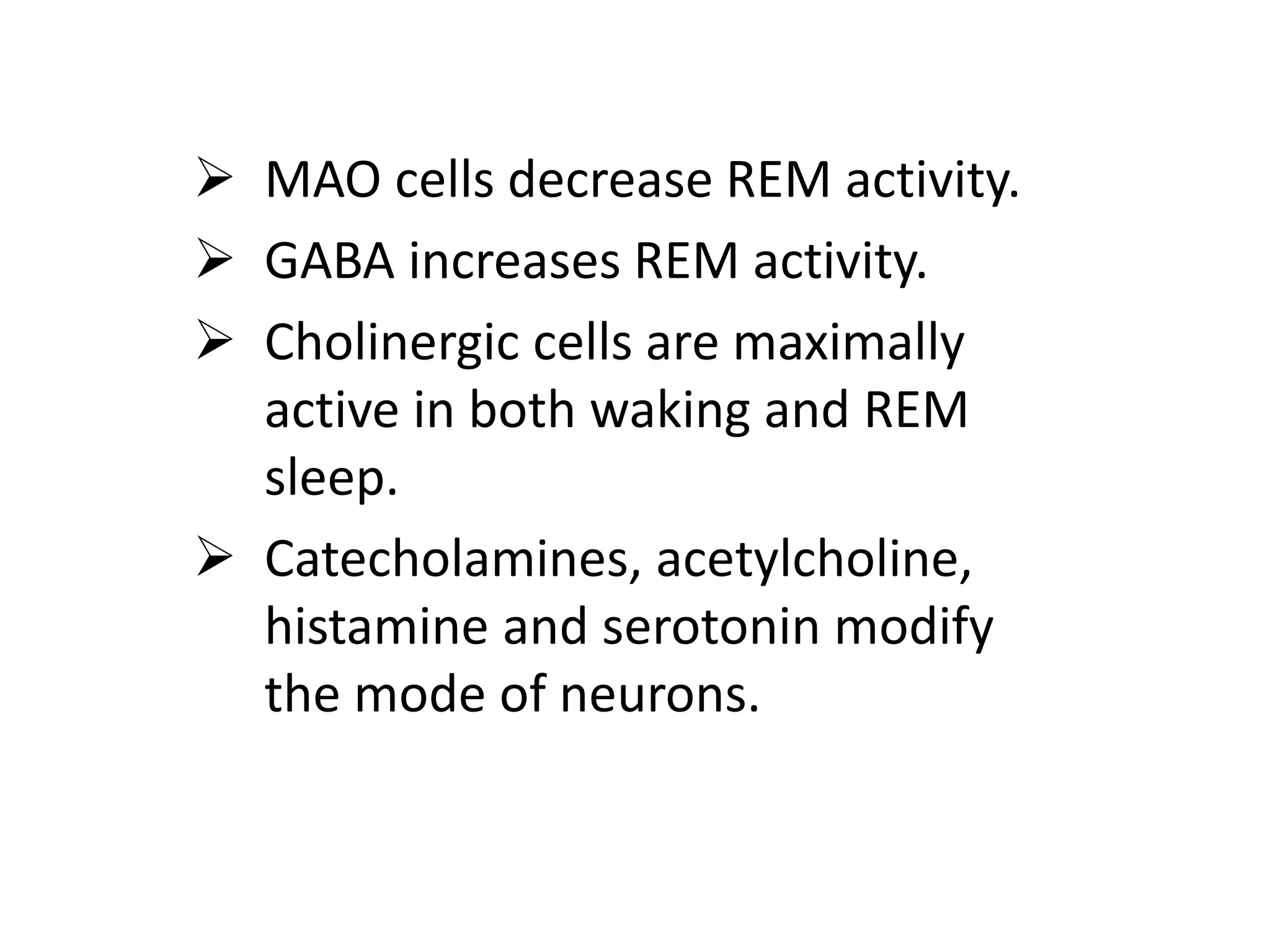
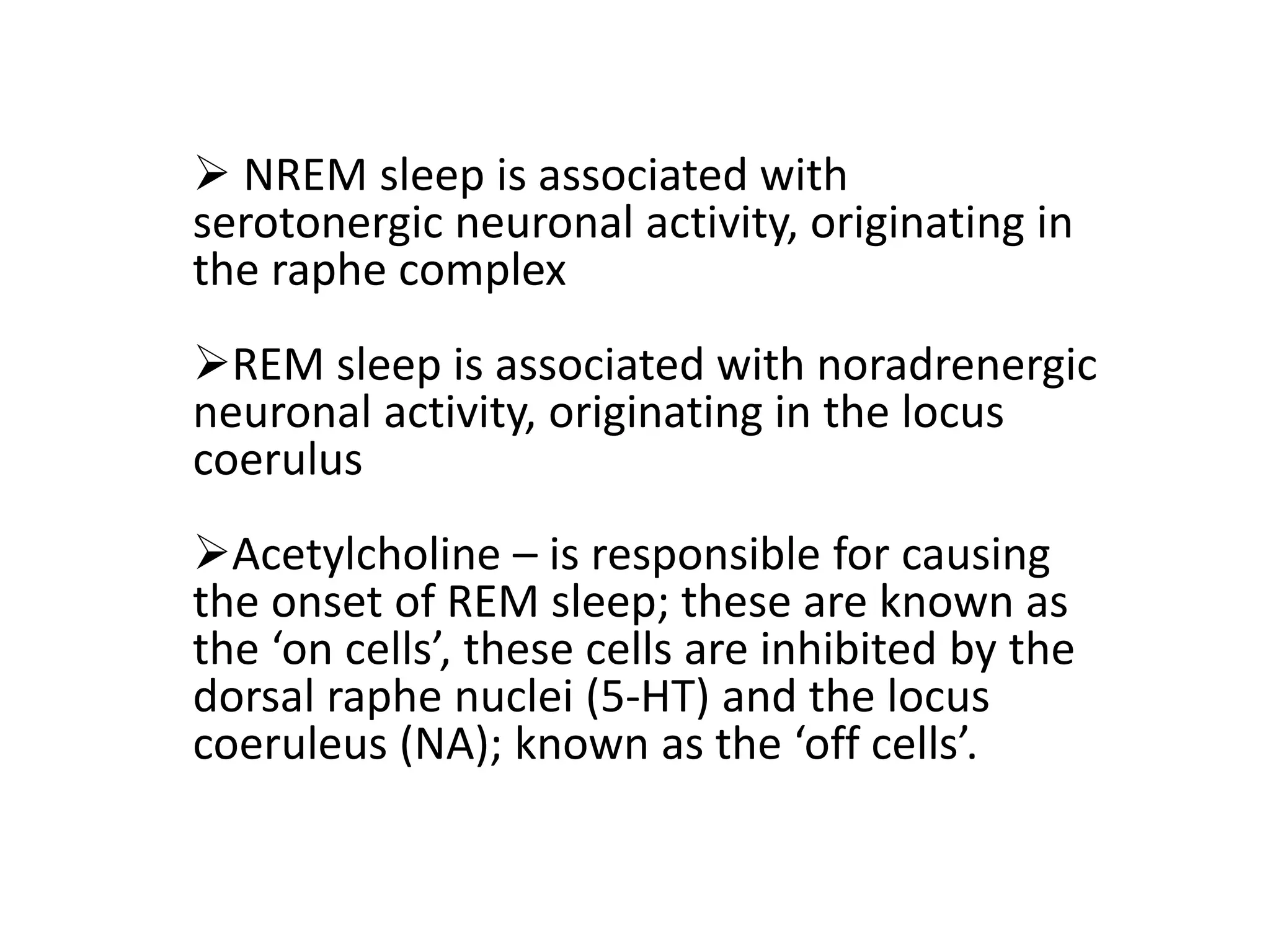
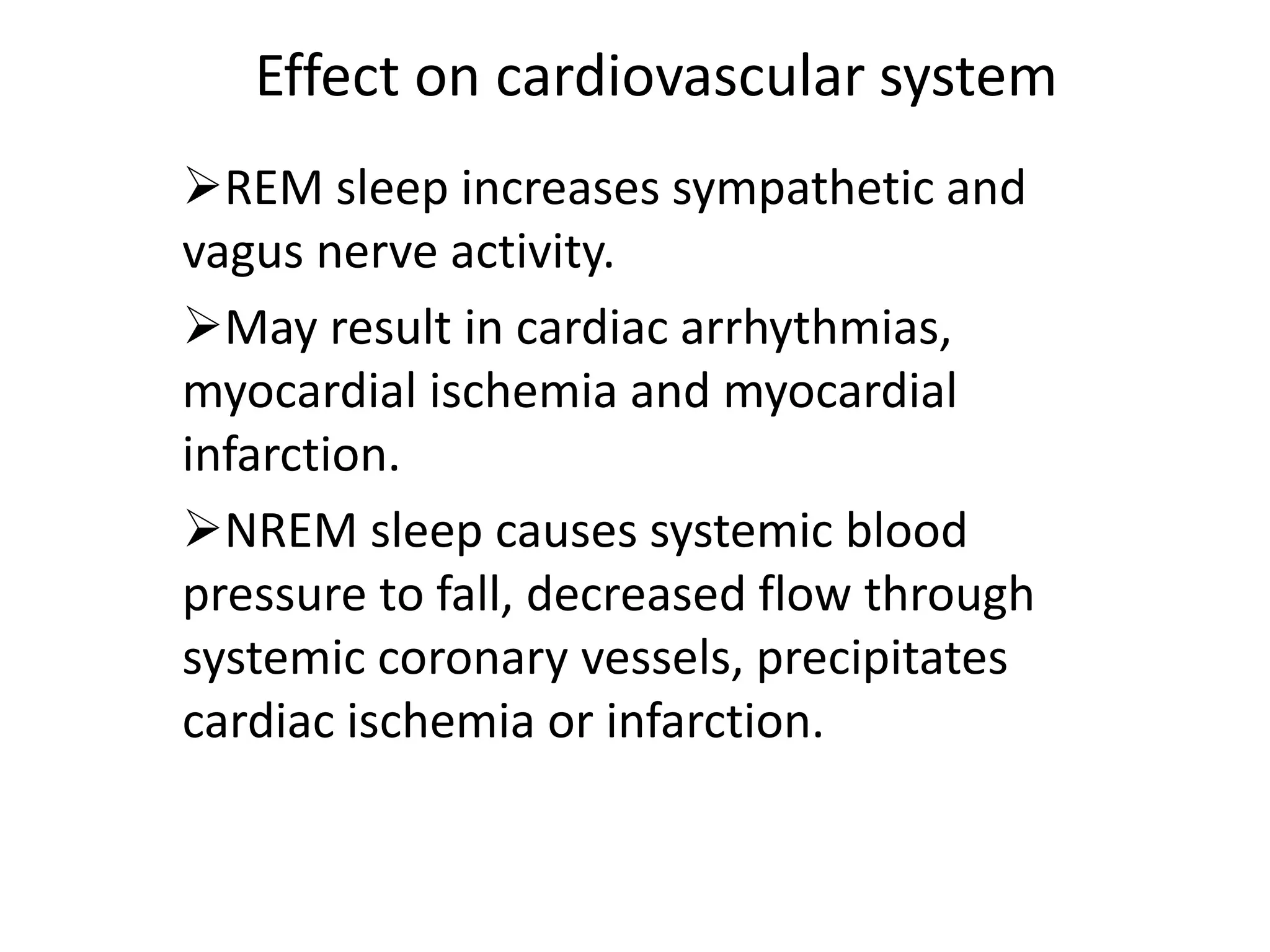
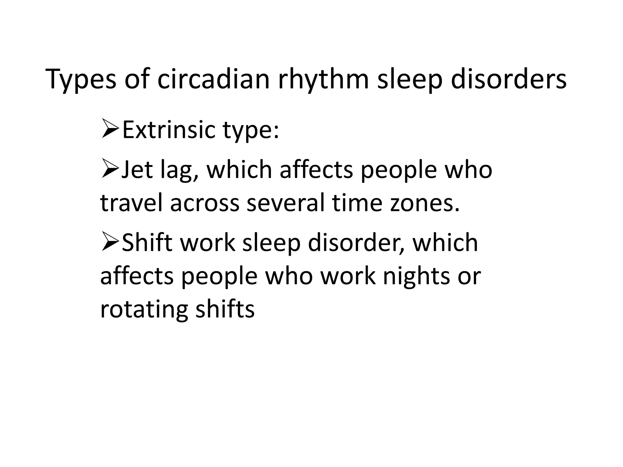
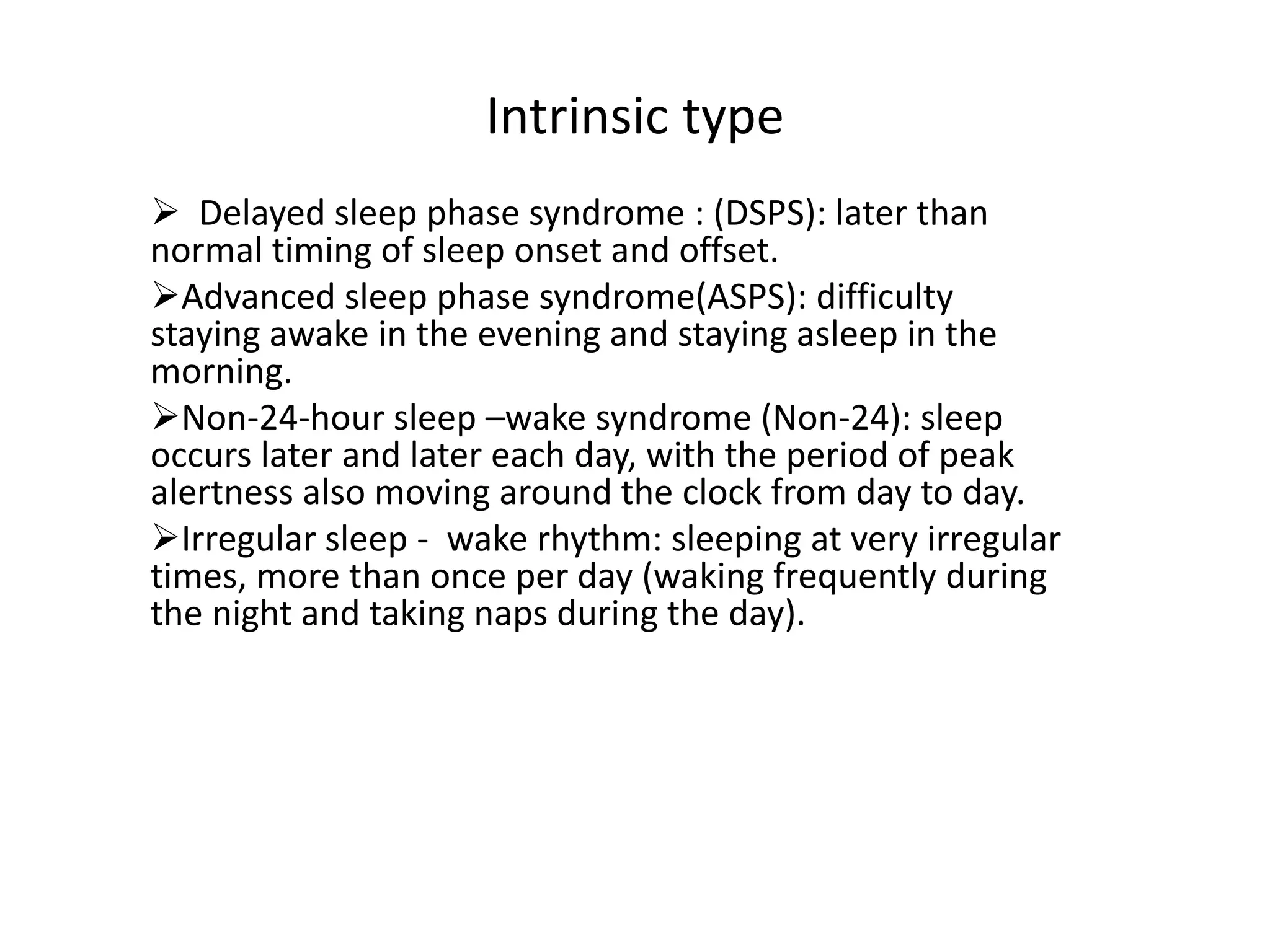
Neurology of sleep
Dr. Ajith,
Dr. Laxminadh.
Sleep: the intermediate state
between wakefulness and death,
being regarded as the active state of
the animal and intellectual functions,
state of decreased awareness of
environmental stimuli.
-Robert Macnish,
-1834.
-Distinguished by :-
 Rapid reversibility.
 Recognise that they feel sleepy.
 Aware that they have been
asleep.
TYPES OF SLEEP
1. (NREM )Non rapid eye movement,
Orthodox sleep – Quiet sleep
2. (REM)Rapid eye movement sleep.
Paradoxical sleep – Active sleep
NREM sleep:
 Synchronous cortical EEG.
 Low muscle tones.
 Minimal psychological activity.
 Reduced recall of dreaming if
woken.
 Increased parasympathetic activity.
 Upward ocular deviation with few
or no eye movements.
 Abolition of tendon reflexes.
 decreased heart rate
 decreased systolic blood pressure
 decreased respiratory rate
 decreased cerebral blood flow
 penis not usually erect
REM Sleep:
 Discovered by Ascerinsky and
Kleitman in 1953.
 Periodic recurrence of rapid eye
movement.
 EMG: Atonia
 Paradox sleep: EEG: resembles of
waking.
 Vivid dreams
 cortical activation,
 low-voltage desynchronization of
the EEG,
 rapid eye movements.
Physiological characters of REM sleep:
 Ponto geniculo occipital spikes on EEG.
 REM sleep has a parasympathetically mediated
tonic component with constriction of pupil.
 Sympathetically mediated phasic component.
It is characterized by skeletal muscle twitches,
increased heart rate and respiratory rate
variability, pupil dilatation, and increased
respiratory rate.
 Muscle atonia. It results from inhibition of
alpha motor neurons by clusters of peri–locus
ceruleus neurons.
NREM is maintained partly through
oscillations between the thalamus and
the cortex.
The 3 major oscillation systems are
1.sleep spindles,
2.delta oscillations, and
3.slow cortical oscillations.
Sleep spindles, a hallmark of stage N2
sleep, are generated by bursts of
hyperpolarizing GABAergic neurons in the
reticular nucleus of the thalamus
Generalizations:
 Sleep is entered through NREM sleep.
 NREM sleep and REM sleep alternate with a
period near 90 minutes.
 Sleep wake schedule predominates in the
first 1/3rd of the night and is linked to the
initiation of sleep.
 REM sleep predominates in the last 1/3rd of
the light and is linked to the circadian
rhythm of body temperature.
 Wakefulness was less than 5%.
 NREM sleep 80% and REM sleep 20%.
Stages of sleep
NREM sleep is further divided into
progressively deeper stages of sleep: stage
N1, stage N2, and stage N3 (deep or delta-
wave sleep).
REM sleep follows NREM sleep and occurs
4-5 times during a normal 8-hour sleep
period.
The NREM-REM cycles vary in length from
70-100 minutes initially to 90-120 minutes
later in the night.
Stage N1 is transition between wake and
sleep. It occurs upon falling asleep usually
accounts for 2-5% of total sleep time.
Stage N2 occurs throughout the sleep
period and represents 45-55% of total sleep
time.
Stage N3 (delta or slow wave sleep) occurs
mostly in the first third of the night and
constitutes 5-15% of total sleep time.
 REM represents 20-25% of total sleep time
and occurs in 4-5 episodes throughout the
night.
EEG EOG EMG
AWAKE LOW VOLTAGE
ALPHA WAVES 8-13 HZ
EYES CLOSED
EYE MOVEMENTS
EYE BLINKS
HIGH TONIC ACTIVITY
VOLUNTARY
MOVEMENTS
NREM N 1 LOW VOLTAGE
THETA WAVES 4-7 HZ
MAY BE PRESENT
SLOW TONIC ACTIVITY
REDUCED
N 2 LOW VOLTAGE MIXED
FREQUENCY WITH
SPINDLES AND K
COMPLEXES
NONE LOW TONIC ACTIVITY
N 3 HIGH AMPLITUDE
SLOW WAVES < 2 HZ
NONE LOW TONIC ACTIVITY
REM LOW VOLTAGE MIXED
FREQ.
SAW TOOTH WAVES
RAPID EYE
MOVEMENTS
ATONIA WITH PHASIC
TWITCHES
Centers of sleep:
 Pons is crucial region for generation
of REM sleep and ponto geniculo
occipital spikes.
 Connections between pons and
medulla responsible for atonia, REM
movements.
 Forebrain responsible for vivid
dreams.
In the pons regions ventral to locus
coeruleus called the nucleus reticularis
pontis oralis or subcoeruleus region
responsible for REM sleep activity.
NREM sleep originating in the raphe
complex.
Neurons mainly involved in
maintaining activation are
concentrated in the oral pontine and
mid brain central tegmentum and
posterior hypothalamus.
Neurons sleep promoting are:
dorsolateral medullary reticular
formation, anterior hypothalamus-
pre optic area.
Neuroanatomy of sleep
Wakefulness :
-Reticular formation
-Thalamic Nuclei (intra laminar & midline)
-Subthalamus
-Hypothalamus
SWS(SLEEP WAKE SCHEDULE) :
-Raphe Nuclei of Brain stem (sleep promotion)
-Thalamic Nuclei (non specific)
- Hypothalamus
Neurotransmitters of wakefulness:
-AcetylCholine (cortex)
-Noradrenaline (cortex)
-peptides
-Histamine
-Corticotrophin releasing hormone
-Thyrotrophin releasing hormone
-Vasoactive intestinal polypeptide
-
Neurotransmitters of SWS:
- GABA
- 5HT
- Alpha-melanocyte secreting hormone
- Somatostatin
 MAO cells decrease REM activity.
 GABA increases REM activity.
 Cholinergic cells are maximally
active in both waking and REM
sleep.
 Catecholamines, acetylcholine,
histamine and serotonin modify
the mode of neurons.
 NREM sleep is associated with
serotonergic neuronal activity, originating in
the raphe complex
REM sleep is associated with noradrenergic
neuronal activity, originating in the locus
coerulus
Acetylcholine – is responsible for causing
the onset of REM sleep; these are known as
the ‘on cells’, these cells are inhibited by the
dorsal raphe nuclei (5-HT) and the locus
coeruleus (NA); known as the ‘off cells’.
Loss of natural sleep: lesions in
lower brain stem, pre opticarea, in
the fore brain.
Loss of wakefulness: lesions in oral
pontine, midbrain tegmentum,
lesions in posterior hypothalamus
and lesions in the basal forebrain.
Effect on cardiovascular system
REM sleep increases sympathetic and
vagus nerve activity.
May result in cardiac arrhythmias,
myocardial ischemia and myocardial
infarction.
NREM sleep causes systemic blood
pressure to fall, decreased flow through
systemic coronary vessels, precipitates
cardiac ischemia or infarction.
Effect on respiratory system
NREM allows occlusive collapse of
the extra thoracic airway in patient
with obstructive sleep apnea.
REM sleep causes rapid, irregular
breathing and hypoventilation.
Atonia of intercostals and accessory
respiratory muscles.
REM sleep causes decreased
ventilatory responses to hypoxia,
hypercapnia and respiratory resistance
during sleep.
Functions of sleep:
1. Consolidation of memory,
2. Stimulation of brain,
3. decreased metabolic demand and
facilitates replenishment of glycogen
stores.
4. REM sleep causes periodic activation
of brain.
Effects of Sleep Deprivation
Glucose-PET studies in individuals deprived of sleep
have shown that after 24 hours of sustained
wakefulness, the metabolic activity of the brain
decreases significantly (up to 6% for the whole brain
and up to 11% for specific cortical and basal
ganglionic areas).
1. decrease in core body temperature,
2. decrease in immune system function ,
3. decrease in the release of growth hormone,
4. cause of increased heart rate variability,
5. Impairment of cognitive work requiring
simultaneous focus on several tasks.
Circadian rhythm:
Primary circadian clock in mammals is
located in the suprachiasmatic nucleus
in the hypothalamus.
Destruction of the suprachiasmatic
nucleus results in the complete absence
of a regular sleep – wake rhythm.
a
Pathway of circadian rhythm
Retina of eye – ganglion cells (photosensitive cells)
– melanopsin – retino hypothalamic tracts –
suprachiasmatic nucleus – pineal gland -
melatonin
PHASE MARKERS FOR MEASURING THE TIMING OF A
CIRCADIAN RHYTHM:
Body temperature cycles are also under hypothalamic control.
An increase in body temperature is seen during the course of the
day and a decrease is observed during the night. The
temperature peaks and troughs are thought to mirror the sleep
rhythm.
Melatonin has been implicated as a modulator of light
entrainment. It is secreted maximally during the night by the
pineal gland.
Prolactin, testosterone, and growth hormone also demonstrate
circadian rhythms, with maximal secretion during the night.
Precise estimation of the periods of the
endogenous circadian rhythms of
melatonin, core body temperature, and
cortisol in healthy young and older
individuals has revealed that the intrinsic
period of the human circadian pacemaker
averages 24.18 hours in both age groups.
Types of circadian rhythm sleep disorders
Extrinsic type:
Jet lag, which affects people who
travel across several time zones.
Shift work sleep disorder, which
affects people who work nights or
rotating shifts
Intrinsic type
 Delayed sleep phase syndrome : (DSPS): later than
normal timing of sleep onset and offset.
Advanced sleep phase syndrome(ASPS): difficulty
staying awake in the evening and staying asleep in the
morning.
Non-24-hour sleep –wake syndrome (Non-24): sleep
occurs later and later each day, with the period of peak
alertness also moving around the clock from day to day.
Irregular sleep - wake rhythm: sleeping at very irregular
times, more than once per day (waking frequently during
the night and taking naps during the day).
Diagnostic tools
 Sleep log: This diary allows identification
of sleep-wake cycles in the patient's normal
environment and allows subjective
assessment of alertness over a 2-week time
period.
Actigraphy: An Actigraph is a small motion-
sensing device worn on the nondominant
wrist, generally for 1 week. It is based on the
principle that wrist motion is decreased during
sleep. This allows a very gross measure of
sleep-wake cycles over time.
Overnight polysomnography: This study is
of value in identifying the patient with
obstructive sleep apnea(OSA), periodic limb
movements of sleep (PLMS).
Multiple sleep latency test (MSLT): This
test allows for objective measurement of
sleepiness. Sleep latencies of less than 5
minutes indicate abnormal sleepiness.
Morningness eveningness questionnaire
(MEQ): It is used for evaluation of circadian
rhythm disturbances.
Treatment options
Behavioural and light therapy are the mainstays of
treatment of circadian rhythm disturbances.
Timed bright light therapy: bright room light over
time may be sufficient; however, a higher intensity of
light (>6000 lux over 30-60 min) is often necessary to
accomplish acute phase shifts.
Light therapy applied in the early evening and night
time delays the cycle in patients with advanced sleep-
phase syndrome [ASPS]), early morning stimulates
morning alertness and an earlier bedtime in patients
with delayed sleep-phase syndrome [DSPS].
Chronotherapy: This behavioural treatment consists
of a gradual shift in sleep time in accordance with the
patient's tendency.
Melatonin: Used to enhance natural
sleep process and for resetting body's
internal time clock when travelling
through different time zones and in
circadian rhythm sleep disorders in blind
people with no light perception.
Non benzodiazepine hypnotics: useful
for insomnia characterized by difficulties
with sleep onset and/or sleep
maintenance.
Narcolepsy:
Disease of the REM sleep,
Features: cataplexy, high amplitude of
activity in the hippocampus.
Medullary inhibition and noradrenergic
disfacilitation are linked to loss of muscle
tone. Histamine activity is responsible for
consciousness.
Loss of hypothalamic cells containing the
peptide hypo cretin (orexin) is the probable
cause.

SLEEP1.ppt

  • 1.
    Neurology of sleep Dr.Ajith, Dr. Laxminadh.
  • 2.
    Sleep: the intermediatestate between wakefulness and death, being regarded as the active state of the animal and intellectual functions, state of decreased awareness of environmental stimuli. -Robert Macnish, -1834.
  • 3.
    -Distinguished by :- Rapid reversibility.  Recognise that they feel sleepy.  Aware that they have been asleep.
  • 4.
    TYPES OF SLEEP 1.(NREM )Non rapid eye movement, Orthodox sleep – Quiet sleep 2. (REM)Rapid eye movement sleep. Paradoxical sleep – Active sleep
  • 5.
    NREM sleep:  Synchronouscortical EEG.  Low muscle tones.  Minimal psychological activity.  Reduced recall of dreaming if woken.  Increased parasympathetic activity.  Upward ocular deviation with few or no eye movements.  Abolition of tendon reflexes.
  • 6.
     decreased heartrate  decreased systolic blood pressure  decreased respiratory rate  decreased cerebral blood flow  penis not usually erect
  • 7.
    REM Sleep:  Discoveredby Ascerinsky and Kleitman in 1953.  Periodic recurrence of rapid eye movement.  EMG: Atonia  Paradox sleep: EEG: resembles of waking.  Vivid dreams
  • 8.
     cortical activation, low-voltage desynchronization of the EEG,  rapid eye movements.
  • 9.
    Physiological characters ofREM sleep:  Ponto geniculo occipital spikes on EEG.  REM sleep has a parasympathetically mediated tonic component with constriction of pupil.  Sympathetically mediated phasic component. It is characterized by skeletal muscle twitches, increased heart rate and respiratory rate variability, pupil dilatation, and increased respiratory rate.  Muscle atonia. It results from inhibition of alpha motor neurons by clusters of peri–locus ceruleus neurons.
  • 10.
    NREM is maintainedpartly through oscillations between the thalamus and the cortex. The 3 major oscillation systems are 1.sleep spindles, 2.delta oscillations, and 3.slow cortical oscillations. Sleep spindles, a hallmark of stage N2 sleep, are generated by bursts of hyperpolarizing GABAergic neurons in the reticular nucleus of the thalamus
  • 11.
    Generalizations:  Sleep isentered through NREM sleep.  NREM sleep and REM sleep alternate with a period near 90 minutes.  Sleep wake schedule predominates in the first 1/3rd of the night and is linked to the initiation of sleep.  REM sleep predominates in the last 1/3rd of the light and is linked to the circadian rhythm of body temperature.  Wakefulness was less than 5%.  NREM sleep 80% and REM sleep 20%.
  • 12.
    Stages of sleep NREMsleep is further divided into progressively deeper stages of sleep: stage N1, stage N2, and stage N3 (deep or delta- wave sleep). REM sleep follows NREM sleep and occurs 4-5 times during a normal 8-hour sleep period. The NREM-REM cycles vary in length from 70-100 minutes initially to 90-120 minutes later in the night.
  • 13.
    Stage N1 istransition between wake and sleep. It occurs upon falling asleep usually accounts for 2-5% of total sleep time. Stage N2 occurs throughout the sleep period and represents 45-55% of total sleep time. Stage N3 (delta or slow wave sleep) occurs mostly in the first third of the night and constitutes 5-15% of total sleep time.  REM represents 20-25% of total sleep time and occurs in 4-5 episodes throughout the night.
  • 16.
    EEG EOG EMG AWAKELOW VOLTAGE ALPHA WAVES 8-13 HZ EYES CLOSED EYE MOVEMENTS EYE BLINKS HIGH TONIC ACTIVITY VOLUNTARY MOVEMENTS NREM N 1 LOW VOLTAGE THETA WAVES 4-7 HZ MAY BE PRESENT SLOW TONIC ACTIVITY REDUCED N 2 LOW VOLTAGE MIXED FREQUENCY WITH SPINDLES AND K COMPLEXES NONE LOW TONIC ACTIVITY N 3 HIGH AMPLITUDE SLOW WAVES < 2 HZ NONE LOW TONIC ACTIVITY REM LOW VOLTAGE MIXED FREQ. SAW TOOTH WAVES RAPID EYE MOVEMENTS ATONIA WITH PHASIC TWITCHES
  • 17.
    Centers of sleep: Pons is crucial region for generation of REM sleep and ponto geniculo occipital spikes.  Connections between pons and medulla responsible for atonia, REM movements.  Forebrain responsible for vivid dreams.
  • 18.
    In the ponsregions ventral to locus coeruleus called the nucleus reticularis pontis oralis or subcoeruleus region responsible for REM sleep activity. NREM sleep originating in the raphe complex.
  • 19.
    Neurons mainly involvedin maintaining activation are concentrated in the oral pontine and mid brain central tegmentum and posterior hypothalamus. Neurons sleep promoting are: dorsolateral medullary reticular formation, anterior hypothalamus- pre optic area.
  • 20.
    Neuroanatomy of sleep Wakefulness: -Reticular formation -Thalamic Nuclei (intra laminar & midline) -Subthalamus -Hypothalamus SWS(SLEEP WAKE SCHEDULE) : -Raphe Nuclei of Brain stem (sleep promotion) -Thalamic Nuclei (non specific) - Hypothalamus
  • 21.
    Neurotransmitters of wakefulness: -AcetylCholine(cortex) -Noradrenaline (cortex) -peptides -Histamine -Corticotrophin releasing hormone -Thyrotrophin releasing hormone -Vasoactive intestinal polypeptide -
  • 22.
    Neurotransmitters of SWS: -GABA - 5HT - Alpha-melanocyte secreting hormone - Somatostatin
  • 23.
     MAO cellsdecrease REM activity.  GABA increases REM activity.  Cholinergic cells are maximally active in both waking and REM sleep.  Catecholamines, acetylcholine, histamine and serotonin modify the mode of neurons.
  • 24.
     NREM sleepis associated with serotonergic neuronal activity, originating in the raphe complex REM sleep is associated with noradrenergic neuronal activity, originating in the locus coerulus Acetylcholine – is responsible for causing the onset of REM sleep; these are known as the ‘on cells’, these cells are inhibited by the dorsal raphe nuclei (5-HT) and the locus coeruleus (NA); known as the ‘off cells’.
  • 25.
    Loss of naturalsleep: lesions in lower brain stem, pre opticarea, in the fore brain. Loss of wakefulness: lesions in oral pontine, midbrain tegmentum, lesions in posterior hypothalamus and lesions in the basal forebrain.
  • 26.
    Effect on cardiovascularsystem REM sleep increases sympathetic and vagus nerve activity. May result in cardiac arrhythmias, myocardial ischemia and myocardial infarction. NREM sleep causes systemic blood pressure to fall, decreased flow through systemic coronary vessels, precipitates cardiac ischemia or infarction.
  • 27.
    Effect on respiratorysystem NREM allows occlusive collapse of the extra thoracic airway in patient with obstructive sleep apnea. REM sleep causes rapid, irregular breathing and hypoventilation. Atonia of intercostals and accessory respiratory muscles.
  • 28.
    REM sleep causesdecreased ventilatory responses to hypoxia, hypercapnia and respiratory resistance during sleep.
  • 29.
    Functions of sleep: 1.Consolidation of memory, 2. Stimulation of brain, 3. decreased metabolic demand and facilitates replenishment of glycogen stores. 4. REM sleep causes periodic activation of brain.
  • 30.
    Effects of SleepDeprivation Glucose-PET studies in individuals deprived of sleep have shown that after 24 hours of sustained wakefulness, the metabolic activity of the brain decreases significantly (up to 6% for the whole brain and up to 11% for specific cortical and basal ganglionic areas). 1. decrease in core body temperature, 2. decrease in immune system function , 3. decrease in the release of growth hormone, 4. cause of increased heart rate variability, 5. Impairment of cognitive work requiring simultaneous focus on several tasks.
  • 31.
    Circadian rhythm: Primary circadianclock in mammals is located in the suprachiasmatic nucleus in the hypothalamus. Destruction of the suprachiasmatic nucleus results in the complete absence of a regular sleep – wake rhythm.
  • 32.
  • 33.
    Retina of eye– ganglion cells (photosensitive cells) – melanopsin – retino hypothalamic tracts – suprachiasmatic nucleus – pineal gland - melatonin
  • 34.
    PHASE MARKERS FORMEASURING THE TIMING OF A CIRCADIAN RHYTHM: Body temperature cycles are also under hypothalamic control. An increase in body temperature is seen during the course of the day and a decrease is observed during the night. The temperature peaks and troughs are thought to mirror the sleep rhythm. Melatonin has been implicated as a modulator of light entrainment. It is secreted maximally during the night by the pineal gland. Prolactin, testosterone, and growth hormone also demonstrate circadian rhythms, with maximal secretion during the night.
  • 36.
    Precise estimation ofthe periods of the endogenous circadian rhythms of melatonin, core body temperature, and cortisol in healthy young and older individuals has revealed that the intrinsic period of the human circadian pacemaker averages 24.18 hours in both age groups.
  • 38.
    Types of circadianrhythm sleep disorders Extrinsic type: Jet lag, which affects people who travel across several time zones. Shift work sleep disorder, which affects people who work nights or rotating shifts
  • 39.
    Intrinsic type  Delayedsleep phase syndrome : (DSPS): later than normal timing of sleep onset and offset. Advanced sleep phase syndrome(ASPS): difficulty staying awake in the evening and staying asleep in the morning. Non-24-hour sleep –wake syndrome (Non-24): sleep occurs later and later each day, with the period of peak alertness also moving around the clock from day to day. Irregular sleep - wake rhythm: sleeping at very irregular times, more than once per day (waking frequently during the night and taking naps during the day).
  • 40.
    Diagnostic tools  Sleeplog: This diary allows identification of sleep-wake cycles in the patient's normal environment and allows subjective assessment of alertness over a 2-week time period. Actigraphy: An Actigraph is a small motion- sensing device worn on the nondominant wrist, generally for 1 week. It is based on the principle that wrist motion is decreased during sleep. This allows a very gross measure of sleep-wake cycles over time.
  • 41.
    Overnight polysomnography: Thisstudy is of value in identifying the patient with obstructive sleep apnea(OSA), periodic limb movements of sleep (PLMS). Multiple sleep latency test (MSLT): This test allows for objective measurement of sleepiness. Sleep latencies of less than 5 minutes indicate abnormal sleepiness. Morningness eveningness questionnaire (MEQ): It is used for evaluation of circadian rhythm disturbances.
  • 42.
    Treatment options Behavioural andlight therapy are the mainstays of treatment of circadian rhythm disturbances. Timed bright light therapy: bright room light over time may be sufficient; however, a higher intensity of light (>6000 lux over 30-60 min) is often necessary to accomplish acute phase shifts. Light therapy applied in the early evening and night time delays the cycle in patients with advanced sleep- phase syndrome [ASPS]), early morning stimulates morning alertness and an earlier bedtime in patients with delayed sleep-phase syndrome [DSPS]. Chronotherapy: This behavioural treatment consists of a gradual shift in sleep time in accordance with the patient's tendency.
  • 43.
    Melatonin: Used toenhance natural sleep process and for resetting body's internal time clock when travelling through different time zones and in circadian rhythm sleep disorders in blind people with no light perception. Non benzodiazepine hypnotics: useful for insomnia characterized by difficulties with sleep onset and/or sleep maintenance.
  • 44.
    Narcolepsy: Disease of theREM sleep, Features: cataplexy, high amplitude of activity in the hippocampus. Medullary inhibition and noradrenergic disfacilitation are linked to loss of muscle tone. Histamine activity is responsible for consciousness. Loss of hypothalamic cells containing the peptide hypo cretin (orexin) is the probable cause.