

Recommended
Recommended
More Related Content
Similar to chronic pathological changes
Similar to chronic pathological changes (20)
More from Mohamed Alashram
More from Mohamed Alashram (20)
Recently uploaded
Recently uploaded (20)
chronic pathological changes
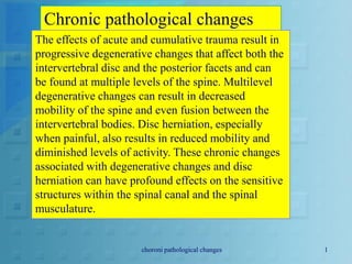
- 1. Chronic pathological changes The effects of acute and cumulative trauma result in progressive degenerative changes that affect both the intervertebral disc and the posterior facets and can be found at multiple levels of the spine. Multilevel degenerative changes can result in decreased mobility of the spine and even fusion between the intervertebral bodies. Disc herniation, especially when painful, also results in reduced mobility and diminished levels of activity. These chronic changes associated with degenerative changes and disc herniation can have profound effects on the sensitive structures within the spinal canal and the spinal musculature. choroni pathological changes 1
- 2. SPINAL STENOSIS The expansion of the facet joints as a result of degenerative changes can encroach on the central canal and the lateral foramina. This encroachment can choroni pathological changes 2
- 3. This transverse section of L5 shows marked stenosis of both the central canal and the lateral recesses due to osteophytic growth of the posterior facets and the vertebral endplates. choroni pathological changes 3
- 4. T2 weighted MR sagittal image of the lumbar spine (a), demonstrating high-grade spinal stenosis at L2– L3, L3–L4 and L4–L5. The spinal fluid has a bright signal intensity and the compression of the intrathecal rootlets is apparent. On the axial T2 MR image (b), the central canal stenosis is caused by thickening of the posterior neural arch and ligamentum flavum, and overgrowth of the posterior facet joints. This causes significant flattening of the normally ovoid- appearing thecal sac a b choroni pathological changes 4
- 5. Longitudinal section through the lumbar spine shows marked degeneration and fusion of the bodies of L4–L5 and L5–S1. There is stenosis or narrowing of the central canal at both levels due to osteophytes protruding into the canal at the level of the disc. choroni pathological changes 5
- 6. This CT transverse section through the lumbar spine shows marked central canal stenosis. The posterior muscle has been partially replaced by fibrofatty tissue. choroni pathological changes 6
- 7. These images are from the same patient. Anteroposterior (a) and lateral (b) views of the lumbar spine following a myelogram, demonstrating a complete block of the contrast at the L2–L3 level choroni pathological changes 7
- 8. become quite marked, especially in the presence of large osteophytes from the vertebral bodies, and can result in significant stenosis of the central canal and lateral foramina. These changes can be visualized on MRI and CT scanning, and, when severe, can disrupt function within the spinal cord and nerve roots. Such disruption can be intermittent and associated with pain or numbness in the legs on activity and which is relieved with rest, known as neurogenic claudication, or it can become permanent, leading to neurologic deficits as a result of encroachment on the spinal cord or cauda equina choroni pathological changes 8
- 9. The degree of spinal stenosis can be measured on CT and MRI imaging. Hypertrophy of the posterior facets encroaching on the neuroforamen is also evident in this type of study. The effect of compression on the spinal cord, cauda equina and/or nerve roots is determined by electrodiagnostic studies. choroni pathological changes 9
- 10. MUSCLE TRAUMA, IMMOBILIZATION AND ATROPHY As degenerative changes progress in the spine or following disc herniation, the mobility of the spine is greatly reduced and patient activity is limited as a result of pain. This immobilization has profound effects on paraspinal muscles. Within 3–4 weeks, atrophy of the muscle fibers can be seen on microscopy. choroni pathological changes 10
- 11. The cells become smaller, the number of nuclei decreases and the spaces between muscle fibers increase in size. Within 7 weeks, the spaces between muscle fibers become large and filled with fibrous collagen and the degeneration of muscle fibers becomes prominent. During exercise and remobilization of the spine, regeneration can be seen in the muscle fibers. Prominent myoblast chains are formed centrally in the empty sheath of damaged choroni pathological changes 11
- 12. These images are from the same patient. Intrathecally enhanced axial computed tomogram reveals central canal stenosis secondary to posterior facet joint hypertrophy and vertebral body osteophyte formation and disc bulging (c). Sagittal proton density MR image (d) demonstrates multiple level spondylotic changes and central canal stenosis at L2–L3 and L3– L4. Axial MR image (e) reveals central canal stenosis c e d choroni pathological changes 12
- 13. Somatosensory evoked responses from the posterior tibial and pudendal nerves are blocked as they travel through the spinal cord. The reflex studies from the bulbocavernosus and urethra to the rectal sphincter are intact below the level of the injury. Cystometrogram shows hyperreflexia. The spinal cord injury could be due to fracture, severe central stenosis or tumor encroaching on the neural canal choroni pathological changes 13
- 14. Axial CT image shows a large left- sided disc protrusion (arrow) at the L5–S1 level. The posterior muscle is replaced by fibrofatty tissue due to prolonged inactivity choroni pathological changes 14
- 15. CT axial image of the L5–S1 segment in a patient with a large disc protrusion leading to prolonged inactivity. This has resulted in atrophy and replacement of the posterior musculature with fibrofatty tissue choroni pathological changes 15
- 16. Light microscopy of muscle tissue after 4 weeks of immobilization. The muscle fibers are much smaller than usual and there are a number of empty muscle sheaths. There are empty spaces between muscle fibers and few nuclei in the remaining muscles choroni pathological changes 16
- 17. Light microscopy of muscle fibers following 7 weeks of immobilization. Note the larger spaces between muscle fibers, sparse nuclei and empty muscle sheaths choroni pathological changes 17
- 18. Light microscopy of muscle fibers (human) showing regeneration. There is extensive replacement of muscle fibers with fibrous tissue. There are multiple thin myoblastic chains and muscle fibers with prominent central myoblastic nuclei choroni pathological changes 18
- 19. Light microscopy of muscle fibers (monkey) showing regeneration after direct trauma. The transverse band of myoblast nuclei is noted to be central in a new muscle fibe choroni pathological changes 19
- 20. Light microscopy of muscle (cat) showing muscle regeneration. There is a central band of myoblast nuclei, each with two small dark nucleoli. choroni pathological changes 20
- 21. Light microscopy of muscle (monkey) 3 months after injury. The upper field shows new muscle fibers (red). The lower field shows primarily collagen (yellow) with a few muscle fibers (red) choroni pathological changes 21
- 22. Electron microscopy of muscle (cat) after 3 months of immobilization; the arrow in the upper field points to a completely empty sarcolemmal sheath. In the lower field, there are almost normal muscle fibers with visible mitochondria choroni pathological changes 22
- 23. Electron microscopy of muscle (cat) 2 months after immobilization. There is degeneration of muscle with a few transverse Z-lines in a sea of debris choroni pathological changes 23