

Recommended
More Related Content
Similar to spine degeneration spine degeneration
Similar to spine degeneration spine degeneration (20)
More from Mohamed Alashram
More from Mohamed Alashram (20)
Recently uploaded
Recently uploaded (20)
spine degeneration spine degeneration
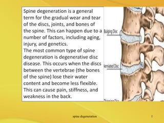
- 1. Spine degeneration is a general term for the gradual wear and tear of the discs, joints, and bones of the spine. This can happen due to a number of factors, including aging, injury, and genetics. The most common type of spine degeneration is degenerative disc disease. This occurs when the discs between the vertebrae (the bones of the spine) lose their water content and become less flexible. This can cause pain, stiffness, and weakness in the back. spine degeneration 1
- 2. Other types of spine degeneration include: 1. Spinal stenosis: This is a narrowing of the spinal canal, which can put pressure on the spinal cord and nerves. 2. Spondylosis: This is a general term for the wear and tear of the joints in the spine. 3. Kyphosis: This is an abnormal rounding of the upper spine. 4. Lordosis: This is an abnormal inward curvature of the lower spine spine degeneration 2
- 3. Symptoms of spine degeneration can vary depending on the type and severity of the condition. Some common symptoms include: •Pain in the back, neck, or arms •Stiffness in the back or neck •Weakness in the back or legs •Numbness or tingling in the arms or legs Loss of balance spine degeneration 3
- 4. Spinal degeneration Degenerative changes within the spine are the most common pathological finding noted on autopsy and on imaging of the spine. Sagittal T2 weighed MR image of lumbar spine demonstrating a normal-appearing signal of all discs except the L5 disc where there is a high signal intensity zone in the posterior aspect of the disc space (arrow). This represents nuclear material that has extended through a confluence of annular tears, leading to a radial fissure in the disc spine degeneration 4
- 5. The speed and extent of the degenerative changes appear to be impacted by hereditary factors as well as specific and continuous traumatic events that occur through a person’s life. Even the most severe degenerative changes can occur in the absence of symptomatology, but back pain is more common in individuals who demonstrate these degenerative changes. It appears that the degenerative changes in the spine make one more vulnerable to the inflammatory effects of trauma. spine degeneration 5
- 6. Degenerative changes are most evident in the intervertebral discs and the facet joints, usually at the same time, but often to varying degrees. It is useful to visualize the vertebral motion-segment as a ‘three joint complex’ in which degenerative changes in the posterior facets impact the intervertebral disc, and pathological changes within the intervertebral disc will create greater stressors upon the posterior facet joints. spine degeneration 6
- 7. THE INTERVERTEBRAL DISC Degenerative changes within the intervertebral disc usually start as small circumferential tears in the annulus fibrosus. These annular tears increase in size and coalesce to form radial fissures. The radial fissures then expand and extend into the nucleus pulposus, disrupting the disc structure internally. There is a loss of proteoglycans and water content from the nucleus which results in a loss of the height of the disc. As degeneration continues, the disc collapses, shortening the distance between the two vertebral bodies. This re-absorption can progress to the point where the vertebral bodies are eventually separated only by dense sclerotic fibrous tissue which is all that remains of the original disc structure spine degeneration 7
- 8. At the same time as the disc is being reabsorbed, the vertebral bodies on either side of the disc become dense and sclerotic. Osteophytes extend from the vertebral bodies around its circumference, presumably in an attempt to stabilize the three-joint complex and reduce motion. Occasionally, the osteophytes may join and fuse, resulting in bony ankylosis of the joint spine degeneration 8
- 9. THE FACET JOINTS Degenerative changes within the posterior facet usually begin with an inflammatory synovitis, which can lead to the formation of a synovial fold, projecting into the joint between the cartilage surfaces. There is gradual thinning of the cartilage, which starts in the periphery with progressive loss of cartilage tissue. Subperiosteal osteophytes begin to form which enlarge both the inferior and superior facets. spine degeneration 9
- 10. This breakdown continues until there is almost total loss of articular cartilage with marked periarticular fibrosis and the formation of subperiosteal new bone expanding the volume of the superior and inferior facets. During the early phases of these degenerative changes, the facet capsule can become very lax, allowing increased movement. It is probably this period of increased mobility of the joint which leads to further degeneration within the posterior facets, and puts further stress on the intervertebral discs spine degeneration 10
- 11. There are two small circumferential tears in the posterior annulus (arrows). This represents stage one of the degenerative process in the discs. The posterior facets are enlarged and the facets show degenerative changes. This demonstrates the interaction between the discs and the posterior joints. spine degeneration 11
- 12. This cross-section of the lumbar spine shows degenerative changes in the intervertebral disc and the posterior facet joints. The extensive degenerative changes in the posterior joints have resulted in enlargement of the facets. On the left side of the disc near the back of the vertebral body, there is a small circumferential tear in the annulus fibrosus. This tear has enlarged and spread to the center of the disc. spine degeneration 12
- 13. this cross-section through a lumbar disc shows very marked degenerative changes. There is complete disintegration of the nucleus pulposus. These changes have resulted in instability of the three- joint complex at this level. spine degeneration 13
- 14. Longitudinal section through the lumbar spine at L4–L5. There is almost complete resorption of the disc which is seen as a small slit between the vertebral bodies. There is sclerosis of the bone in the vertebral bodies on either side of the disc. spine degeneration 14
- 15. Longitudinal sagittal section of the lumbar spine showing marked degeneration at four levels. There is a Schmorl’s node at the upper level at L2–L3, with herniation of the nucleus pulposus into the vertebral body. There is disintegration of the L3–L4 and L4–L5 discs. At the L5–S1 level, there is disc resorption, with sclerotic bone on either side of the remnants of the disc. spine degeneration 15