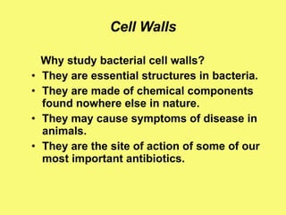
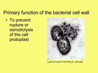
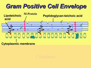
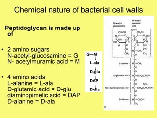
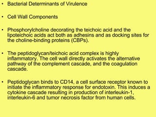
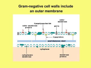
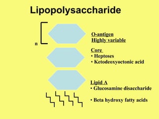
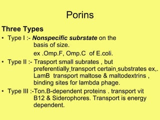
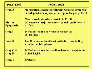
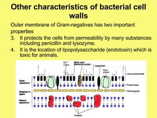
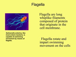
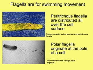
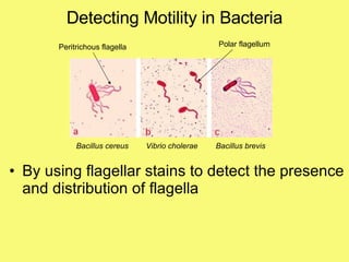
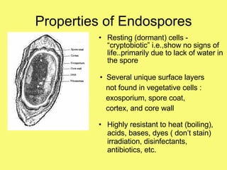
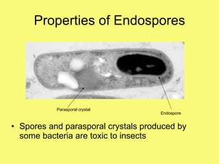
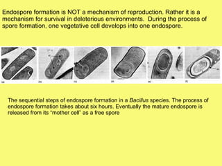
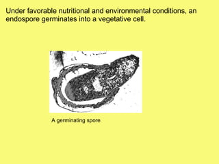
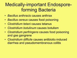
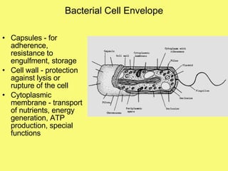
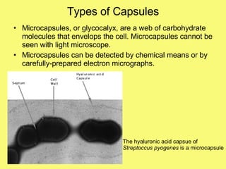
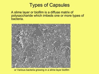
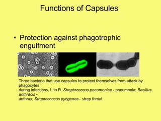
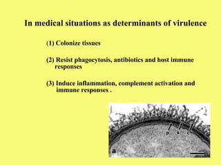
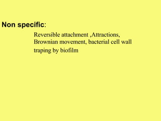
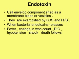
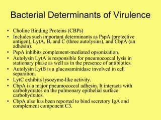
Bacterial cell wall and applied importance Dr.Raghu prakash
Cell Walls Why study bacterial cell walls? They are essential structures in bacteria. They are made of chemical components found nowhere else in nature. They may cause symptoms of disease in animals. They are the site of action of some of our most important antibiotics.
Primary function of the bacterial cell wall To prevent rupture or osmoticlysis of the cell protoplast Lysis of a pair of dividing  E. coli  cells
Chemical nature of bacterial cell walls Bacterial cell walls always contain murein, which is a type of peptidoglycan Chemical nature of murein accounts for the function of the cell wall Murein is only found in the cell walls of bacteria
Gram Positive Cell Envelope Cytoplasm Lipoteichoic acid Peptidoglycan-teichoic acid  Cytoplasmic membrane M-Protein r r r r r r r r r r
Chemical nature of bacterial cell walls Peptidoglycan is made up of 2 amino sugars  N-acetyl-glucosamine = G  N- acetylmuramic acid = M  4 amino acids L-alanine = L-ala D-glutamic acid = D-glu diaminopimelic acid = DAP D-alanine = D-ala G—M L-ala D-glu DAP D-ala
Peptidoglycan Synthesis
UMP UDP UTP UTP + Glc NAc-1-p UDP-Glc NAc UDP-Glc NAc Enolpyruvate UDP – Mur NAc UDP – Mur NAc-L-Ala UDP – Mur NAc-L-Ala- D -Glu UDP – Mur NAc-L-Ala- D -Glu-L-R3 UDP – Mur NAc-L-Ala - D -Glu-L-R 3 -(DAla) 2 D-Ala-DAla 2-L-Ala Cycloserine   inhibits 9MurF + L-R3 +D-Glu +L-Ala +NADPH +PEP - PP 1 Phosphonomycin inhibits D -ALa D-Ala transport Undecaprenyl-PP -Mur NAc-  pentapeptide Undecaprenyl-P Cytoplasm Membrane Wall Undecaprenyl-PP Disaccaride -pentapeptide Bacitracin inhibits Disaccaride -pentapeptide Nascent  Peptidoglycan Expanding cross  linked wall Peptidoglycan 3 Lactum Antibiotic inhibits Vancomycin inhibits
Neuraminidase - cleaves sialic (neuraminic) acid, possibly to expose receptors or to dissolve interstitial cement  IgA protease - cleaves and inactivates secretory IgA Surface Proteins •  Five penicillin binding proteins (PBPs)  •  Two neuraminidases •  IgA protease •  Twelve choline-binding proteins (CBPs) - include important determinants of virulence such as PspA (protective antigen), LytA, B, and C (three autolysins), and CbpA (an adhesin)
Bacterial Determinants of Virulence Cell Wall Components Phosphorylcholine decorating the teichoic acid and the lipoteichoic acids act both as adhesins and as docking sites for the choline-binding proteins (CBPs).  The peptidoglycan/teichoic acid complex is highly inflammatory. The cell wall directly activates the alternative pathway of the complement cascade, and the coagulation cascade. Peptidoglycan binds to CD14, a cell surface receptor known to initiate the inflammatory response for endotoxin. This induces a cytokine cascade resulting in production of interleukin-1, interleukin-6 and tumor necrosis factor from human cells.
Peptidoglycan hydrolases Three types – Glycan-strands hydrolyzing Endo-N-acetylmuramidases Endo-N-acetylgucosaminidases Endopeptidase hydrolyzing  Peptide bonds in the interior of the peptide bridges  Bonds involving the C-terminal D-alanine residue  N-acetylmuramoyl –L-alanine amidase Acting at the junction b/w glycan strands and the  peptide units This enzymes appear to play an imp role in number of cellular activities septum and wall extension during cell growth ,cell separation,turn over of wall components,sporulation competency for transformation, excretion of toxins and exoenzymes
Gram-negative cell walls include  an outer membrane
Lipopolysaccharide Lipid A Glucosamine disaccharide Beta hydroxy fatty acids Core  Heptoses Ketodeoxyoctonic acid O-antigen Highly variable n
Porins Three Types  Type I :-  Nonspecific subrstate  on the  basis of size.   ex .Omp.F, Omp.C  of E.coli. Type II :- Trasport small subrates , but  preferentially   transport certain   substrates ex,.  LamB  transport maltose & maltodextrins ,  binding sites for lambda phage. Type III :-Ton.B-dependent proteins . transport vit  B12 & Siderophores. Transport is energy  dependent.
Stabilization of outer membrane &mating aggregates in F-dependent conjugation;receptor for phage Tu11. Most abundant surface protein in E.coli &S.enterica ;major structural protein ;stabilizes cell surface. Diffusion channel for various metabolies  ex: maltose .  LamB  transport maltose&maltodextrins,binding sites for lambda phages .  Diffusion channel for small molecules ;receptors for Tulb&T3,T4. Protease. Omp.A  Murien lipoprotein OmpB Lam.B Omp.C & Omp.F Omp.T FUNCTIONS PROTEINS
Anion-selective diffusion channel under phosphate limitation Anion-selective diffusion channel in  P.aeruginosainduced under phosphate . Maintenance of OM integrity;activity of group a colicins Ferrichrome siderophore uptake;receptor for phages T1,T5,80,&COLICIN M. Dependent proteins . transport vit B12 &Siderophores  PhoE Protein P TolA TonA Ton.B
Other characteristics of bacterial cell walls Outer membrane of Gram-negatives has two important properties It protects the cells from permeability by many substances including penicillin and lysozyme. It is the location of lipopolysaccharide (endotoxin) which is toxic for animals.
S-layer composed of identical proteins or glycoproteins  sub units  s-layer is quiet different in many organisms  In Gram + sub units it is linked to peptidoglycan layer or secondary cell wall polymers  In Gram – it is linked to lipopolysaccharides of outer membrane S-layer contain acidic and hydrophobic A.A  Functions / attachment to surfaces and to host tissues Campylobacter and Aeromonas S-layer serves as virulence factors  In archaea S-layer is outer most layer ,next to cell membrane  it must contribute to the shape of the cell
Bacterial Surface Appendages Flagella - organelles for swimming motility Pili (or fimbriae) - for attachment or adherence to surfaces; sex pilus used during some genetic exchange processes
Flagella Flagella are long whiplike filaments composed of protein that originate in the cell membrane. Flagella rotate and impart swimming movement on the cells Salmonella enterica,  like most enteric bacteria, is capable of swimming movement by means of flagella.
Flagella are for swimming movement Peritrichous flagella are distributed all over the cell surface Polar flagella originate at the pole of a cell Proteus mirabilis  swims by means of peritrichous flagella Vibrio cholerae  has a single polar flagellum
Detecting Motility in Bacteria By using flagellar stains to detect the presence and distribution of flagella Bacillus cereus Vibrio cholerae Bacillus brevis Polar flagellum Peritrichous flagella
 
Detecting Motility in Bacteria By inoculation of the bacteria into motility test medium (SIM).   Staphylococcus epidermidis Non motile E. coli motile
Ecological Advantages to Swimming Survival: escape predatory protozoa and white blood cells (phagocytes) Swim towards nutrients or away from harmful substances (chemotaxis) Swim towards or away from O 2  (aerotaxis) Swim towards light (phototaxis) Swim toward the North Pole or the South Pole (magnetotaxis)
The Structure of the Bacterial Surface: Flagella Summary Flagella  filamentous protein structures attached to the cell surface that provide swimming movement for most motile bacterial cells. The flagellar filament is rotated by a motor apparatus in the plasma membrane allowing the cell to swim in fluid environments. tactic behavior or motility is the ability to move (swim) in response to environmental stimuli. Chemotaxis: a bacterium can sense the quality and quantity of certain chemicals in its environment and swim towards them (if they are useful nutrients) or away from them (if they are harmful substances).  Aerotaxis: bacteria swim toward or away from O 2 motility as a determinant of virulence: e.g.  Vibrio cholerae, Campylobacter, Helicobacter, Pseudomonas
Functions of Pili and Fimbriae Attachment to a surface or substrate Shigella dysenteriae  uses its  fimbriae to attach to the intestine and then produces a toxin that  causes diarrhea. Neissera gonorrhoeae,  the cause of the gonorrhea, uses pili to attach to the urogenital and cervical epithelium when it causes disease Pili (also called fimbriae) are short hair-like structures composed of protein on the cell surface.
Functions of Pili and Fimbriae Resistance to Phagocytic engulfment Phagocytosis of streptococci by a macrophage Chain of streptococci protected from engulfment by fimbrial (M) protein
Functions of Pili and Fimbriae A special type of pilus called the sex pilus is used in mating between bacteria E. coli  uses its sex pilus (called the F-pilus) to transfer DNA between mating bacteria during conjugation.
How Flagella Work Filament is rotated by a protein “motor” in the cell membrane Motor is powered by proton motive force (pmf) on the outside of membrane motor basal body filament pmf  on this  side of membrane
Endospores are produced as intracellular structures within the cytoplasm of certain bacteria, most notably  Bacillus  and  Clostridium  species. Endospore forming bacteria left to right:  Clostridium botulinum, Bacillus brevis, Bacillus thuringiensis
Properties of Endospores Resting (dormant) cells - “cryptobiotic” i.e.,show no signs of life..primarily due to lack of water in the spore Several unique surface layers not found in vegetative cells :  exosporium, spore coat, cortex, and core wall Highly resistant to heat (boiling), acids, bases, dyes ( don’t stain) irradiation, disinfectants, antibiotics, etc.
Properties of Endospores Spores and parasporal crystals produced by some bacteria are toxic to insects Parasporal crystal Endospore
Endospore formation is NOT a mechanism of reproduction. Rather it is a mechanism for survival in deleterious environments.  During the process of spore formation, one vegetative cell develops into one endospore. The sequential steps of endospore formation in a  Bacillus  species. The process of endospore formation takes about six hours. Eventually the mature endospore is released from its “mother cell” as a free spore
Under favorable nutritional and environmental conditions, an endospore germinates into a vegetative cell. A germinating spore
Medically-important Endospore-forming Bacteria Bacillus anthracis  causes anthrax Bacillus cereus  causes food poisoning Clostridium tetani  causes tetanus Clostridium botulinum  causes botulism Clostridium perfringens  causes food poisoning and gas gangrene Clostridium difficile  causes antibiotic-induced diarrhea and pseudomembranous colitis
Bacterial Cell Envelope Capsules - for adherence, resistance to engulfment, storage Cell wall - protection against lysis or rupture of the cell Cytoplasmic membrane - transport of nutrients, energy generation, ATP production, special functions
Capsules Capsules are composed of polysaccharides (occasionally polypeptides) deposited outside the cell wall. Using special staining techniques,  some capsules can be demonstrated  as a halo surrounding the bacterial cells. Bacterial cell Capsular material
Types of Capsules True capsules are discrete layers enclosing a cell or group of cells that can be readily visualized microscopically. Negative stain of  Streptococcus pneumoniae  outlining its notorious polysaccharide capsule Usually, if a bacterium forms a capsule, it will  grow on certain media with a gummy or mucoid type of colony, such as these colonies of  Bacillus anthracis .
Types of Capsules Microcapsules, or glycocalyx, are a web of carbohydrate molecules that envelops the cell. Microcapsules cannot be seen with light microscope. Microcapsules can be detected by chemical means or by carefully-prepared electron micrographs. The hyaluronic acid capsue of  Streptoccus pyogenes  is a microcapsule
Types of Capsules A slime layer or biofilm is a diffuse matrix of polysaccharide which imbeds one or more types of bacteria. or Various bacteria growing in a slime layer biofilm
Functions of Capsules Protection against phagotrophic engulfment Mediate adherence to surfaces Protection against drying Reserve of nutrients Biofilms for protection and metabolic communication among microbes
Functions of Capsules Protection against phagotrophic engulfment Three bacteria that use capsules to protect themselves from attack by phagocytes during infections. L to R.  Streptococcus pneumoniae  - pneumonia;  Bacillus anthracis  -  anthrax;  Streptococcus pyogenes  - strep throat.
Functions of Capsules Mediate adherence to surfaces Oral streptococci use their capsular slime to adhere to the the surfaces of the teeth and gums.
Functions of Capsules Reserve of nutrients Colonies of oral streptococci growing on mitis-salivarius agar. The medium contains 5% sucrose.  Streptococcus salivarius  (left) stores excess sugar as “levan” polymer;  Streptococcus mutans  (right) stores the carbohydrate as a dextran polymer. The polysaccharide polymers give the colonies there glistening, sugary appearance.
Functions of Capsules Biofilms for protection and metabolic communication among microbes Biofilm development by  Pseudomonas aeruginosa . Figure from: Kolter, R. and R. Losick. 1998. One for all and all for one. Science 280:226-227. After the bacteria form the biofilm, they are protected from antibiotics, detergents, disinfectants, etc., which cannot penetrate the slime.
The Importance of the Bacterial Surface Possible natural functions of bacterial surface components   Permeability barriers that allow selective passage of nutrients and exclusion of harmful  substances (e.g. antimicrobial agents) Adhesins used to attach or adhere to specific surfaces or tissues (3) Enzymes to mediate specific reactions on the cell surface important in the survival of the organism (4) Protective structures against phagocytic engulfment or killing (5) Antigenic disguises  (6) “Sensing proteins" that can respond to temperature, osmolarity, salinity, light, oxygen, nutrients, cell density (quorum sensing), etc. Cell surface of a  Bacillus
In medical situations as determinants of virulence ( 1) Colonize tissues  (2) Resist phagocytosis, antibiotics and host immune    responses (3) Induce inflammation, complement activation and    immune responses .
 
Colonization Colonization is a firs step of infection. Establishment of  pathogen at a specific body site frequently followed after entry to the host tissue,. Colonization occurs in body systems intact with external environment,eg:- urogenital tract , digestive tract, respiratory tract and peritoneum in females through the fallopian tubes.  Adherence to Surface : 1)  Specific 2)  Non specific
Adherence to Surface Specific : Reversible or permanent ,specific covalent bonds between  adhesion and receptor molecules. Species specific tropism E.g:-N.gonorehea ,N meningitis, group A strepto ,E.coli  (CFA-1 and CFA-2) Eg:-E.coli,uropathogenic pattern are determined by binding specificity of the PapG adhesion  Pap G  alleles of E.coli exists in three typesClass 1, 2, 3 Tissue host specific complementery
Non specific : Reversible attachment ,Attractions,  Brownian movement, bacterial cell wall  traping by biofilm
Endotoxin Cell envelop component shed as a membrane blebs or vesicles . They  are exemplified by LOS and LPS . When bacterial endotoxins releases  Fever  change in wbc count  DIC ,  hypotension  shock  death follows
Bacterial Determinants of Virulence Choline Binding Proteins (CBPs) Includes such important determinants as PspA (protective antigen), LytA, B, and C (three autolysins), and CbpA (an adhesin). PspA inhibits complement-mediated opsonization. Autolysin LytA is responsible for pneumococcal lysis in stationary phase as well as in the presence of antibiotics.  Autolysin LytB is a glucosaminidase involved in cell separation.  LytC exhibits lysozyme-like activity. CbpA is a major pneumococcal adhesin. It interacts with carbohydrates on the pulmonary epithelial surface carbohydrates.  CbpA also has been reported to bind secretory IgA and  complement component C3.
THANK  U

China Cell

  • 1.
    Bacterial cell walland applied importance Dr.Raghu prakash
  • 2.
    Cell Walls Whystudy bacterial cell walls? They are essential structures in bacteria. They are made of chemical components found nowhere else in nature. They may cause symptoms of disease in animals. They are the site of action of some of our most important antibiotics.
  • 3.
    Primary function ofthe bacterial cell wall To prevent rupture or osmoticlysis of the cell protoplast Lysis of a pair of dividing E. coli cells
  • 4.
    Chemical nature ofbacterial cell walls Bacterial cell walls always contain murein, which is a type of peptidoglycan Chemical nature of murein accounts for the function of the cell wall Murein is only found in the cell walls of bacteria
  • 5.
    Gram Positive CellEnvelope Cytoplasm Lipoteichoic acid Peptidoglycan-teichoic acid Cytoplasmic membrane M-Protein r r r r r r r r r r
  • 6.
    Chemical nature ofbacterial cell walls Peptidoglycan is made up of 2 amino sugars N-acetyl-glucosamine = G N- acetylmuramic acid = M 4 amino acids L-alanine = L-ala D-glutamic acid = D-glu diaminopimelic acid = DAP D-alanine = D-ala G—M L-ala D-glu DAP D-ala
  • 7.
  • 8.
    UMP UDP UTPUTP + Glc NAc-1-p UDP-Glc NAc UDP-Glc NAc Enolpyruvate UDP – Mur NAc UDP – Mur NAc-L-Ala UDP – Mur NAc-L-Ala- D -Glu UDP – Mur NAc-L-Ala- D -Glu-L-R3 UDP – Mur NAc-L-Ala - D -Glu-L-R 3 -(DAla) 2 D-Ala-DAla 2-L-Ala Cycloserine inhibits 9MurF + L-R3 +D-Glu +L-Ala +NADPH +PEP - PP 1 Phosphonomycin inhibits D -ALa D-Ala transport Undecaprenyl-PP -Mur NAc- pentapeptide Undecaprenyl-P Cytoplasm Membrane Wall Undecaprenyl-PP Disaccaride -pentapeptide Bacitracin inhibits Disaccaride -pentapeptide Nascent Peptidoglycan Expanding cross linked wall Peptidoglycan 3 Lactum Antibiotic inhibits Vancomycin inhibits
  • 9.
    Neuraminidase - cleavessialic (neuraminic) acid, possibly to expose receptors or to dissolve interstitial cement IgA protease - cleaves and inactivates secretory IgA Surface Proteins • Five penicillin binding proteins (PBPs) • Two neuraminidases • IgA protease • Twelve choline-binding proteins (CBPs) - include important determinants of virulence such as PspA (protective antigen), LytA, B, and C (three autolysins), and CbpA (an adhesin)
  • 10.
    Bacterial Determinants ofVirulence Cell Wall Components Phosphorylcholine decorating the teichoic acid and the lipoteichoic acids act both as adhesins and as docking sites for the choline-binding proteins (CBPs). The peptidoglycan/teichoic acid complex is highly inflammatory. The cell wall directly activates the alternative pathway of the complement cascade, and the coagulation cascade. Peptidoglycan binds to CD14, a cell surface receptor known to initiate the inflammatory response for endotoxin. This induces a cytokine cascade resulting in production of interleukin-1, interleukin-6 and tumor necrosis factor from human cells.
  • 11.
    Peptidoglycan hydrolases Threetypes – Glycan-strands hydrolyzing Endo-N-acetylmuramidases Endo-N-acetylgucosaminidases Endopeptidase hydrolyzing Peptide bonds in the interior of the peptide bridges Bonds involving the C-terminal D-alanine residue N-acetylmuramoyl –L-alanine amidase Acting at the junction b/w glycan strands and the peptide units This enzymes appear to play an imp role in number of cellular activities septum and wall extension during cell growth ,cell separation,turn over of wall components,sporulation competency for transformation, excretion of toxins and exoenzymes
  • 12.
    Gram-negative cell wallsinclude an outer membrane
  • 13.
    Lipopolysaccharide Lipid AGlucosamine disaccharide Beta hydroxy fatty acids Core Heptoses Ketodeoxyoctonic acid O-antigen Highly variable n
  • 14.
    Porins Three Types Type I :- Nonspecific subrstate on the basis of size. ex .Omp.F, Omp.C of E.coli. Type II :- Trasport small subrates , but preferentially transport certain substrates ex,. LamB transport maltose & maltodextrins , binding sites for lambda phage. Type III :-Ton.B-dependent proteins . transport vit B12 & Siderophores. Transport is energy dependent.
  • 15.
    Stabilization of outermembrane &mating aggregates in F-dependent conjugation;receptor for phage Tu11. Most abundant surface protein in E.coli &S.enterica ;major structural protein ;stabilizes cell surface. Diffusion channel for various metabolies ex: maltose . LamB transport maltose&maltodextrins,binding sites for lambda phages . Diffusion channel for small molecules ;receptors for Tulb&T3,T4. Protease. Omp.A Murien lipoprotein OmpB Lam.B Omp.C & Omp.F Omp.T FUNCTIONS PROTEINS
  • 16.
    Anion-selective diffusion channelunder phosphate limitation Anion-selective diffusion channel in P.aeruginosainduced under phosphate . Maintenance of OM integrity;activity of group a colicins Ferrichrome siderophore uptake;receptor for phages T1,T5,80,&COLICIN M. Dependent proteins . transport vit B12 &Siderophores PhoE Protein P TolA TonA Ton.B
  • 17.
    Other characteristics ofbacterial cell walls Outer membrane of Gram-negatives has two important properties It protects the cells from permeability by many substances including penicillin and lysozyme. It is the location of lipopolysaccharide (endotoxin) which is toxic for animals.
  • 18.
    S-layer composed ofidentical proteins or glycoproteins sub units s-layer is quiet different in many organisms In Gram + sub units it is linked to peptidoglycan layer or secondary cell wall polymers In Gram – it is linked to lipopolysaccharides of outer membrane S-layer contain acidic and hydrophobic A.A Functions / attachment to surfaces and to host tissues Campylobacter and Aeromonas S-layer serves as virulence factors In archaea S-layer is outer most layer ,next to cell membrane it must contribute to the shape of the cell
  • 19.
    Bacterial Surface AppendagesFlagella - organelles for swimming motility Pili (or fimbriae) - for attachment or adherence to surfaces; sex pilus used during some genetic exchange processes
  • 20.
    Flagella Flagella arelong whiplike filaments composed of protein that originate in the cell membrane. Flagella rotate and impart swimming movement on the cells Salmonella enterica, like most enteric bacteria, is capable of swimming movement by means of flagella.
  • 21.
    Flagella are forswimming movement Peritrichous flagella are distributed all over the cell surface Polar flagella originate at the pole of a cell Proteus mirabilis swims by means of peritrichous flagella Vibrio cholerae has a single polar flagellum
  • 22.
    Detecting Motility inBacteria By using flagellar stains to detect the presence and distribution of flagella Bacillus cereus Vibrio cholerae Bacillus brevis Polar flagellum Peritrichous flagella
  • 23.
  • 24.
    Detecting Motility inBacteria By inoculation of the bacteria into motility test medium (SIM). Staphylococcus epidermidis Non motile E. coli motile
  • 25.
    Ecological Advantages toSwimming Survival: escape predatory protozoa and white blood cells (phagocytes) Swim towards nutrients or away from harmful substances (chemotaxis) Swim towards or away from O 2 (aerotaxis) Swim towards light (phototaxis) Swim toward the North Pole or the South Pole (magnetotaxis)
  • 26.
    The Structure ofthe Bacterial Surface: Flagella Summary Flagella filamentous protein structures attached to the cell surface that provide swimming movement for most motile bacterial cells. The flagellar filament is rotated by a motor apparatus in the plasma membrane allowing the cell to swim in fluid environments. tactic behavior or motility is the ability to move (swim) in response to environmental stimuli. Chemotaxis: a bacterium can sense the quality and quantity of certain chemicals in its environment and swim towards them (if they are useful nutrients) or away from them (if they are harmful substances). Aerotaxis: bacteria swim toward or away from O 2 motility as a determinant of virulence: e.g. Vibrio cholerae, Campylobacter, Helicobacter, Pseudomonas
  • 27.
    Functions of Piliand Fimbriae Attachment to a surface or substrate Shigella dysenteriae uses its fimbriae to attach to the intestine and then produces a toxin that causes diarrhea. Neissera gonorrhoeae, the cause of the gonorrhea, uses pili to attach to the urogenital and cervical epithelium when it causes disease Pili (also called fimbriae) are short hair-like structures composed of protein on the cell surface.
  • 28.
    Functions of Piliand Fimbriae Resistance to Phagocytic engulfment Phagocytosis of streptococci by a macrophage Chain of streptococci protected from engulfment by fimbrial (M) protein
  • 29.
    Functions of Piliand Fimbriae A special type of pilus called the sex pilus is used in mating between bacteria E. coli uses its sex pilus (called the F-pilus) to transfer DNA between mating bacteria during conjugation.
  • 30.
    How Flagella WorkFilament is rotated by a protein “motor” in the cell membrane Motor is powered by proton motive force (pmf) on the outside of membrane motor basal body filament pmf on this side of membrane
  • 31.
    Endospores are producedas intracellular structures within the cytoplasm of certain bacteria, most notably Bacillus and Clostridium species. Endospore forming bacteria left to right: Clostridium botulinum, Bacillus brevis, Bacillus thuringiensis
  • 32.
    Properties of EndosporesResting (dormant) cells - “cryptobiotic” i.e.,show no signs of life..primarily due to lack of water in the spore Several unique surface layers not found in vegetative cells : exosporium, spore coat, cortex, and core wall Highly resistant to heat (boiling), acids, bases, dyes ( don’t stain) irradiation, disinfectants, antibiotics, etc.
  • 33.
    Properties of EndosporesSpores and parasporal crystals produced by some bacteria are toxic to insects Parasporal crystal Endospore
  • 34.
    Endospore formation isNOT a mechanism of reproduction. Rather it is a mechanism for survival in deleterious environments. During the process of spore formation, one vegetative cell develops into one endospore. The sequential steps of endospore formation in a Bacillus species. The process of endospore formation takes about six hours. Eventually the mature endospore is released from its “mother cell” as a free spore
  • 35.
    Under favorable nutritionaland environmental conditions, an endospore germinates into a vegetative cell. A germinating spore
  • 36.
    Medically-important Endospore-forming BacteriaBacillus anthracis causes anthrax Bacillus cereus causes food poisoning Clostridium tetani causes tetanus Clostridium botulinum causes botulism Clostridium perfringens causes food poisoning and gas gangrene Clostridium difficile causes antibiotic-induced diarrhea and pseudomembranous colitis
  • 37.
    Bacterial Cell EnvelopeCapsules - for adherence, resistance to engulfment, storage Cell wall - protection against lysis or rupture of the cell Cytoplasmic membrane - transport of nutrients, energy generation, ATP production, special functions
  • 38.
    Capsules Capsules arecomposed of polysaccharides (occasionally polypeptides) deposited outside the cell wall. Using special staining techniques, some capsules can be demonstrated as a halo surrounding the bacterial cells. Bacterial cell Capsular material
  • 39.
    Types of CapsulesTrue capsules are discrete layers enclosing a cell or group of cells that can be readily visualized microscopically. Negative stain of Streptococcus pneumoniae outlining its notorious polysaccharide capsule Usually, if a bacterium forms a capsule, it will grow on certain media with a gummy or mucoid type of colony, such as these colonies of Bacillus anthracis .
  • 40.
    Types of CapsulesMicrocapsules, or glycocalyx, are a web of carbohydrate molecules that envelops the cell. Microcapsules cannot be seen with light microscope. Microcapsules can be detected by chemical means or by carefully-prepared electron micrographs. The hyaluronic acid capsue of Streptoccus pyogenes is a microcapsule
  • 41.
    Types of CapsulesA slime layer or biofilm is a diffuse matrix of polysaccharide which imbeds one or more types of bacteria. or Various bacteria growing in a slime layer biofilm
  • 42.
    Functions of CapsulesProtection against phagotrophic engulfment Mediate adherence to surfaces Protection against drying Reserve of nutrients Biofilms for protection and metabolic communication among microbes
  • 43.
    Functions of CapsulesProtection against phagotrophic engulfment Three bacteria that use capsules to protect themselves from attack by phagocytes during infections. L to R. Streptococcus pneumoniae - pneumonia; Bacillus anthracis - anthrax; Streptococcus pyogenes - strep throat.
  • 44.
    Functions of CapsulesMediate adherence to surfaces Oral streptococci use their capsular slime to adhere to the the surfaces of the teeth and gums.
  • 45.
    Functions of CapsulesReserve of nutrients Colonies of oral streptococci growing on mitis-salivarius agar. The medium contains 5% sucrose. Streptococcus salivarius (left) stores excess sugar as “levan” polymer; Streptococcus mutans (right) stores the carbohydrate as a dextran polymer. The polysaccharide polymers give the colonies there glistening, sugary appearance.
  • 46.
    Functions of CapsulesBiofilms for protection and metabolic communication among microbes Biofilm development by Pseudomonas aeruginosa . Figure from: Kolter, R. and R. Losick. 1998. One for all and all for one. Science 280:226-227. After the bacteria form the biofilm, they are protected from antibiotics, detergents, disinfectants, etc., which cannot penetrate the slime.
  • 47.
    The Importance ofthe Bacterial Surface Possible natural functions of bacterial surface components Permeability barriers that allow selective passage of nutrients and exclusion of harmful substances (e.g. antimicrobial agents) Adhesins used to attach or adhere to specific surfaces or tissues (3) Enzymes to mediate specific reactions on the cell surface important in the survival of the organism (4) Protective structures against phagocytic engulfment or killing (5) Antigenic disguises (6) “Sensing proteins" that can respond to temperature, osmolarity, salinity, light, oxygen, nutrients, cell density (quorum sensing), etc. Cell surface of a Bacillus
  • 48.
    In medical situationsas determinants of virulence ( 1) Colonize tissues (2) Resist phagocytosis, antibiotics and host immune responses (3) Induce inflammation, complement activation and immune responses .
  • 49.
  • 50.
    Colonization Colonization isa firs step of infection. Establishment of pathogen at a specific body site frequently followed after entry to the host tissue,. Colonization occurs in body systems intact with external environment,eg:- urogenital tract , digestive tract, respiratory tract and peritoneum in females through the fallopian tubes. Adherence to Surface : 1) Specific 2) Non specific
  • 51.
    Adherence to SurfaceSpecific : Reversible or permanent ,specific covalent bonds between adhesion and receptor molecules. Species specific tropism E.g:-N.gonorehea ,N meningitis, group A strepto ,E.coli (CFA-1 and CFA-2) Eg:-E.coli,uropathogenic pattern are determined by binding specificity of the PapG adhesion Pap G alleles of E.coli exists in three typesClass 1, 2, 3 Tissue host specific complementery
  • 52.
    Non specific :Reversible attachment ,Attractions, Brownian movement, bacterial cell wall traping by biofilm
  • 53.
    Endotoxin Cell envelopcomponent shed as a membrane blebs or vesicles . They are exemplified by LOS and LPS . When bacterial endotoxins releases Fever change in wbc count DIC , hypotension shock death follows
  • 54.
    Bacterial Determinants ofVirulence Choline Binding Proteins (CBPs) Includes such important determinants as PspA (protective antigen), LytA, B, and C (three autolysins), and CbpA (an adhesin). PspA inhibits complement-mediated opsonization. Autolysin LytA is responsible for pneumococcal lysis in stationary phase as well as in the presence of antibiotics. Autolysin LytB is a glucosaminidase involved in cell separation. LytC exhibits lysozyme-like activity. CbpA is a major pneumococcal adhesin. It interacts with carbohydrates on the pulmonary epithelial surface carbohydrates. CbpA also has been reported to bind secretory IgA and complement component C3.
  • 55.