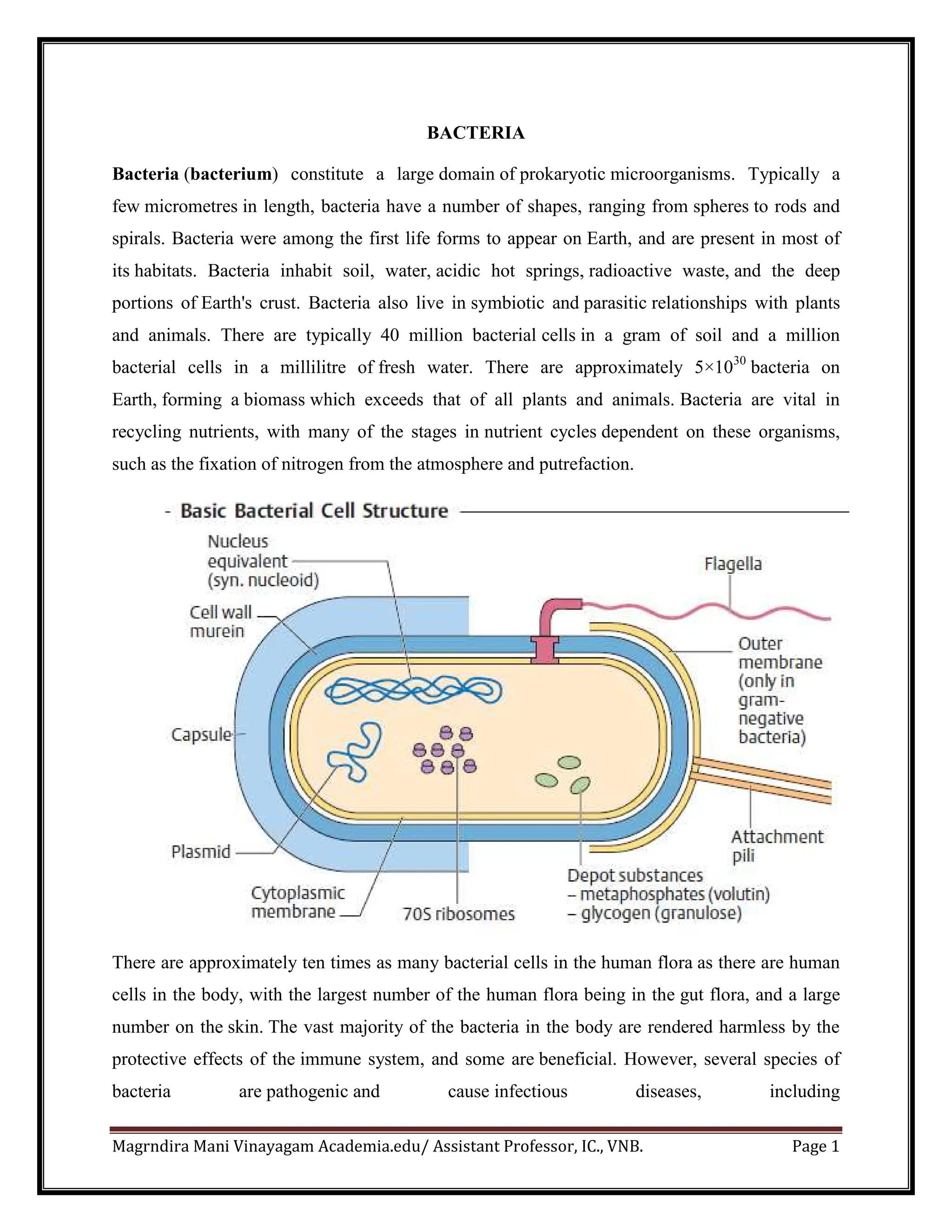
Magrndira Mani Vinayagam Academia.edu/ Assistant Professor, IC., VNB. Page 1
BACTERIA
Bacteria (bacterium) constitute a large domain of prokaryotic microorganisms. Typically a
few micrometres in length, bacteria have a number of shapes, ranging from spheres to rods and
spirals. Bacteria were among the first life forms to appear on Earth, and are present in most of
its habitats. Bacteria inhabit soil, water, acidic hot springs, radioactive waste, and the deep
portions of Earth's crust. Bacteria also live in symbiotic and parasitic relationships with plants
and animals. There are typically 40 million bacterial cells in a gram of soil and a million
bacterial cells in a millilitre of fresh water. There are approximately 5×1030
bacteria on
Earth, forming a biomass which exceeds that of all plants and animals. Bacteria are vital in
recycling nutrients, with many of the stages in nutrient cycles dependent on these organisms,
such as the fixation of nitrogen from the atmosphere and putrefaction.
There are approximately ten times as many bacterial cells in the human flora as there are human
cells in the body, with the largest number of the human flora being in the gut flora, and a large
number on the skin. The vast majority of the bacteria in the body are rendered harmless by the
protective effects of the immune system, and some are beneficial. However, several species of
bacteria are pathogenic and cause infectious diseases, including
Magrndira Mani Vinayagam Academia.edu/ Assistant Professor, IC., VNB. Page 2
cholera, syphilis, anthrax, leprosy, and bubonic plague. The most common fatal bacterial
diseases are respiratory infections, with tuberculosis alone killing about 2 million people a year,
mostly in sub-Saharan Africa. In developed countries, antibiotics are used to treat bacterial
infections and are also used in farming, making antibiotic resistance a growing problem. In
industry, bacteria are important in sewage treatment and the breakdown of oil spills, the
production of cheese and yogurt through fermentation, and the recovery of gold, palladium,
copper and other metals in the mining sector, as well as in biotechnology, and the manufacture of
antibiotics and other chemicals.
Bacterial Morphology
Bacteria display a wide diversity of shapes
and sizes, called morphologies. Bacterial cells
are about one-tenth the size of eukaryotic
cells and are typically 0.5–5.0 micrometres in
length. Most bacterial species are either
spherical, called cocci (single coccus,), or
rod-shaped, called bacilli (sing. bacillus).
Elongation is associated with
swimming. Some bacteria, called vibrio, are
shaped like slightly curved rods or comma-
shaped; others can be spiral-shaped,
called spirilla, or tightly coiled,
called spirochaetes. A small number of
species even have tetrahedral or cuboidal
shapes. More recently, bacteria were
discovered deep under Earth's crust that
grows as branching filamentous types with a
star-shaped cross-section. The large surface area to volume ratio of this morphology may give
these bacteria an advantage in nutrient-poor environments. This wide variety of shapes is
determined by the bacterial cell wall and cytoskeleton, and is important because it can influence
the ability of bacteria to acquire nutrients, attach to surfaces, swim through liquids and
escape predators.
Magrndira Mani Vinayagam Academia.edu/ Assistant Professor, IC., VNB. Page 3
Cell wall
The cell envelope is
composed of the plasma
membrane and cell wall. As
in other organisms, the
bacterial cell wall provides
structural integrity to the cell
and determination of cell
shape. The bacterial cell wall
differs from that of all other
organisms by the presence
of peptidoglycan .
Peptidoglycan is made up of
a polysaccharide backbone
consisting of alternating N-
Acetylmuramic acid (NAMA) and N-acetylglucosamine (NAGA) residues in equal
amounts. While all bacterial cell walls contain peptidoglycan, not all cell walls have the same
overall structures. Since the cell wall is required for bacterial survival, but is absent in
eukaryotes, several antibiotics notably the (penicillins and cephalosporins) stop bacterial
infections by interfering with cell wall synthesis, while having no effects on human cells which
have no cell wall only a cell membrane. There are two main types of bacterial cell walls, those
of Gram-positive bacteria and those of Gram-negative bacteria, which are differentiated by
their Gram staining characteristics. For both these types of bacteria, particles of approximately
2 nm can pass through the peptidoglycan. β-Lactam antibiotics such as penicillin inhibit the
formation of peptidoglycan cross-links in the bacterial cell wall. The enzyme lysozyme, found in
human tears, also digests the cell wall of bacteria and is the body's main defense against eye
infections.
Magrndira Mani Vinayagam Academia.edu/ Assistant Professor, IC., VNB. Page 4
The Gram-positive cell wall
Gram-positive cell walls are thick and
the peptidoglycan (also known
as murein) layer constitutes almost 95%
of the cell wall in some Gram-positive
bacteria and as little as 5-10% of the cell
wall in Gram-negative bacteria. The cell
wall of some Gram-positive bacteria can
be completely dissolved by lysozyme, as
this enzyme attacks the bonds between
GA and MA. In other Gram-positive
bacteria, such as Staphylococcus aureus,
the walls are resistant to the action of
lysozyme. They have O-acetyl groups on
carbon-6 of some MA residues. The
matrix substances in the walls of Gram-
positive bacteria may be polysaccharides
or teichoic acids. There are two main
types of teichoic acid: ribitol teichoic
acids and glycerol teichoic acids. A
major component of the gram-positive
cell wall is lipoteichoic acid. One of its
purposes is providing an antigenic
function.
The Gram-negative cell wall
Gram-negative cell walls are thin and
unlike the Gram-positive cell walls, they
contain a thin peptidoglycan layer
adjacent to the cytoplasmic membrane. The chemical structure of the outer membrane's
lipopolysaccharides is often unique to specific bacterial sub-species and is responsible for many
Magrndira Mani Vinayagam Academia.edu/ Assistant Professor, IC., VNB. Page 5
of the antigenic properties of these strains. Lipopolysaccharides, also called endotoxins, are
composed of polysaccharides and lipid A which are responsible for much of the toxicity of
Gram-negative bacteria. It consists of characteristic lipopolysaccarides embedded in the
membrane.
Plasma membrane
The plasma membrane or bacterial cytoplasmic membrane is composed of a phospholipid
bilayer and thus has all of the general functions of a cell membrane such as acting as a
permeability barrier for most molecules and serving as the location for the transport of molecules
into the cell and energy conservation .
As a phospholipid bilayer, the lipid portion of the outer membrane is impermeable to charged
molecules. However, channels called porins are present in the outer membrane that allow
for passive transport of many ions, sugars and amino acids across the outer membrane. These
molecules are therefore present in the periplasm, the region between the cytoplasmic and outer
membranes. The periplasm contains the peptidoglycan layer and many proteins responsible for
substrate binding or hydrolysis and reception of extracellular signals.
Extracellular (external) structures
Fimbrae and pili
Fimbrae (sometimes called "attachment pili") are protein tubes that extend out from the outer
membrane in many members of the Proteobacteria. They are generally short in length and
present in high numbers about the entire bacterial cell surface. Fimbrae usually function to
Magrndira Mani Vinayagam Academia.edu/ Assistant Professor, IC., VNB. Page 6
facilitate the attachment of
a bacterium to a surface (e.g. to form
a biofilm) or to other cells (e.g.
animal cells during pathogenesis). A
few organisms (e.g. Myxococcus) use
fimbrae for motility to facilitate the
assembly of multicellular structures
such as fruiting bodies. Pili are similar
in structure to fimbrae but are much longer and present on the bacterial cell in low
numbers. Pili are involved in the process of bacterial conjugation where they are
called conjugation pili or "sex pili". Type IV pili (non-sex pili) also aid bacteria in gripping
surfaces.
S-layers
An S-layer (surface layer) is a cell
surface protein layer found in
many different bacteria and in
some archaea, where it serves as
the cell wall. The exact function
of S-layers is unknown, but it has
been suggested that they act as a
partial permeability barrier for
large substrates. For example,
an S-layer could conceivably keep extracellular proteins near the cell membrane by preventing
their diffusion away from the cell. In some pathogenic species, an S-layer may help to facilitate
survival within the host by conferring protection against host defence mechanisms.
Glycocalyx
Many bacteria secrete extracellular polymers outside of their cell walls called glycocalyx. These
polymers are usually composed of polysaccharides and some timesprotein. Capsules are
relatively impermeable structures that cannot be stained with dyes such as India ink. They are
structures that help protect bacteria from phagocytosis and desiccation. Slime layer is involved in
Magrndira Mani Vinayagam Academia.edu/ Assistant Professor, IC., VNB. Page 7
attachment of bacteria to other cells or inanimate surfaces to form biofilms. Slime layers can also
be used as a food reserve for the cell.
Flagella
Flagella are whip-like structures protruding from the bacterial cell wall
and are responsible for bacterial motility (i.e. movement). The
arrangement of flagella about the bacterial cell is unique to the species
observed. Common forms include:
A. Monotrichous - Single flagellum
B. Lophotrichous - A tuft of flagella found at one of the cell pole
C. Amphitrichous - Single flagellum found at each of two opposite poles
D. Peritrichous - Multiple flagella found at several locations about the cell
The bacterial flagellum consists of three basic components: a whip-like filament, a motor
complex, and a hook that connects them. The filament is approximately 20 nm in diameter and
consists of several protofilaments, each made up of thousands of flagellin subunits. The bundle is
held together by a cap and may or may not be encapsulated. The motor complex consists of a
series of rings anchoring the flagellum in the inner and outer membranes, followed by a proton-
driven motor that drives rotational movement in the filament.
Intracellular (intenal) structures
Bacteria do not contain organelles in the same sense as eukaryotes. Instead, the chromosome and
perhaps ribosomes are the only easily observable intracellular structures found in all bacteria.
They do exist, however, specialized groups of bacteria that contain more complex intracellular
structures, some of which are discussed below.
The bacterial DNA and plasmids
Unlike eukaryotes, the bacterial DNA is not enclosed inside of a membrane-bound nucleus but
instead resides inside the bacterial cytoplasm. This means that the transfer of cellular information
through the processes of translation, transcription and DNA replication all occur within the same
compartment and can interact with other cytoplasmic structures, most notably ribosomes. The
bacterial DNA is not packaged using histones to form chromatin as in eukaryotes but instead
Magrndira Mani Vinayagam Academia.edu/ Assistant Professor, IC., VNB. Page 8
exists as a highly compact supercoiled structure,
the precise nature of which remains unclear. Most
bacterial DNA are circular although some
examples of linear DNA exist (e.g. Borrelia
burgdorferi). Along with chromosomal DNA, most
bacteria also contain small independent pieces of
DNA called plasmids that often encode for traits
that are advantageous but not essential to their bacterial host. Plasmids can be easily gained or
lost by a bacterium and can be transferred between bacteria as a form of horizontal gene
transfer.So plasmids can be described as an extra chromosomal DNA in a bacterial cell.
Ribosomes and other multiprotein complexes
In most bacteria the most numerous intracellular structure is the ribosome, the site of protein
synthesis in all living organisms. All prokaryotes have 70S ribosomes while eukaryotes contain
larger 80S ribosomes in their cytosol. The 70S ribosome is made up of a 50S and 30S subunits.
The 50S subunit contains the 23S and 5S rRNA while the 30S subunit contains the 16S rRNA.
Intracellular membranes
Some microbes contain intracellular membranes in addition to their cytoplasmic
membranes. Examples are phototrophs, nitrifying bacteria and methane-oxidising bacteria. These
membranes more closely resemble organellar membranes in eukaryotes and are currently of
unknown function.
Cytoskeleton
The prokaryotic cytoskeleton is the collective name for all structural filaments in prokaryotes.
Cytoskeletal elements play essential roles in cell division, protection, shape determination, and
polarity determination in various prokaryotes.
Nutrient storage structures
Most bacteria do not live in environments that contain large amounts of nutrients at all times. To
accommodate these transient levels of nutrients bacteria contain several different methods of
nutrient storage in times of plenty for use in times of want. For example, many bacteria store
excess carbon in the form of polyhydroxyalkanoates or glycogen. Some microbes store soluble
Magrndira Mani Vinayagam Academia.edu/ Assistant Professor, IC., VNB. Page 9
nutrients such as nitrate in vacuoles. Sulfur is most often stored as elemental (S0
) granules which
can be deposited either intra- or extracellularly. Sulfur granules are especially common
in bacteria that use hydrogen sulfide as an electron source.
Inclusions
Inclusions are considered to be nonliving components of the cell that do not possess metabolic
activity and are not bounded by membranes. The most common inclusions are glycogen, lipid
droplets, crystals, and pigments. Volutin granules are cytoplasmic inclusions of complexed
inorganic polyphosphate. These granules are called metachromatic granules due to their
displaying the metachromatic effect; they appear red or blue when stained with the blue dyes
methylene blue or toluidine blue.
Gas vacuoles
Gas vacuoles are membrane-bound, spindle-shaped vesicles, found in some planktonic bacteria
and Cyanobacteria. They are made up of a shell of protein that has a highly hydrophobic inner
surface, making it impermeable to water but permeable to most gases. The diameter of the gas
vesicle will also help determine which species survive in different bodies of water. Deep lakes
that experience winter mixing expose the cells to the hydrostatic pressure generated by the full
water column.
Microcompartments
Bacterial microcompartments are widespread, membrane-bound organelles that are made of a
protein shell that surrounds and encloses various enzymes.
Carboxysomes
Carboxysomes are bacterial microcompartments found in many autotrophic bacteria such as
Cyanobacteria, Knallgasbacteria, Nitroso- and Nitrobacteria. They are proteinaceous structures
resembling phage heads in their morphology and contain the enzymes of carbon dioxide fixation
in these organisms (especially ribulose bisphosphate carboxylase/oxygenase, RuBisCO, and
carbonic anhydrase). It is thought that the high local concentration of the enzymes along with the
fast conversion of bicarbonate to carbon dioxide by carbonic anhydrase allows faster and more
efficient carbon dioxide fixation than possible inside the cytoplasm.
Magrndira Mani Vinayagam Academia.edu/ Assistant Professor, IC., VNB. Page 10
Magnetosomes
Magnetosomes are bacterial microcompartments found
in magnetotactic bacteria that allow them to sense and align
themselves along a magnetic field (magnetotaxis). The
ecological role of magnetotaxis is unknown but is thought
to be involved in the determination of optimal oxygen
concentrations. Magnetosomes are composed of the
mineral magnetite or greigite and are surrounded by a lipid
bilayer membrane. The morphology of magnetosomes is species-specific.
Endospores
Perhaps, the most well-known bacterial adaptation to stress is
the formation of endospores. Endospores are bacterial survival
structures that are highly resistant to many different types of
chemical and environmental stresses and therefore enable the
survival of bacteria in environments that would be lethal for
these cells in their normal vegetative form. It has been proposed
that endospore formation has allowed for the survival of some bacteria for hundreds of millions
of years (e.g. in salt crystals). Endospore formation is limited to several genera of Gram-
positive bacteria such as Bacillus and Clostridium.
ALL THE BEST
VMM

Ultra_structure_of_Bacteria in 1 unit of botany

  • 1.
    Magrndira Mani VinayagamAcademia.edu/ Assistant Professor, IC., VNB. Page 1 BACTERIA Bacteria (bacterium) constitute a large domain of prokaryotic microorganisms. Typically a few micrometres in length, bacteria have a number of shapes, ranging from spheres to rods and spirals. Bacteria were among the first life forms to appear on Earth, and are present in most of its habitats. Bacteria inhabit soil, water, acidic hot springs, radioactive waste, and the deep portions of Earth's crust. Bacteria also live in symbiotic and parasitic relationships with plants and animals. There are typically 40 million bacterial cells in a gram of soil and a million bacterial cells in a millilitre of fresh water. There are approximately 5×1030 bacteria on Earth, forming a biomass which exceeds that of all plants and animals. Bacteria are vital in recycling nutrients, with many of the stages in nutrient cycles dependent on these organisms, such as the fixation of nitrogen from the atmosphere and putrefaction. There are approximately ten times as many bacterial cells in the human flora as there are human cells in the body, with the largest number of the human flora being in the gut flora, and a large number on the skin. The vast majority of the bacteria in the body are rendered harmless by the protective effects of the immune system, and some are beneficial. However, several species of bacteria are pathogenic and cause infectious diseases, including
  • 2.
    Magrndira Mani VinayagamAcademia.edu/ Assistant Professor, IC., VNB. Page 2 cholera, syphilis, anthrax, leprosy, and bubonic plague. The most common fatal bacterial diseases are respiratory infections, with tuberculosis alone killing about 2 million people a year, mostly in sub-Saharan Africa. In developed countries, antibiotics are used to treat bacterial infections and are also used in farming, making antibiotic resistance a growing problem. In industry, bacteria are important in sewage treatment and the breakdown of oil spills, the production of cheese and yogurt through fermentation, and the recovery of gold, palladium, copper and other metals in the mining sector, as well as in biotechnology, and the manufacture of antibiotics and other chemicals. Bacterial Morphology Bacteria display a wide diversity of shapes and sizes, called morphologies. Bacterial cells are about one-tenth the size of eukaryotic cells and are typically 0.5–5.0 micrometres in length. Most bacterial species are either spherical, called cocci (single coccus,), or rod-shaped, called bacilli (sing. bacillus). Elongation is associated with swimming. Some bacteria, called vibrio, are shaped like slightly curved rods or comma- shaped; others can be spiral-shaped, called spirilla, or tightly coiled, called spirochaetes. A small number of species even have tetrahedral or cuboidal shapes. More recently, bacteria were discovered deep under Earth's crust that grows as branching filamentous types with a star-shaped cross-section. The large surface area to volume ratio of this morphology may give these bacteria an advantage in nutrient-poor environments. This wide variety of shapes is determined by the bacterial cell wall and cytoskeleton, and is important because it can influence the ability of bacteria to acquire nutrients, attach to surfaces, swim through liquids and escape predators.
  • 3.
    Magrndira Mani VinayagamAcademia.edu/ Assistant Professor, IC., VNB. Page 3 Cell wall The cell envelope is composed of the plasma membrane and cell wall. As in other organisms, the bacterial cell wall provides structural integrity to the cell and determination of cell shape. The bacterial cell wall differs from that of all other organisms by the presence of peptidoglycan . Peptidoglycan is made up of a polysaccharide backbone consisting of alternating N- Acetylmuramic acid (NAMA) and N-acetylglucosamine (NAGA) residues in equal amounts. While all bacterial cell walls contain peptidoglycan, not all cell walls have the same overall structures. Since the cell wall is required for bacterial survival, but is absent in eukaryotes, several antibiotics notably the (penicillins and cephalosporins) stop bacterial infections by interfering with cell wall synthesis, while having no effects on human cells which have no cell wall only a cell membrane. There are two main types of bacterial cell walls, those of Gram-positive bacteria and those of Gram-negative bacteria, which are differentiated by their Gram staining characteristics. For both these types of bacteria, particles of approximately 2 nm can pass through the peptidoglycan. β-Lactam antibiotics such as penicillin inhibit the formation of peptidoglycan cross-links in the bacterial cell wall. The enzyme lysozyme, found in human tears, also digests the cell wall of bacteria and is the body's main defense against eye infections.
  • 4.
    Magrndira Mani VinayagamAcademia.edu/ Assistant Professor, IC., VNB. Page 4 The Gram-positive cell wall Gram-positive cell walls are thick and the peptidoglycan (also known as murein) layer constitutes almost 95% of the cell wall in some Gram-positive bacteria and as little as 5-10% of the cell wall in Gram-negative bacteria. The cell wall of some Gram-positive bacteria can be completely dissolved by lysozyme, as this enzyme attacks the bonds between GA and MA. In other Gram-positive bacteria, such as Staphylococcus aureus, the walls are resistant to the action of lysozyme. They have O-acetyl groups on carbon-6 of some MA residues. The matrix substances in the walls of Gram- positive bacteria may be polysaccharides or teichoic acids. There are two main types of teichoic acid: ribitol teichoic acids and glycerol teichoic acids. A major component of the gram-positive cell wall is lipoteichoic acid. One of its purposes is providing an antigenic function. The Gram-negative cell wall Gram-negative cell walls are thin and unlike the Gram-positive cell walls, they contain a thin peptidoglycan layer adjacent to the cytoplasmic membrane. The chemical structure of the outer membrane's lipopolysaccharides is often unique to specific bacterial sub-species and is responsible for many
  • 5.
    Magrndira Mani VinayagamAcademia.edu/ Assistant Professor, IC., VNB. Page 5 of the antigenic properties of these strains. Lipopolysaccharides, also called endotoxins, are composed of polysaccharides and lipid A which are responsible for much of the toxicity of Gram-negative bacteria. It consists of characteristic lipopolysaccarides embedded in the membrane. Plasma membrane The plasma membrane or bacterial cytoplasmic membrane is composed of a phospholipid bilayer and thus has all of the general functions of a cell membrane such as acting as a permeability barrier for most molecules and serving as the location for the transport of molecules into the cell and energy conservation . As a phospholipid bilayer, the lipid portion of the outer membrane is impermeable to charged molecules. However, channels called porins are present in the outer membrane that allow for passive transport of many ions, sugars and amino acids across the outer membrane. These molecules are therefore present in the periplasm, the region between the cytoplasmic and outer membranes. The periplasm contains the peptidoglycan layer and many proteins responsible for substrate binding or hydrolysis and reception of extracellular signals. Extracellular (external) structures Fimbrae and pili Fimbrae (sometimes called "attachment pili") are protein tubes that extend out from the outer membrane in many members of the Proteobacteria. They are generally short in length and present in high numbers about the entire bacterial cell surface. Fimbrae usually function to
  • 6.
    Magrndira Mani VinayagamAcademia.edu/ Assistant Professor, IC., VNB. Page 6 facilitate the attachment of a bacterium to a surface (e.g. to form a biofilm) or to other cells (e.g. animal cells during pathogenesis). A few organisms (e.g. Myxococcus) use fimbrae for motility to facilitate the assembly of multicellular structures such as fruiting bodies. Pili are similar in structure to fimbrae but are much longer and present on the bacterial cell in low numbers. Pili are involved in the process of bacterial conjugation where they are called conjugation pili or "sex pili". Type IV pili (non-sex pili) also aid bacteria in gripping surfaces. S-layers An S-layer (surface layer) is a cell surface protein layer found in many different bacteria and in some archaea, where it serves as the cell wall. The exact function of S-layers is unknown, but it has been suggested that they act as a partial permeability barrier for large substrates. For example, an S-layer could conceivably keep extracellular proteins near the cell membrane by preventing their diffusion away from the cell. In some pathogenic species, an S-layer may help to facilitate survival within the host by conferring protection against host defence mechanisms. Glycocalyx Many bacteria secrete extracellular polymers outside of their cell walls called glycocalyx. These polymers are usually composed of polysaccharides and some timesprotein. Capsules are relatively impermeable structures that cannot be stained with dyes such as India ink. They are structures that help protect bacteria from phagocytosis and desiccation. Slime layer is involved in
  • 7.
    Magrndira Mani VinayagamAcademia.edu/ Assistant Professor, IC., VNB. Page 7 attachment of bacteria to other cells or inanimate surfaces to form biofilms. Slime layers can also be used as a food reserve for the cell. Flagella Flagella are whip-like structures protruding from the bacterial cell wall and are responsible for bacterial motility (i.e. movement). The arrangement of flagella about the bacterial cell is unique to the species observed. Common forms include: A. Monotrichous - Single flagellum B. Lophotrichous - A tuft of flagella found at one of the cell pole C. Amphitrichous - Single flagellum found at each of two opposite poles D. Peritrichous - Multiple flagella found at several locations about the cell The bacterial flagellum consists of three basic components: a whip-like filament, a motor complex, and a hook that connects them. The filament is approximately 20 nm in diameter and consists of several protofilaments, each made up of thousands of flagellin subunits. The bundle is held together by a cap and may or may not be encapsulated. The motor complex consists of a series of rings anchoring the flagellum in the inner and outer membranes, followed by a proton- driven motor that drives rotational movement in the filament. Intracellular (intenal) structures Bacteria do not contain organelles in the same sense as eukaryotes. Instead, the chromosome and perhaps ribosomes are the only easily observable intracellular structures found in all bacteria. They do exist, however, specialized groups of bacteria that contain more complex intracellular structures, some of which are discussed below. The bacterial DNA and plasmids Unlike eukaryotes, the bacterial DNA is not enclosed inside of a membrane-bound nucleus but instead resides inside the bacterial cytoplasm. This means that the transfer of cellular information through the processes of translation, transcription and DNA replication all occur within the same compartment and can interact with other cytoplasmic structures, most notably ribosomes. The bacterial DNA is not packaged using histones to form chromatin as in eukaryotes but instead
  • 8.
    Magrndira Mani VinayagamAcademia.edu/ Assistant Professor, IC., VNB. Page 8 exists as a highly compact supercoiled structure, the precise nature of which remains unclear. Most bacterial DNA are circular although some examples of linear DNA exist (e.g. Borrelia burgdorferi). Along with chromosomal DNA, most bacteria also contain small independent pieces of DNA called plasmids that often encode for traits that are advantageous but not essential to their bacterial host. Plasmids can be easily gained or lost by a bacterium and can be transferred between bacteria as a form of horizontal gene transfer.So plasmids can be described as an extra chromosomal DNA in a bacterial cell. Ribosomes and other multiprotein complexes In most bacteria the most numerous intracellular structure is the ribosome, the site of protein synthesis in all living organisms. All prokaryotes have 70S ribosomes while eukaryotes contain larger 80S ribosomes in their cytosol. The 70S ribosome is made up of a 50S and 30S subunits. The 50S subunit contains the 23S and 5S rRNA while the 30S subunit contains the 16S rRNA. Intracellular membranes Some microbes contain intracellular membranes in addition to their cytoplasmic membranes. Examples are phototrophs, nitrifying bacteria and methane-oxidising bacteria. These membranes more closely resemble organellar membranes in eukaryotes and are currently of unknown function. Cytoskeleton The prokaryotic cytoskeleton is the collective name for all structural filaments in prokaryotes. Cytoskeletal elements play essential roles in cell division, protection, shape determination, and polarity determination in various prokaryotes. Nutrient storage structures Most bacteria do not live in environments that contain large amounts of nutrients at all times. To accommodate these transient levels of nutrients bacteria contain several different methods of nutrient storage in times of plenty for use in times of want. For example, many bacteria store excess carbon in the form of polyhydroxyalkanoates or glycogen. Some microbes store soluble
  • 9.
    Magrndira Mani VinayagamAcademia.edu/ Assistant Professor, IC., VNB. Page 9 nutrients such as nitrate in vacuoles. Sulfur is most often stored as elemental (S0 ) granules which can be deposited either intra- or extracellularly. Sulfur granules are especially common in bacteria that use hydrogen sulfide as an electron source. Inclusions Inclusions are considered to be nonliving components of the cell that do not possess metabolic activity and are not bounded by membranes. The most common inclusions are glycogen, lipid droplets, crystals, and pigments. Volutin granules are cytoplasmic inclusions of complexed inorganic polyphosphate. These granules are called metachromatic granules due to their displaying the metachromatic effect; they appear red or blue when stained with the blue dyes methylene blue or toluidine blue. Gas vacuoles Gas vacuoles are membrane-bound, spindle-shaped vesicles, found in some planktonic bacteria and Cyanobacteria. They are made up of a shell of protein that has a highly hydrophobic inner surface, making it impermeable to water but permeable to most gases. The diameter of the gas vesicle will also help determine which species survive in different bodies of water. Deep lakes that experience winter mixing expose the cells to the hydrostatic pressure generated by the full water column. Microcompartments Bacterial microcompartments are widespread, membrane-bound organelles that are made of a protein shell that surrounds and encloses various enzymes. Carboxysomes Carboxysomes are bacterial microcompartments found in many autotrophic bacteria such as Cyanobacteria, Knallgasbacteria, Nitroso- and Nitrobacteria. They are proteinaceous structures resembling phage heads in their morphology and contain the enzymes of carbon dioxide fixation in these organisms (especially ribulose bisphosphate carboxylase/oxygenase, RuBisCO, and carbonic anhydrase). It is thought that the high local concentration of the enzymes along with the fast conversion of bicarbonate to carbon dioxide by carbonic anhydrase allows faster and more efficient carbon dioxide fixation than possible inside the cytoplasm.
  • 10.
    Magrndira Mani VinayagamAcademia.edu/ Assistant Professor, IC., VNB. Page 10 Magnetosomes Magnetosomes are bacterial microcompartments found in magnetotactic bacteria that allow them to sense and align themselves along a magnetic field (magnetotaxis). The ecological role of magnetotaxis is unknown but is thought to be involved in the determination of optimal oxygen concentrations. Magnetosomes are composed of the mineral magnetite or greigite and are surrounded by a lipid bilayer membrane. The morphology of magnetosomes is species-specific. Endospores Perhaps, the most well-known bacterial adaptation to stress is the formation of endospores. Endospores are bacterial survival structures that are highly resistant to many different types of chemical and environmental stresses and therefore enable the survival of bacteria in environments that would be lethal for these cells in their normal vegetative form. It has been proposed that endospore formation has allowed for the survival of some bacteria for hundreds of millions of years (e.g. in salt crystals). Endospore formation is limited to several genera of Gram- positive bacteria such as Bacillus and Clostridium. ALL THE BEST VMM