Download as PDF, PPTX


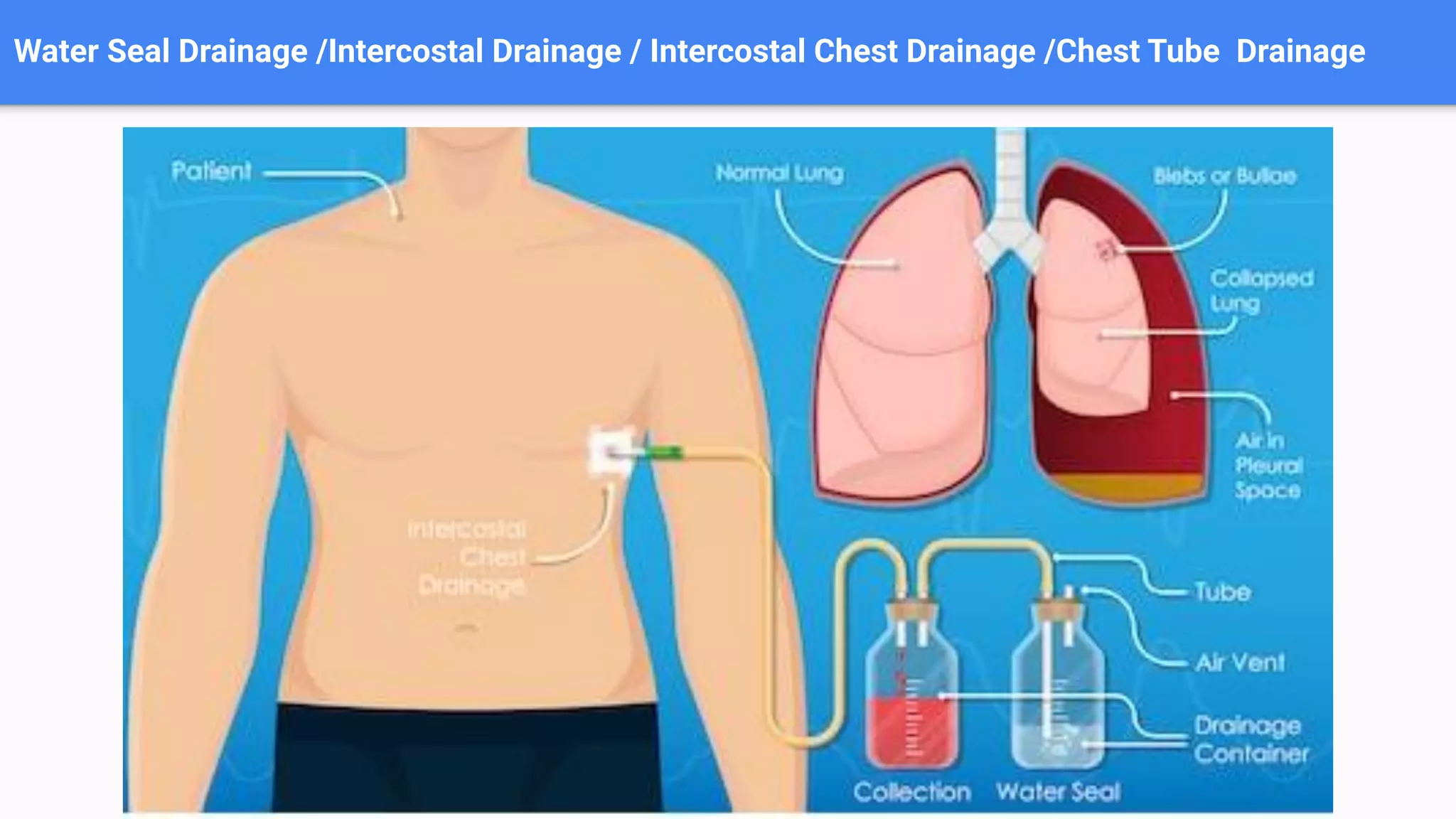


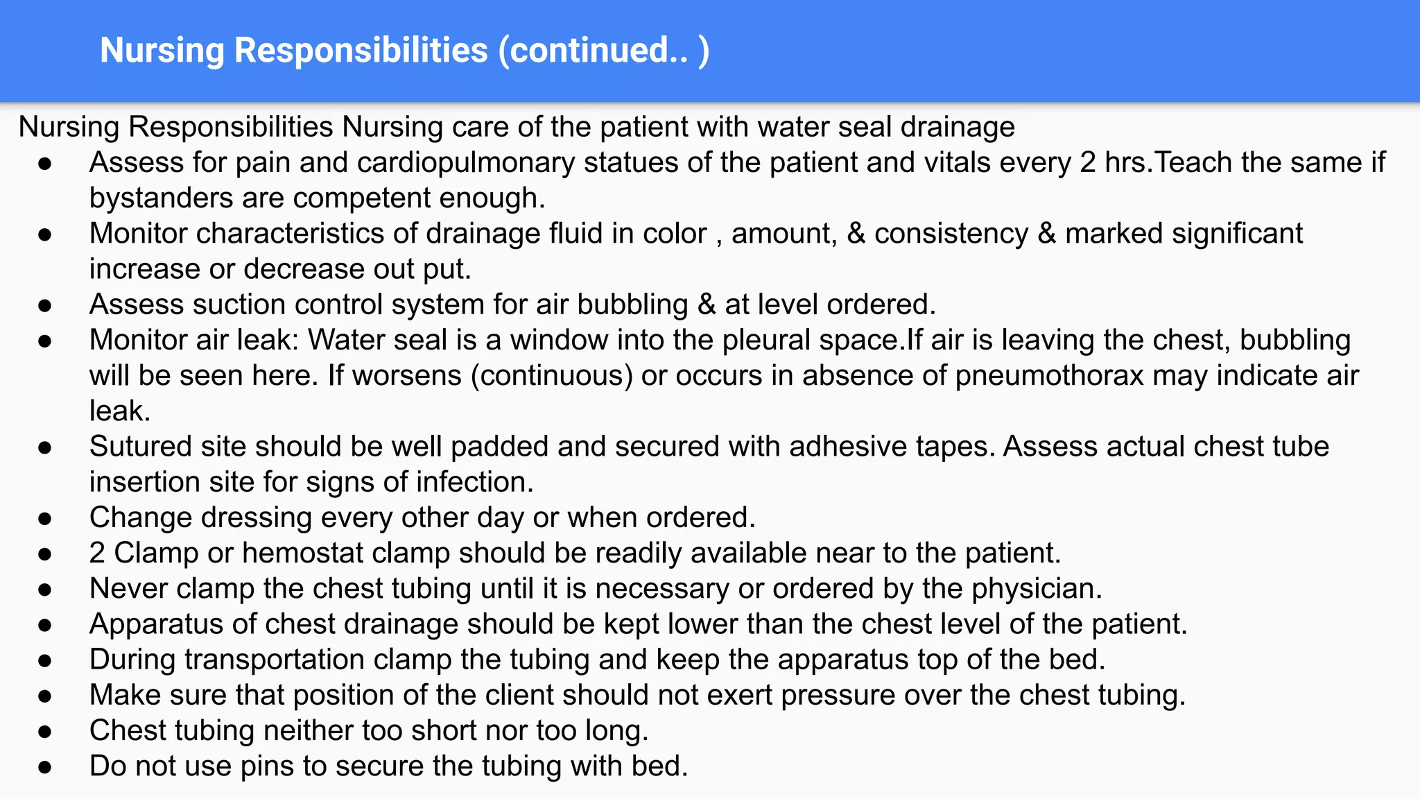
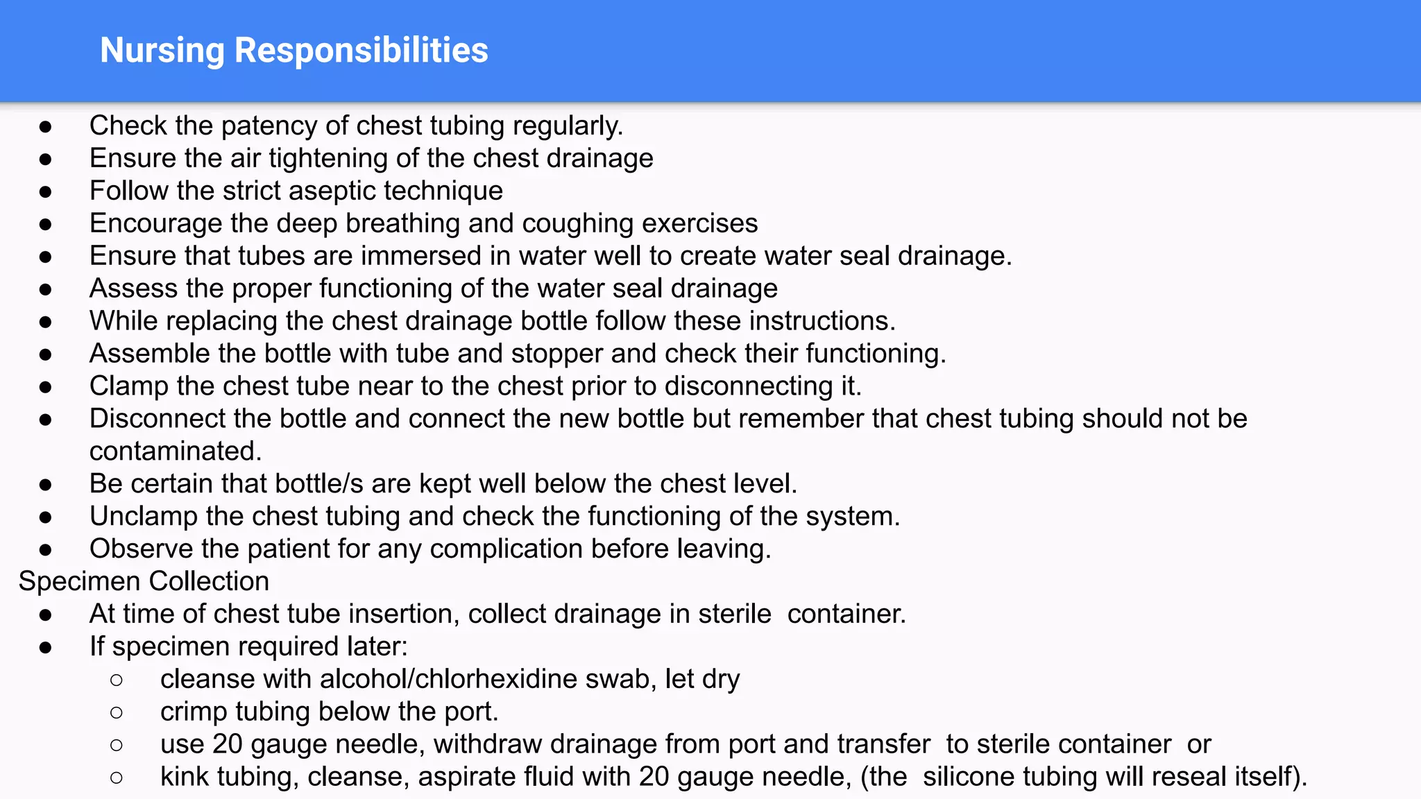

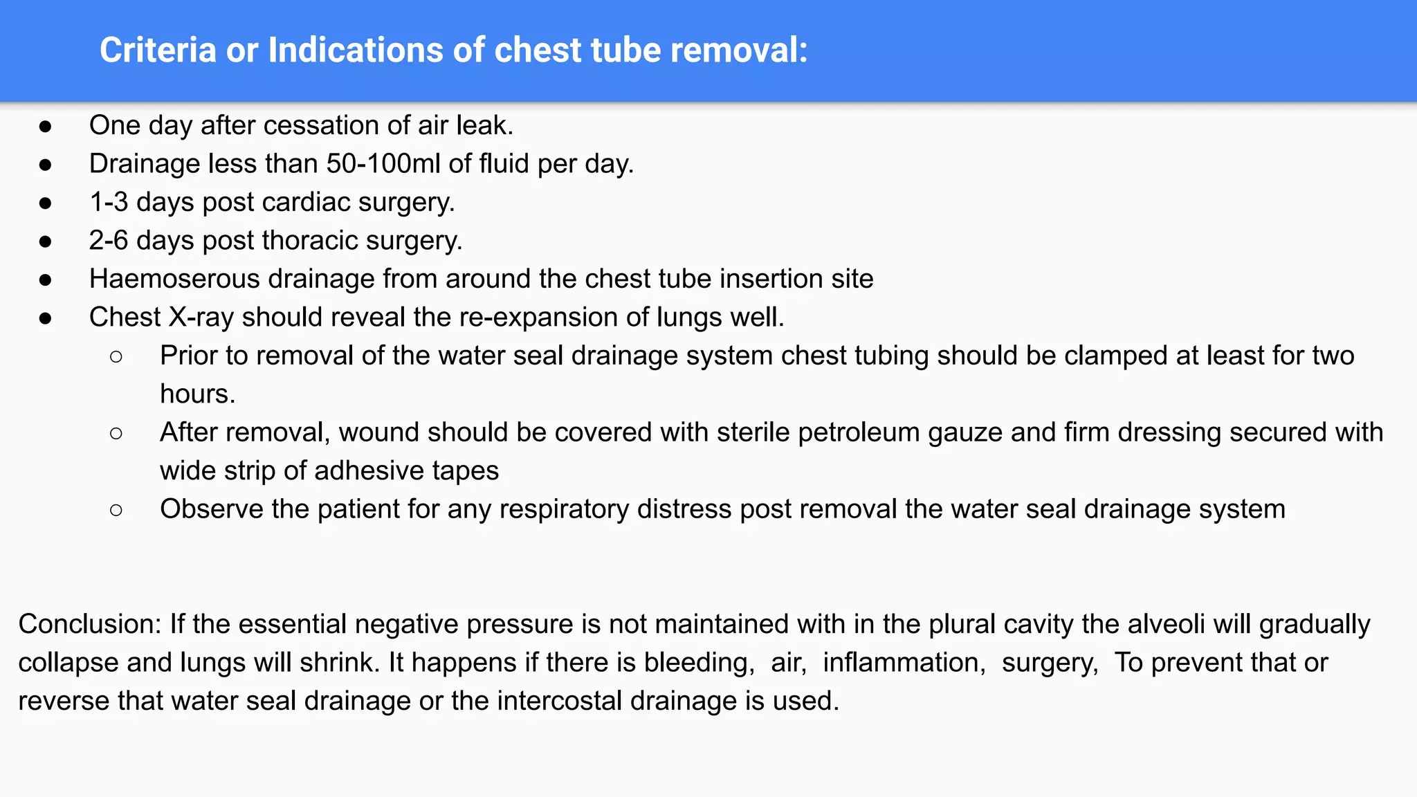


The document presents a comprehensive overview of water seal/chest tube drainage, including its purpose, indications, contraindications, insertion sites, types, and nursing responsibilities. It emphasizes the importance of maintaining an airtight drainage system and outlines procedures for effective patient care, including monitoring, handling complications, and specimen collection. The presentation aims to equip nursing students with the necessary knowledge and skills to confidently manage patients with water seal drainage systems.


