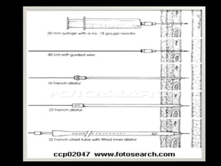
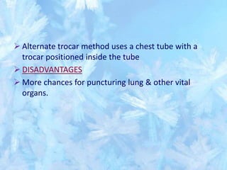
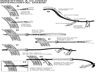
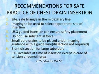
INTERCOSTAL
DRAINAGE
Points to discuss…
 When to put a drain ( indications)
 Site of insertion
 Choosing the drain
 Drainage system
 Clamping the chest drain
 Time & method of removal
INDICATIONS
Pneumothorax
Primary spontaneous ptx
Secondary spontaneous ptx
Tension ptx(after initial needle aspiration)
Malignant pleural effusion
Empyema
Traumatic haemopneumothorax
Postoperative, eg: after esophageal, cardiac or
pleural surgery.
Post pneumonectomy bronchopleural fistula
CONTRAINDICATION
NO ABSOLUTE CONTRAINDICATION
RELATIVE C/I ARE-
Infection at the site of insertion
Uncorrected coagulopathy
SITE OF INSERTION ?
 Exact site depends on the location of abnormality.
 5th ICS in mid axillary line is the site used most often.
 Earlier it was believed that air can be drained only
through anteriorly placed tube in 2nd ICS in mid-
clavicular line.A tube placed too medially can injure
internal mammary artery causing serious
haemorrhage.
 Experience has shown that a tube of proper
size,inserted through 5th ICS in mid axillary line can
drain effectively.
TRIANGLE OF SAFETY
Area bordered by the anterior
border of latissmus dorsi,the
lateral border of the pectoralis
major, a line superior to the
horizontal level of nipple,with
its apex towards axilla
This is the usual site which
corresponds to the 5th or 6th ICS
in mid-axillary line
POSITION OF THE PATIENT
A chest tube can be inserted in supine,sitting
or lateral position.
Most preferred is supine position, in which
patient lies flat on the bed, slightly rotated to
the opposite side,with ipsilateral arm behind
her/his head.
Patients who are breathless may be asked to
sit upright in the bed,leaning over a cardiac
trolley with a pillow to place their arms
GUIDEWIRE TUBE THORACOSTOMY
 Easiest way to insert a chest tube.
 Usually done under the guidance of either USG or CT.
 This procedure uses the Seldinger technique with guide
wires & dilators.
 Skin,periosteum and parietal pleura are anesthetized and
incision is made in skin
 18 gauge needle attached to a syringe is introduced into
the pleural space. Fluid or air is aspirated to confirm the
diagnosis.
 Syringe is removed and J wire is threaded through the
needle in desired direction into the pleural space
 Needle is then removed and smallest dilator is been
inserted with a rotating movement, it is advanced into
pleural space.
 The first dilator is removed leaving the wire in place.
 Then the next size dilator is advanced over the guidewire
into the pleural space and removed.
 Finally chest tube containing the inserter is been threaded
over the guide wire.
 Once tube is in place inserter & guide wire are withdrawn.
 Tube is then clamped until it is attached to chest drainage
system.
 Tube is been anchored in place by means of purse string
suture.
 Incision is sutured without tension to avoid necrosis of skin
 Sterile dressing applied.
TROCAR TUBE THORACOSTOMY
 Initially requires a 2-4cm incision parallel to superior
border of the rib through the skin and subcutaneous
tissues after LA.
 Trocar is inserted between the ribs into the pleural
cavity,with flat edge cephalad to prevent damage to
intercostal vessels
 The hand not applying force should be placed next to the
chest wall to control depth of penetration
 Once trocar is in pleural space,stylet is removed and
chest tube with its distal end clamped is inserted into
the pleural space.
 Tocar is then removed.
 Alternate trocar method uses a chest tube with a
trocar positioned inside the tube
 DISADVANTAGES
 More chances for puncturing lung & other vital
organs.
OPERATING TUBE THORACOSTOMY
 Most commonly practiced
 Patient should be given anxiolytic 10-15mins before the
procedure and liberal doses of LA be used.
 3-4cm incision is made in the skin parallel to the chosen
intercostal space. The incision should be made down to
the fascia overlying the intercostal muscle.
 Once fascia has been incised the muscle fibers are
spread with a blunt tipped hemostat
 Incision is then made in the intercostal fascia just above
the superior border of the inferior rib over which tube
will pass
 Parietal pleura is then penetrated using blunt tipped
hemostat
 Hole in the parietal pleura is then enlarged with
operators index finger
 Operator should then palpate adjacent pleural space to
detect any adhesions.
 Chest tube with its distal end clamped is then introduced
with help of a hemostat into the pleural space
 Tube is sutured in place(mattress sutures) as per BTS
guidelines
 Site is cleaned and sterile dressing applied
 ADVANTAGES
 Safer then other methods
 Adhesions between lung & chest wall can be
removed.
 DISADVANTAGES
 Insertion of tube ectopically
CHOOSING THE CHEST DRAIN.
SIZE MATTERS ??
MALIGNANT EFFUSION FOR
PLEURODESIS
SMALL BORE TUBES 10-14F
HEMOTHORAX 28-32F
PNEUMOTHORAX 8-14F(SUCCESS RATE OF 84-97%)
EMPYEMAS 24-28F
ONE BOTTLE COLLECTION SYSTEM
 Consists of one bottle that serves as both a
collection container and a water seal.
 Chest tube is connected to a rigid straw
inserted through a stopper into a sterile
bottle
 Enough sterile solution is instilled into the
bottle so that tip of the rigid straw is
approximately 2cms below the surface of
saline solution.
 Bottles stopper must have a vent to prevent
pressure from building up when air or fluid
coming from pleural space enters the
bottle.
When pleural pressure is positive,the pressure in
the rigid straw becomes positive,and if the
pressure inside the rigid straw is greater than the
depth to which straw is inserted into the saline
solution,air(or liquid)will enter the bottle and will
be vented to the atmosphere(or collect in the
bottle).
If the pleural pressure is negative,fluid will be
drawn from the bottle into the rigid straw and no
extra air will enter the system.
Thus water in the bottle seals the pleural space
from air or fluid from outside the body
• ADVANTAGES:
• Easy to carry & works well for uncomplicated
pneumothorax
• DISADVANTAGES:
• If large amounts of fluid is draining from patients
pleural space level of fluid will rise in one bottle system
and therefore pressure will have to be higher & higher
in the rigid straw to allow additional air or fluid to exit
from pleural space
• If the bottle is inadverently placed above level of the
patients chest,fluid can flow back into the pleural
cavity.
TWO BOTTLE COLLECTION SYSTEM
 Preferred when substantial amounts of fluid is draining
from pleural space
 With this system, bottle adjacent to the patient acts as a
collection bottle for drainage, and second bottle
provides the water seal and the air vent.
 Degree of water seal does not increase as the drainage
accumulates.
SUCTION & THREE BOTTLE
COLLECTION SYSTEMS
Applying negative pressure to the pleural
space helps in facilitating reexpansion of the
underlying lung or to expedite removal of air
or fluid from pleural space
Suction at a fixed level, usually -15 to -20cm
H2o,can be applied to the vent on a one or
two bottle collection system with an Emerson
pump
In this system, suction is poorly controlled.
 Controlled amount of
suction can be applied to a
three bottle collection
system.
 A vent on suction control
bottle is connected to a
vent on the water seal
bottle.
 Suction control bottle has
a rigid straw. Suction is
connected to a second
vent on the suction control
bottle.
 When suction is applied to suction
control bottle, air enters this bottle
through it rigid straw if the
pressure in the bottle is more
negative than the depth to which
straw is submerged.
 Amount of negative pressure in
the system is equal to the depth to
which rigid straw in the suction
control bottle is submerged below
the surface as long as bubbles are
entering suction control bottle
through its rigid straw.
 Air enters the suction control bottle from the atmosphere
while its rigid straw is submerged at 20cm H2o.Thwe
pressure in the suction control bottle is -20cm H20.
 Same pressure exists in water seal bottle,since these two
bottles are in direct communication.The pressure in
drainage bottle is less negative than in other bottles
 In this case depth of water seal is 2cm,so pressure in the
drainage collection bottle and pleural space is --18cm H20.
 Amount of negative pressure in the system can be adjusted
by changing the position of rigid straw or depth of water in
suction bottle.
 If the distance from patients chest to the top of collection
apparatus is 50cms and tube is filled with liquid, there will
be a negative pressure of 50cm H2o in the pleural space if
no suction applied
 Actual negative pressure applied to the pleural space from
the entire system is the net vertical distance that the liquid
occupies the tube(A-B)minus the level of fluid in the water
seal(C) plus negative pressure applied through
suction(D).negative pressure in chest is A-B-C+D
 If there is no liquid in the tube, actual applied pressure will
be suction pressure minus the depth of the water seal.
PLEUR EVAC UNIT
• The drainage system is
disposable,molded plastic
unit with three chambers
duplicating the classic three
bottle system
ADVANTAGE
 It is a simple & renders freedom of the patient from a
bulky drainage apparatus
 Patients can be sent home with the flutter valve in place
Newer compect drainage system
• Large reservior for fluid collection, analysis and
keeping records
• Compect to permit patient ambulation
• Reuseble
• Inexpensive
• Detect size of airleak
• Various alarms
• Flushes the tube intermittently to prevent
blockadge
Advantages
• Early ambulation
• Shorter duration of chest tube usage
• Decreased cost by decreasing hospital stay
CLAMPING
 A bubbling chest tube should never be
clamped.
 Drainage of a large pleural effusion should be
controlled to prevent the potential
complication of re-expansion pulmonary
oedema.
 In cases of pneumothorax, clamping of the
chest tube should usually be avoided.
COMPLICATIONS
 Injury to the neurovascular bundle in the ICS
 Injury to lung parenchyma
 Injury to diaphragm and consequent injury to
intraperitoneal structures may occur
 Injury to heart and other vessels
 Massive bleeding
 Re expansion pulmonary oedema due to rapid
evacuation of fluid from the pleural cavity
 Empyema
 Skin excoriation and inflammation
 Subcutaneous emphysema & subcutaneous haematoma
REMOVAL OF THE DRAIN
 Original indication
 Clinical progress
 Daily drainage should be <100ml.
 There should be no air leak
 No fresh or altered blood should be draining from
chest tube
 Radiological status-lung should be fully expanded.
 End expiration/valsalva
Procedure for chest tube removal
 Gather supplies and explain procedure to patient
 The clinician will remove the dressing and sutures
 During peak exhalation, the clinician will remove the chest tube in one
quick movement
 Immediately apply a sterile gauze dressing containing petroleum to
prevent air from entering pleural space
 Monitor patient’s respiratory status
 Arrange for chest X-ray to confirm lung reexpansion
 Monitor patient’s respiratory status and SpO2 for 1-2 hours after
removal
PATIENTS REQUIRING ASSISTED
VENTILATION
 During the insertion of a chest tube in a patient on a high
pressure ventilator (especially with positive end
expiratory pressure (PEEP), it is essential to disconnect
from the ventilator at the time of insertion to avoid the
potentially serious complication of lung
penetration, although as long as blunt dissection is
carried out and no sharp instruments are used, this risk is
reduced.
RECOMMENDATIONS FOR SAFE
PRACTICE OF CHEST DRAIN INSERTION
1. Site: safe triangle in the midaxillary line
2. Imaging to be used to select appropriate site of
insertion
3. USG guided insertion can ensure safety placement
4. Do not use substantial force
5. Small bore drains to be placed under imaging
guidance with a guide wire(dissection not required)
6. Blunt dissection for large tube bore
7. CXR available at time of insertion except in case of
tension pneumothorax
BTS GUIDELINESS
THANK YOU

Chesttubedrainage

  • 1.
  • 2.
    Points to discuss… When to put a drain ( indications)  Site of insertion  Choosing the drain  Drainage system  Clamping the chest drain  Time & method of removal
  • 3.
    INDICATIONS Pneumothorax Primary spontaneous ptx Secondaryspontaneous ptx Tension ptx(after initial needle aspiration) Malignant pleural effusion Empyema Traumatic haemopneumothorax Postoperative, eg: after esophageal, cardiac or pleural surgery. Post pneumonectomy bronchopleural fistula
  • 4.
    CONTRAINDICATION NO ABSOLUTE CONTRAINDICATION RELATIVEC/I ARE- Infection at the site of insertion Uncorrected coagulopathy
  • 5.
    SITE OF INSERTION?  Exact site depends on the location of abnormality.  5th ICS in mid axillary line is the site used most often.  Earlier it was believed that air can be drained only through anteriorly placed tube in 2nd ICS in mid- clavicular line.A tube placed too medially can injure internal mammary artery causing serious haemorrhage.  Experience has shown that a tube of proper size,inserted through 5th ICS in mid axillary line can drain effectively.
  • 6.
    TRIANGLE OF SAFETY Areabordered by the anterior border of latissmus dorsi,the lateral border of the pectoralis major, a line superior to the horizontal level of nipple,with its apex towards axilla This is the usual site which corresponds to the 5th or 6th ICS in mid-axillary line
  • 7.
    POSITION OF THEPATIENT A chest tube can be inserted in supine,sitting or lateral position. Most preferred is supine position, in which patient lies flat on the bed, slightly rotated to the opposite side,with ipsilateral arm behind her/his head. Patients who are breathless may be asked to sit upright in the bed,leaning over a cardiac trolley with a pillow to place their arms
  • 10.
    GUIDEWIRE TUBE THORACOSTOMY Easiest way to insert a chest tube.  Usually done under the guidance of either USG or CT.  This procedure uses the Seldinger technique with guide wires & dilators.  Skin,periosteum and parietal pleura are anesthetized and incision is made in skin  18 gauge needle attached to a syringe is introduced into the pleural space. Fluid or air is aspirated to confirm the diagnosis.  Syringe is removed and J wire is threaded through the needle in desired direction into the pleural space  Needle is then removed and smallest dilator is been inserted with a rotating movement, it is advanced into pleural space.
  • 11.
     The firstdilator is removed leaving the wire in place.  Then the next size dilator is advanced over the guidewire into the pleural space and removed.  Finally chest tube containing the inserter is been threaded over the guide wire.  Once tube is in place inserter & guide wire are withdrawn.  Tube is then clamped until it is attached to chest drainage system.  Tube is been anchored in place by means of purse string suture.  Incision is sutured without tension to avoid necrosis of skin  Sterile dressing applied.
  • 13.
    TROCAR TUBE THORACOSTOMY Initially requires a 2-4cm incision parallel to superior border of the rib through the skin and subcutaneous tissues after LA.  Trocar is inserted between the ribs into the pleural cavity,with flat edge cephalad to prevent damage to intercostal vessels  The hand not applying force should be placed next to the chest wall to control depth of penetration  Once trocar is in pleural space,stylet is removed and chest tube with its distal end clamped is inserted into the pleural space.  Tocar is then removed.
  • 14.
     Alternate trocarmethod uses a chest tube with a trocar positioned inside the tube  DISADVANTAGES  More chances for puncturing lung & other vital organs.
  • 16.
    OPERATING TUBE THORACOSTOMY Most commonly practiced  Patient should be given anxiolytic 10-15mins before the procedure and liberal doses of LA be used.  3-4cm incision is made in the skin parallel to the chosen intercostal space. The incision should be made down to the fascia overlying the intercostal muscle.  Once fascia has been incised the muscle fibers are spread with a blunt tipped hemostat  Incision is then made in the intercostal fascia just above the superior border of the inferior rib over which tube will pass  Parietal pleura is then penetrated using blunt tipped hemostat
  • 17.
     Hole inthe parietal pleura is then enlarged with operators index finger  Operator should then palpate adjacent pleural space to detect any adhesions.  Chest tube with its distal end clamped is then introduced with help of a hemostat into the pleural space  Tube is sutured in place(mattress sutures) as per BTS guidelines  Site is cleaned and sterile dressing applied
  • 19.
     ADVANTAGES  Saferthen other methods  Adhesions between lung & chest wall can be removed.  DISADVANTAGES  Insertion of tube ectopically
  • 20.
    CHOOSING THE CHESTDRAIN. SIZE MATTERS ?? MALIGNANT EFFUSION FOR PLEURODESIS SMALL BORE TUBES 10-14F HEMOTHORAX 28-32F PNEUMOTHORAX 8-14F(SUCCESS RATE OF 84-97%) EMPYEMAS 24-28F
  • 21.
    ONE BOTTLE COLLECTIONSYSTEM  Consists of one bottle that serves as both a collection container and a water seal.  Chest tube is connected to a rigid straw inserted through a stopper into a sterile bottle  Enough sterile solution is instilled into the bottle so that tip of the rigid straw is approximately 2cms below the surface of saline solution.  Bottles stopper must have a vent to prevent pressure from building up when air or fluid coming from pleural space enters the bottle.
  • 22.
    When pleural pressureis positive,the pressure in the rigid straw becomes positive,and if the pressure inside the rigid straw is greater than the depth to which straw is inserted into the saline solution,air(or liquid)will enter the bottle and will be vented to the atmosphere(or collect in the bottle). If the pleural pressure is negative,fluid will be drawn from the bottle into the rigid straw and no extra air will enter the system. Thus water in the bottle seals the pleural space from air or fluid from outside the body
  • 23.
    • ADVANTAGES: • Easyto carry & works well for uncomplicated pneumothorax • DISADVANTAGES: • If large amounts of fluid is draining from patients pleural space level of fluid will rise in one bottle system and therefore pressure will have to be higher & higher in the rigid straw to allow additional air or fluid to exit from pleural space • If the bottle is inadverently placed above level of the patients chest,fluid can flow back into the pleural cavity.
  • 24.
    TWO BOTTLE COLLECTIONSYSTEM  Preferred when substantial amounts of fluid is draining from pleural space  With this system, bottle adjacent to the patient acts as a collection bottle for drainage, and second bottle provides the water seal and the air vent.  Degree of water seal does not increase as the drainage accumulates.
  • 25.
    SUCTION & THREEBOTTLE COLLECTION SYSTEMS Applying negative pressure to the pleural space helps in facilitating reexpansion of the underlying lung or to expedite removal of air or fluid from pleural space Suction at a fixed level, usually -15 to -20cm H2o,can be applied to the vent on a one or two bottle collection system with an Emerson pump In this system, suction is poorly controlled.
  • 26.
     Controlled amountof suction can be applied to a three bottle collection system.  A vent on suction control bottle is connected to a vent on the water seal bottle.  Suction control bottle has a rigid straw. Suction is connected to a second vent on the suction control bottle.
  • 27.
     When suctionis applied to suction control bottle, air enters this bottle through it rigid straw if the pressure in the bottle is more negative than the depth to which straw is submerged.  Amount of negative pressure in the system is equal to the depth to which rigid straw in the suction control bottle is submerged below the surface as long as bubbles are entering suction control bottle through its rigid straw.
  • 28.
     Air entersthe suction control bottle from the atmosphere while its rigid straw is submerged at 20cm H2o.Thwe pressure in the suction control bottle is -20cm H20.  Same pressure exists in water seal bottle,since these two bottles are in direct communication.The pressure in drainage bottle is less negative than in other bottles  In this case depth of water seal is 2cm,so pressure in the drainage collection bottle and pleural space is --18cm H20.  Amount of negative pressure in the system can be adjusted by changing the position of rigid straw or depth of water in suction bottle.
  • 29.
     If thedistance from patients chest to the top of collection apparatus is 50cms and tube is filled with liquid, there will be a negative pressure of 50cm H2o in the pleural space if no suction applied  Actual negative pressure applied to the pleural space from the entire system is the net vertical distance that the liquid occupies the tube(A-B)minus the level of fluid in the water seal(C) plus negative pressure applied through suction(D).negative pressure in chest is A-B-C+D  If there is no liquid in the tube, actual applied pressure will be suction pressure minus the depth of the water seal.
  • 30.
    PLEUR EVAC UNIT •The drainage system is disposable,molded plastic unit with three chambers duplicating the classic three bottle system
  • 31.
    ADVANTAGE  It isa simple & renders freedom of the patient from a bulky drainage apparatus  Patients can be sent home with the flutter valve in place
  • 32.
  • 33.
    • Large reserviorfor fluid collection, analysis and keeping records • Compect to permit patient ambulation • Reuseble • Inexpensive • Detect size of airleak • Various alarms • Flushes the tube intermittently to prevent blockadge
  • 34.
    Advantages • Early ambulation •Shorter duration of chest tube usage • Decreased cost by decreasing hospital stay
  • 35.
    CLAMPING  A bubblingchest tube should never be clamped.  Drainage of a large pleural effusion should be controlled to prevent the potential complication of re-expansion pulmonary oedema.  In cases of pneumothorax, clamping of the chest tube should usually be avoided.
  • 36.
    COMPLICATIONS  Injury tothe neurovascular bundle in the ICS  Injury to lung parenchyma  Injury to diaphragm and consequent injury to intraperitoneal structures may occur  Injury to heart and other vessels  Massive bleeding  Re expansion pulmonary oedema due to rapid evacuation of fluid from the pleural cavity  Empyema  Skin excoriation and inflammation  Subcutaneous emphysema & subcutaneous haematoma
  • 37.
    REMOVAL OF THEDRAIN  Original indication  Clinical progress  Daily drainage should be <100ml.  There should be no air leak  No fresh or altered blood should be draining from chest tube  Radiological status-lung should be fully expanded.  End expiration/valsalva
  • 38.
    Procedure for chesttube removal  Gather supplies and explain procedure to patient  The clinician will remove the dressing and sutures  During peak exhalation, the clinician will remove the chest tube in one quick movement  Immediately apply a sterile gauze dressing containing petroleum to prevent air from entering pleural space  Monitor patient’s respiratory status  Arrange for chest X-ray to confirm lung reexpansion  Monitor patient’s respiratory status and SpO2 for 1-2 hours after removal
  • 39.
    PATIENTS REQUIRING ASSISTED VENTILATION During the insertion of a chest tube in a patient on a high pressure ventilator (especially with positive end expiratory pressure (PEEP), it is essential to disconnect from the ventilator at the time of insertion to avoid the potentially serious complication of lung penetration, although as long as blunt dissection is carried out and no sharp instruments are used, this risk is reduced.
  • 40.
    RECOMMENDATIONS FOR SAFE PRACTICEOF CHEST DRAIN INSERTION 1. Site: safe triangle in the midaxillary line 2. Imaging to be used to select appropriate site of insertion 3. USG guided insertion can ensure safety placement 4. Do not use substantial force 5. Small bore drains to be placed under imaging guidance with a guide wire(dissection not required) 6. Blunt dissection for large tube bore 7. CXR available at time of insertion except in case of tension pneumothorax BTS GUIDELINESS
  • 41.