SlideShare a Scribd company logo
1 of 7
Drawings of the Eye
Cross section drawing of the eye - (side view) with major parts labeled.
Cross section drawing of the eye - (rear view).
Cut-away view of the eye in its socket showing the: bony socket, orbital muscles, eyelids and
eyelashes.
The lacrimal system - (tear ducts) produce tears to clean, moisten and lubricate the eyes and then
drains the excess fluid into the nose.
Our Eyes and brain divide what we see into a right and left half. In the drawing above, light gray
represents the left half; dark gray represents the right half. The eyes invert the image and the left
side of what we see ends up in the right side of our brain and visa versa. This all works out
because the right side of our brain controls the left side of our bodies and visa versa.
Anterior Chamber
The space between the cornea and iris filled with Aqueous Humor.
Aqueous Humor
A water like fluid, produced by the ciliary body, it fills the front of the eye
between the lens and cornea and provides the cornea and lens with oxygen and
nutrients. It drains back into the blood stream through the canals of schlemm.
Brain
The brain is where the electrical signals sent from our eyes are processed into
vision. Damage to the brain can lead to vision loss if the visual cortex or optic
pathways are damaged. The majority of nerve fibers in the optic tract connect
to the LGN. Several nerve fibers leave the optic tract before the LGN to
connect to sub cortical structures through out the brain. These parts of the brain
regulate things like: eye and head movements, pupillary light reflex - (pupil
size), and circadian rhythms - (light/dark cycle). Damage to these parts of the
brain often leads to vision disorders too.
Canals of Schlemm
These canals are located around the perimeter of the iris. They allow aqueous
fluid to drain back into the blood stream. The Trabecular Meshwork along with
the Canals of Schlemm regulate the eyes internal pressure. In the eye disease
called glaucoma, these canals become blocked leading to increased pressure.
The increased pressure, from this condition, destroys the optic nerve.
Choroid
The choroid is a layer of blood vessels between the retina and sclera; it supplies
blood to the retina. In the disease called Macular Degeneration, abnormal blood
vessels grow into the space between the retina and choroid damaging the
macula.
Ciliary Body
This is where the Aqueous Humor is produced.
Ciliary Muscle
The eye can bring the fine print in a phone book into focus, or focus in on the
moon over ¼ million miles away. The ciliary muscle changes the shape of the
lens - (this is called accommodation). It relaxes to flatten the lens for distance
vision; for close work it contracts rounding out the lens. Everyone will develop
an eye condition called presbyopia. As we age, the ciliary muscle and
crystalline lens lose their elasticity. This is why most people need reading
glasses by their 40's.
Conjunctiva
The conjunctiva is a thin, clear membrane covering the front of the eye and
inner eyelids. Cells in this lining produce mucous that helps to lubricate the
eye. This is the eyes first layer of protection against infection. Inflammation of
this membrane is called conjunctivitis, or pink eye.
Cornea
The cornea is a clear, dome-shaped surface that covers the front of the eye. It is
the first and most powerful lens in the eye's optical system. To keep it
transparent the cornea contains no blood vessels. Tears that flow over it and
aqueous humor in the chamber behind it keep it nourished. When you hear of
eye banks and eye transplants, it is the cornea that is being replaced. The cornea
can be damaged from: accidents, infections, and genetic defects.
Crystalline Lens
The eye's crystalline lens works like the adjustable lens in a camera. Positioned
just behind the cornea; it is responsible for keeping images in focus on the
retina. It is adjustable for distance and close work. A cataract is the lens
clouding up. This happens to most people as they age. A few people are even
born with cataracts. Modern surgery has all but eliminated cataracts as a cause
of blindness in the developed world.
Eyeball
The eye is like a little video camera measuring about 1 inch or 2.5 cm. in
diameter. If someone's eyeball is larger then this, they will be nearsighted
(myopic); if it is smaller then this, they will be farsighted (hyperopic). Having
two eyes gives us binocular vision - (depth perception). This is due to the
fusing of both images in the visual cortex.
Eyelashes and Eyebrows
These specialized hairs protect the eyes from particles that may injure them.
They form a screen to keep dust and insects out. Anything touching them
triggers the eyelids to blink.
Eyelids
Our eyelids protect and lubricate our eyes. Small oil-producing glands line the
inner edge of our eyelids. These oils mix with tears when we blink, keeping the
eye moist and clean.
Eye Socket
The orbit or eye socket is a cone-shaped bony cavity that protects the eye. The
socket is padded with fatty tissue that allows the eye to move easily.
Fovea - (small pit)
The fovea is an indentation in the center of the macula. Its diameter is only 1.5
mm or about 1/16 inch. This small part of our retina is responsible for our
highest visual acuity. It is the center of our central vision.
Lacrimal Gland - (Tear Duct)
This gland continually releases tears and other protective fluids onto the surface
of the eye. It lubricates and keeps the cornea from becoming dehydrated.
Lacrimal Sac
The lacrimal sac is a tiny pump that drains tears and other debris from the eye.
The fluids flow down the nasolacrimal duct into the nose where they help keep
the nasal linings moist. This is why your nose runs when you cry.
Lateral Geniculate Nucleus - (LGN)
This part of the brain acts as a relay station; it decodes visual information from
the optic tract before sending it to the visual cortex for final processing.
Lens Sack or Capsule
During modern cataract surgery the outer membrane of the lens is left in place.
The artificial intraocular lens is placed in this sack.
Iris
This is the colored part of the eye: brown, green, blue, etc. It is a ring of muscle
fibers located behind the cornea and in front of the lens. It contracts and
expands, opening and closing the pupil, in response to the brightness of
surrounding light. Just as the aperture in a camera protects the film from over
exposure, the iris of the eye helps protect the sensitive retina.
Macula - (yellow spot)
This part of the retina is the most sensitive. Its diameter is only 7 mm or about
1/4 inch. It is responsible for our central, or reading vision. This part of the
retina gives us 20/20 vision. Without the macula, you would be blind - Legally
Blind that is. People with eye diseases like Macular Degeneration have vision
from 20/200 to 20/800.
Optic Chiasm
This is the first part of the brain to receive visual input. Each eye takes a
slightly different picture of the world. At the optic chiasm each picture is
divided in half. The outer left and right halves continue back toward the visual
cortex. The inner left and right halves cross over to the other side of the brain
then continue back toward the visual cortex. See Drawing of optic pathways.
Optic Disk
The optic disk is the spot on the retina where the optic nerve leaves the eye.
There are no sensory cells here, creating a blind spot. Each eye covers for the
blind spot of the other eye and the brain fills in the missing information.
Optic Nerve
Each optic nerve has about 1.2 million nerve fibers. This is the cable
connecting the eye to the brain.
Optic Tract
The nerves that connect the optic chiasm to the LGN are called the optic tract.
If one of these tracts is damaged, vision will be lost in one side of each eye.
Orbital Muscles
Six muscles are in charge of eye movement. Four of these move the eye up,
down, left and right. The other two control the twisting motion of the eye when
we tilt our head. Defects in these muscles and the nerves that control them lead
to conditions like Nystagmus and Amblyopia (Lazy Eye).
Photoreceptor Cells
The retina is composed of two types of photoreceptor cells. When light falls on
one of these cells, it causes a chemical reaction that sends an electrical signal to
the brain.
Cone cells give us our detailed color daytime vision. There are 6 million of
them in each human eye. Most of them are located in the central retina - macula
fovea area. There are three types of cone cells: one sensitive to red light,
another to green light, and the third sensitive to blue light.
Rod cells are about 500 times more sensitive to light then cone cells; they give
us our dim light or night vision. They are also more sensitive to motion then
cone cells. There are 120 million rod cells in the human eye. Most rod cells are
located in our peripheral or side vision.
Posterior Chamber
The space between the iris and the front of the lens filled with Aqueous Humor.
Pupil
The pupil is the hole in the center of the iris that light passes through. The iris
muscles control its size.
Retina
The retina is the film of the eye. It converts light rays into electrical signals and
sends them to the brain through the optic nerve. The sides of the retina are
responsible for our peripheral vision. The center area, called the macula, is used
for our fine central vision and color vision. The retina is where most the
problems leading to vision loss Occur. Three of the leading causes of blindness,
from retina damage, are Retinitis Pigmentosa, Macular Degeneration and
Diabetic Retinopathy.
Retinal Blood Vessels
A doctor can see the blood vessels that supply the retina when he looks into
your eyes. These vessels are in the choroid just beneath the retina. Abnormal
blood vessel growth and leaking blood vessels are the cause of vision loss in
eye conditions like, Diabetic Retinopathy, ROP, and Macular Degeneration.
Retinal Pigment Epithelium - (RPE)
The RPE is a layer of cells between the retina and choroid. The inside of a
camera is panted black to absorb scattered and reflected light. The black
pigment known as melanin in the RPE dose the same job for the eye. The RPE
gets rid of waste products produced by the photoreceptor cells. As we age, the
RPE can sometimes lose its ability to process this waste. Deposits of this waste,
called drusen, can distort and damage the retina leading to an eye condition
called dry macular degeneration.
Sclera
The sclera is the white, tough wall of the eye. It along with internal fluid
pressure keeps the eyes shape and protects its delicate internal parts.
Uvea
The uvea is the middle Vascular layer of the eye. It is made up of three parts:
the iris, ciliary body and chorid. Uveitis is the inflammation (or swelling) of
these parts of the eye.
Visual Axis
The Visual Axis is an imaginary line drawn through the center of the pupil to
the center of the Fovea. The orbital muscles keep the visual axis of both eyes
aligned on the center of what you are looking at (fixation point). An eye
condition called Strabismus - (misaligned eyes) results when the orbital
muscles fail to keep the eyes in alignment. Any damage to eye structures along
this axis leads to severe vision loss.
Visual Cortex
The part of the brain that processes and combines visual information from both
eyes and converts it into sight. Damage to the visual cortex results in a
condition called cortical blindness.
Visual Fields
The retina of each eye has two sections the nasal retina - (nose side) and
temporal retina - (ear side). For example: with your right eye, you see the right
half of the world with your nasal retina; you see the left half of the world with
your temporal retina. The picture your eye takes is flipped left for right and
upside down; its up to the brain to sort things out.
Vitreous Cavity
The space between the lens and retina filled with the gel like Vitreous Humor.
Vitreous Humor
The vitreous humor is a jelly like liquid that fills most of the eye (from the lens
back). As we age it changes from a gel to a liquid and gradually shrinks
separating from the retina. This is when people start seeing floaters, dark specs
in their vision. This is a normal sign of aging, but in a few cases the retina can
become detached as the vitreous separates.
Zonules
Zonules are hundreds of string like fibers that hold the lens suspended in
position and enable it to change shape for near or distant vision.
Top of Page
Site Information

More Related Content

What's hot

Anatomy of the eye presentation
Anatomy of the eye presentationAnatomy of the eye presentation
Anatomy of the eye presentation
Hasan Mokbel
 
Primary eye care
Primary eye carePrimary eye care
Primary eye care
JB Chand
 

What's hot (20)

Anatomy and physiology of the eye
Anatomy and physiology of the eyeAnatomy and physiology of the eye
Anatomy and physiology of the eye
 
The Human Eye
The Human EyeThe Human Eye
The Human Eye
 
Anatomy of the eye presentation
Anatomy of the eye presentationAnatomy of the eye presentation
Anatomy of the eye presentation
 
ppt on Eye anatomy
ppt on Eye anatomy ppt on Eye anatomy
ppt on Eye anatomy
 
Coats of eyeball
Coats  of  eyeballCoats  of  eyeball
Coats of eyeball
 
PHYSIOLOGY OF VISION,Part -II
PHYSIOLOGY OF VISION,Part -IIPHYSIOLOGY OF VISION,Part -II
PHYSIOLOGY OF VISION,Part -II
 
structure of eye ball
structure of eye ballstructure of eye ball
structure of eye ball
 
Physiology of eye
Physiology  of eyePhysiology  of eye
Physiology of eye
 
The human eye
The human eyeThe human eye
The human eye
 
The human eye amatomy
The human eye amatomyThe human eye amatomy
The human eye amatomy
 
Physiology of vision
Physiology of visionPhysiology of vision
Physiology of vision
 
Lens
Lens Lens
Lens
 
Physiology of the eye
Physiology of the eye Physiology of the eye
Physiology of the eye
 
Anatomy and physiology of the Eye-Nursing
Anatomy and physiology of the Eye-NursingAnatomy and physiology of the Eye-Nursing
Anatomy and physiology of the Eye-Nursing
 
Primary eye care
Primary eye carePrimary eye care
Primary eye care
 
anatomy of the eyeball
anatomy of the eyeballanatomy of the eyeball
anatomy of the eyeball
 
Trail box
Trail boxTrail box
Trail box
 
Retina and layers
Retina and layersRetina and layers
Retina and layers
 
Eye Anatomy & Physiology
Eye Anatomy & Physiology Eye Anatomy & Physiology
Eye Anatomy & Physiology
 
Visual pathway ppt
Visual pathway pptVisual pathway ppt
Visual pathway ppt
 

Similar to diagram of eye

The human eye 10 13-2011
The human eye 10 13-2011The human eye 10 13-2011
The human eye 10 13-2011
Varunitya
 
Presentation of eye ths
Presentation of eye thsPresentation of eye ths
Presentation of eye ths
Shubham Gupta
 
VISION IN ANIMALS VETERINARY PHYSIOLOGY.pdf
VISION IN ANIMALS VETERINARY PHYSIOLOGY.pdfVISION IN ANIMALS VETERINARY PHYSIOLOGY.pdf
VISION IN ANIMALS VETERINARY PHYSIOLOGY.pdf
TatendaMageja
 
SHAHID KHAN AZMI.organ of vision and pathway of vision.ppt
SHAHID KHAN AZMI.organ of vision and pathway of vision.pptSHAHID KHAN AZMI.organ of vision and pathway of vision.ppt
SHAHID KHAN AZMI.organ of vision and pathway of vision.ppt
sk2023765
 
Looking Into The Eye
Looking Into The EyeLooking Into The Eye
Looking Into The Eye
DLHarland
 

Similar to diagram of eye (20)

Sense organs
Sense organsSense organs
Sense organs
 
The human eye 10 13-2011
The human eye 10 13-2011The human eye 10 13-2011
The human eye 10 13-2011
 
Human eye
Human eye Human eye
Human eye
 
VISION.pdf
VISION.pdfVISION.pdf
VISION.pdf
 
Presentation of eye ths
Presentation of eye thsPresentation of eye ths
Presentation of eye ths
 
Lv book (1).pdf
Lv book (1).pdfLv book (1).pdf
Lv book (1).pdf
 
14th aSpecial Senses of sight.pptx
14th aSpecial Senses of sight.pptx14th aSpecial Senses of sight.pptx
14th aSpecial Senses of sight.pptx
 
Eyes Anatomy & Physiology with Diagnostic investigation.
Eyes Anatomy & Physiology with Diagnostic investigation.Eyes Anatomy & Physiology with Diagnostic investigation.
Eyes Anatomy & Physiology with Diagnostic investigation.
 
VISION IN ANIMALS VETERINARY PHYSIOLOGY.pdf
VISION IN ANIMALS VETERINARY PHYSIOLOGY.pdfVISION IN ANIMALS VETERINARY PHYSIOLOGY.pdf
VISION IN ANIMALS VETERINARY PHYSIOLOGY.pdf
 
Anatomy of the eye and Layers of eyeball
Anatomy of the eye and Layers of eyeball Anatomy of the eye and Layers of eyeball
Anatomy of the eye and Layers of eyeball
 
SENSORY ORGANS...(EYE).pptx
SENSORY ORGANS...(EYE).pptxSENSORY ORGANS...(EYE).pptx
SENSORY ORGANS...(EYE).pptx
 
SHAHID KHAN AZMI.organ of vision and pathway of vision.ppt
SHAHID KHAN AZMI.organ of vision and pathway of vision.pptSHAHID KHAN AZMI.organ of vision and pathway of vision.ppt
SHAHID KHAN AZMI.organ of vision and pathway of vision.ppt
 
structure and fuction of eyes and ears,types of memory,sharpe memory,attention
structure and fuction of eyes and ears,types of memory,sharpe memory,attentionstructure and fuction of eyes and ears,types of memory,sharpe memory,attention
structure and fuction of eyes and ears,types of memory,sharpe memory,attention
 
Looking Into The Eye
Looking Into The EyeLooking Into The Eye
Looking Into The Eye
 
Presentation on eye
Presentation on eyePresentation on eye
Presentation on eye
 
Eye structure.pptx
Eye structure.pptxEye structure.pptx
Eye structure.pptx
 
Chapter 8 - Special Senses
Chapter 8 - Special SensesChapter 8 - Special Senses
Chapter 8 - Special Senses
 
Chapter 8 special senses
Chapter 8   special sensesChapter 8   special senses
Chapter 8 special senses
 
Eye ball
Eye ballEye ball
Eye ball
 
Assessment of eye.pptx
Assessment of eye.pptxAssessment of eye.pptx
Assessment of eye.pptx
 

More from Yoshasri kommineni (6)

Metabolic disorders of carbohydrates ,protiens,lipids
Metabolic disorders of carbohydrates ,protiens,lipidsMetabolic disorders of carbohydrates ,protiens,lipids
Metabolic disorders of carbohydrates ,protiens,lipids
 
Metabolic disorders
Metabolic disordersMetabolic disorders
Metabolic disorders
 
Heart sounds
Heart soundsHeart sounds
Heart sounds
 
Deglutition
DeglutitionDeglutition
Deglutition
 
endocrine system
endocrine systemendocrine system
endocrine system
 
drugs and cosmetics act -1940
drugs and cosmetics act -1940drugs and cosmetics act -1940
drugs and cosmetics act -1940
 

Recently uploaded

Beyond the EU: DORA and NIS 2 Directive's Global Impact
Beyond the EU: DORA and NIS 2 Directive's Global ImpactBeyond the EU: DORA and NIS 2 Directive's Global Impact
Beyond the EU: DORA and NIS 2 Directive's Global Impact
PECB
 
Gardella_Mateo_IntellectualProperty.pdf.
Gardella_Mateo_IntellectualProperty.pdf.Gardella_Mateo_IntellectualProperty.pdf.
Gardella_Mateo_IntellectualProperty.pdf.
MateoGardella
 
Making and Justifying Mathematical Decisions.pdf
Making and Justifying Mathematical Decisions.pdfMaking and Justifying Mathematical Decisions.pdf
Making and Justifying Mathematical Decisions.pdf
Chris Hunter
 
An Overview of Mutual Funds Bcom Project.pdf
An Overview of Mutual Funds Bcom Project.pdfAn Overview of Mutual Funds Bcom Project.pdf
An Overview of Mutual Funds Bcom Project.pdf
SanaAli374401
 

Recently uploaded (20)

Advance Mobile Application Development class 07
Advance Mobile Application Development class 07Advance Mobile Application Development class 07
Advance Mobile Application Development class 07
 
Mattingly "AI & Prompt Design: Structured Data, Assistants, & RAG"
Mattingly "AI & Prompt Design: Structured Data, Assistants, & RAG"Mattingly "AI & Prompt Design: Structured Data, Assistants, & RAG"
Mattingly "AI & Prompt Design: Structured Data, Assistants, & RAG"
 
Advanced Views - Calendar View in Odoo 17
Advanced Views - Calendar View in Odoo 17Advanced Views - Calendar View in Odoo 17
Advanced Views - Calendar View in Odoo 17
 
Basic Civil Engineering first year Notes- Chapter 4 Building.pptx
Basic Civil Engineering first year Notes- Chapter 4 Building.pptxBasic Civil Engineering first year Notes- Chapter 4 Building.pptx
Basic Civil Engineering first year Notes- Chapter 4 Building.pptx
 
Beyond the EU: DORA and NIS 2 Directive's Global Impact
Beyond the EU: DORA and NIS 2 Directive's Global ImpactBeyond the EU: DORA and NIS 2 Directive's Global Impact
Beyond the EU: DORA and NIS 2 Directive's Global Impact
 
ICT Role in 21st Century Education & its Challenges.pptx
ICT Role in 21st Century Education & its Challenges.pptxICT Role in 21st Century Education & its Challenges.pptx
ICT Role in 21st Century Education & its Challenges.pptx
 
Ecological Succession. ( ECOSYSTEM, B. Pharmacy, 1st Year, Sem-II, Environmen...
Ecological Succession. ( ECOSYSTEM, B. Pharmacy, 1st Year, Sem-II, Environmen...Ecological Succession. ( ECOSYSTEM, B. Pharmacy, 1st Year, Sem-II, Environmen...
Ecological Succession. ( ECOSYSTEM, B. Pharmacy, 1st Year, Sem-II, Environmen...
 
Explore beautiful and ugly buildings. Mathematics helps us create beautiful d...
Explore beautiful and ugly buildings. Mathematics helps us create beautiful d...Explore beautiful and ugly buildings. Mathematics helps us create beautiful d...
Explore beautiful and ugly buildings. Mathematics helps us create beautiful d...
 
Measures of Dispersion and Variability: Range, QD, AD and SD
Measures of Dispersion and Variability: Range, QD, AD and SDMeasures of Dispersion and Variability: Range, QD, AD and SD
Measures of Dispersion and Variability: Range, QD, AD and SD
 
Mixin Classes in Odoo 17 How to Extend Models Using Mixin Classes
Mixin Classes in Odoo 17  How to Extend Models Using Mixin ClassesMixin Classes in Odoo 17  How to Extend Models Using Mixin Classes
Mixin Classes in Odoo 17 How to Extend Models Using Mixin Classes
 
How to Give a Domain for a Field in Odoo 17
How to Give a Domain for a Field in Odoo 17How to Give a Domain for a Field in Odoo 17
How to Give a Domain for a Field in Odoo 17
 
Holdier Curriculum Vitae (April 2024).pdf
Holdier Curriculum Vitae (April 2024).pdfHoldier Curriculum Vitae (April 2024).pdf
Holdier Curriculum Vitae (April 2024).pdf
 
Class 11th Physics NEET formula sheet pdf
Class 11th Physics NEET formula sheet pdfClass 11th Physics NEET formula sheet pdf
Class 11th Physics NEET formula sheet pdf
 
Gardella_Mateo_IntellectualProperty.pdf.
Gardella_Mateo_IntellectualProperty.pdf.Gardella_Mateo_IntellectualProperty.pdf.
Gardella_Mateo_IntellectualProperty.pdf.
 
Making and Justifying Mathematical Decisions.pdf
Making and Justifying Mathematical Decisions.pdfMaking and Justifying Mathematical Decisions.pdf
Making and Justifying Mathematical Decisions.pdf
 
Nutritional Needs Presentation - HLTH 104
Nutritional Needs Presentation - HLTH 104Nutritional Needs Presentation - HLTH 104
Nutritional Needs Presentation - HLTH 104
 
An Overview of Mutual Funds Bcom Project.pdf
An Overview of Mutual Funds Bcom Project.pdfAn Overview of Mutual Funds Bcom Project.pdf
An Overview of Mutual Funds Bcom Project.pdf
 
fourth grading exam for kindergarten in writing
fourth grading exam for kindergarten in writingfourth grading exam for kindergarten in writing
fourth grading exam for kindergarten in writing
 
Application orientated numerical on hev.ppt
Application orientated numerical on hev.pptApplication orientated numerical on hev.ppt
Application orientated numerical on hev.ppt
 
Mattingly "AI & Prompt Design: The Basics of Prompt Design"
Mattingly "AI & Prompt Design: The Basics of Prompt Design"Mattingly "AI & Prompt Design: The Basics of Prompt Design"
Mattingly "AI & Prompt Design: The Basics of Prompt Design"
 

diagram of eye

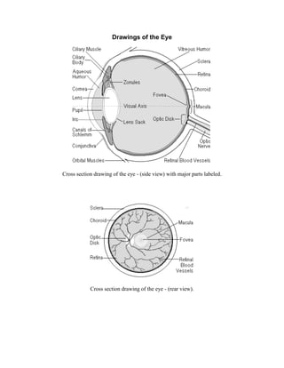
  • 1. Drawings of the Eye Cross section drawing of the eye - (side view) with major parts labeled. Cross section drawing of the eye - (rear view).
  • 2. Cut-away view of the eye in its socket showing the: bony socket, orbital muscles, eyelids and eyelashes. The lacrimal system - (tear ducts) produce tears to clean, moisten and lubricate the eyes and then drains the excess fluid into the nose. Our Eyes and brain divide what we see into a right and left half. In the drawing above, light gray represents the left half; dark gray represents the right half. The eyes invert the image and the left
  • 3. side of what we see ends up in the right side of our brain and visa versa. This all works out because the right side of our brain controls the left side of our bodies and visa versa. Anterior Chamber The space between the cornea and iris filled with Aqueous Humor. Aqueous Humor A water like fluid, produced by the ciliary body, it fills the front of the eye between the lens and cornea and provides the cornea and lens with oxygen and nutrients. It drains back into the blood stream through the canals of schlemm. Brain The brain is where the electrical signals sent from our eyes are processed into vision. Damage to the brain can lead to vision loss if the visual cortex or optic pathways are damaged. The majority of nerve fibers in the optic tract connect to the LGN. Several nerve fibers leave the optic tract before the LGN to connect to sub cortical structures through out the brain. These parts of the brain regulate things like: eye and head movements, pupillary light reflex - (pupil size), and circadian rhythms - (light/dark cycle). Damage to these parts of the brain often leads to vision disorders too. Canals of Schlemm These canals are located around the perimeter of the iris. They allow aqueous fluid to drain back into the blood stream. The Trabecular Meshwork along with the Canals of Schlemm regulate the eyes internal pressure. In the eye disease called glaucoma, these canals become blocked leading to increased pressure. The increased pressure, from this condition, destroys the optic nerve. Choroid The choroid is a layer of blood vessels between the retina and sclera; it supplies blood to the retina. In the disease called Macular Degeneration, abnormal blood vessels grow into the space between the retina and choroid damaging the macula. Ciliary Body This is where the Aqueous Humor is produced. Ciliary Muscle The eye can bring the fine print in a phone book into focus, or focus in on the moon over ¼ million miles away. The ciliary muscle changes the shape of the lens - (this is called accommodation). It relaxes to flatten the lens for distance vision; for close work it contracts rounding out the lens. Everyone will develop an eye condition called presbyopia. As we age, the ciliary muscle and crystalline lens lose their elasticity. This is why most people need reading glasses by their 40's. Conjunctiva The conjunctiva is a thin, clear membrane covering the front of the eye and inner eyelids. Cells in this lining produce mucous that helps to lubricate the
  • 4. eye. This is the eyes first layer of protection against infection. Inflammation of this membrane is called conjunctivitis, or pink eye. Cornea The cornea is a clear, dome-shaped surface that covers the front of the eye. It is the first and most powerful lens in the eye's optical system. To keep it transparent the cornea contains no blood vessels. Tears that flow over it and aqueous humor in the chamber behind it keep it nourished. When you hear of eye banks and eye transplants, it is the cornea that is being replaced. The cornea can be damaged from: accidents, infections, and genetic defects. Crystalline Lens The eye's crystalline lens works like the adjustable lens in a camera. Positioned just behind the cornea; it is responsible for keeping images in focus on the retina. It is adjustable for distance and close work. A cataract is the lens clouding up. This happens to most people as they age. A few people are even born with cataracts. Modern surgery has all but eliminated cataracts as a cause of blindness in the developed world. Eyeball The eye is like a little video camera measuring about 1 inch or 2.5 cm. in diameter. If someone's eyeball is larger then this, they will be nearsighted (myopic); if it is smaller then this, they will be farsighted (hyperopic). Having two eyes gives us binocular vision - (depth perception). This is due to the fusing of both images in the visual cortex. Eyelashes and Eyebrows These specialized hairs protect the eyes from particles that may injure them. They form a screen to keep dust and insects out. Anything touching them triggers the eyelids to blink. Eyelids Our eyelids protect and lubricate our eyes. Small oil-producing glands line the inner edge of our eyelids. These oils mix with tears when we blink, keeping the eye moist and clean. Eye Socket The orbit or eye socket is a cone-shaped bony cavity that protects the eye. The socket is padded with fatty tissue that allows the eye to move easily. Fovea - (small pit) The fovea is an indentation in the center of the macula. Its diameter is only 1.5 mm or about 1/16 inch. This small part of our retina is responsible for our highest visual acuity. It is the center of our central vision. Lacrimal Gland - (Tear Duct) This gland continually releases tears and other protective fluids onto the surface of the eye. It lubricates and keeps the cornea from becoming dehydrated. Lacrimal Sac
  • 5. The lacrimal sac is a tiny pump that drains tears and other debris from the eye. The fluids flow down the nasolacrimal duct into the nose where they help keep the nasal linings moist. This is why your nose runs when you cry. Lateral Geniculate Nucleus - (LGN) This part of the brain acts as a relay station; it decodes visual information from the optic tract before sending it to the visual cortex for final processing. Lens Sack or Capsule During modern cataract surgery the outer membrane of the lens is left in place. The artificial intraocular lens is placed in this sack. Iris This is the colored part of the eye: brown, green, blue, etc. It is a ring of muscle fibers located behind the cornea and in front of the lens. It contracts and expands, opening and closing the pupil, in response to the brightness of surrounding light. Just as the aperture in a camera protects the film from over exposure, the iris of the eye helps protect the sensitive retina. Macula - (yellow spot) This part of the retina is the most sensitive. Its diameter is only 7 mm or about 1/4 inch. It is responsible for our central, or reading vision. This part of the retina gives us 20/20 vision. Without the macula, you would be blind - Legally Blind that is. People with eye diseases like Macular Degeneration have vision from 20/200 to 20/800. Optic Chiasm This is the first part of the brain to receive visual input. Each eye takes a slightly different picture of the world. At the optic chiasm each picture is divided in half. The outer left and right halves continue back toward the visual cortex. The inner left and right halves cross over to the other side of the brain then continue back toward the visual cortex. See Drawing of optic pathways. Optic Disk The optic disk is the spot on the retina where the optic nerve leaves the eye. There are no sensory cells here, creating a blind spot. Each eye covers for the blind spot of the other eye and the brain fills in the missing information. Optic Nerve Each optic nerve has about 1.2 million nerve fibers. This is the cable connecting the eye to the brain. Optic Tract The nerves that connect the optic chiasm to the LGN are called the optic tract. If one of these tracts is damaged, vision will be lost in one side of each eye. Orbital Muscles Six muscles are in charge of eye movement. Four of these move the eye up, down, left and right. The other two control the twisting motion of the eye when we tilt our head. Defects in these muscles and the nerves that control them lead to conditions like Nystagmus and Amblyopia (Lazy Eye).
  • 6. Photoreceptor Cells The retina is composed of two types of photoreceptor cells. When light falls on one of these cells, it causes a chemical reaction that sends an electrical signal to the brain. Cone cells give us our detailed color daytime vision. There are 6 million of them in each human eye. Most of them are located in the central retina - macula fovea area. There are three types of cone cells: one sensitive to red light, another to green light, and the third sensitive to blue light. Rod cells are about 500 times more sensitive to light then cone cells; they give us our dim light or night vision. They are also more sensitive to motion then cone cells. There are 120 million rod cells in the human eye. Most rod cells are located in our peripheral or side vision. Posterior Chamber The space between the iris and the front of the lens filled with Aqueous Humor. Pupil The pupil is the hole in the center of the iris that light passes through. The iris muscles control its size. Retina The retina is the film of the eye. It converts light rays into electrical signals and sends them to the brain through the optic nerve. The sides of the retina are responsible for our peripheral vision. The center area, called the macula, is used for our fine central vision and color vision. The retina is where most the problems leading to vision loss Occur. Three of the leading causes of blindness, from retina damage, are Retinitis Pigmentosa, Macular Degeneration and Diabetic Retinopathy. Retinal Blood Vessels A doctor can see the blood vessels that supply the retina when he looks into your eyes. These vessels are in the choroid just beneath the retina. Abnormal blood vessel growth and leaking blood vessels are the cause of vision loss in eye conditions like, Diabetic Retinopathy, ROP, and Macular Degeneration. Retinal Pigment Epithelium - (RPE) The RPE is a layer of cells between the retina and choroid. The inside of a camera is panted black to absorb scattered and reflected light. The black pigment known as melanin in the RPE dose the same job for the eye. The RPE gets rid of waste products produced by the photoreceptor cells. As we age, the RPE can sometimes lose its ability to process this waste. Deposits of this waste, called drusen, can distort and damage the retina leading to an eye condition called dry macular degeneration. Sclera The sclera is the white, tough wall of the eye. It along with internal fluid pressure keeps the eyes shape and protects its delicate internal parts. Uvea
  • 7. The uvea is the middle Vascular layer of the eye. It is made up of three parts: the iris, ciliary body and chorid. Uveitis is the inflammation (or swelling) of these parts of the eye. Visual Axis The Visual Axis is an imaginary line drawn through the center of the pupil to the center of the Fovea. The orbital muscles keep the visual axis of both eyes aligned on the center of what you are looking at (fixation point). An eye condition called Strabismus - (misaligned eyes) results when the orbital muscles fail to keep the eyes in alignment. Any damage to eye structures along this axis leads to severe vision loss. Visual Cortex The part of the brain that processes and combines visual information from both eyes and converts it into sight. Damage to the visual cortex results in a condition called cortical blindness. Visual Fields The retina of each eye has two sections the nasal retina - (nose side) and temporal retina - (ear side). For example: with your right eye, you see the right half of the world with your nasal retina; you see the left half of the world with your temporal retina. The picture your eye takes is flipped left for right and upside down; its up to the brain to sort things out. Vitreous Cavity The space between the lens and retina filled with the gel like Vitreous Humor. Vitreous Humor The vitreous humor is a jelly like liquid that fills most of the eye (from the lens back). As we age it changes from a gel to a liquid and gradually shrinks separating from the retina. This is when people start seeing floaters, dark specs in their vision. This is a normal sign of aging, but in a few cases the retina can become detached as the vitreous separates. Zonules Zonules are hundreds of string like fibers that hold the lens suspended in position and enable it to change shape for near or distant vision. Top of Page Site Information