This document describes a case of congenital diaphragmatic hernia (CDH) in a newborn male infant. It provides details of the pregnancy, delivery, initial symptoms of respiratory distress, diagnostic imaging findings confirming CDH, and surgical repair procedure. Post-operatively, the infant experienced further complications including fever, seizures, and respiratory failure, and ultimately expired after 48 hours. CDH is a birth defect involving an abnormal opening in the diaphragm allowing abdominal organs in the chest cavity. It can be diagnosed prenatally by ultrasound and is typically treated with surgery to repair the diaphragmatic defect along with respiratory support.
Congenital Diaphragmatic Hernia (CDH) is a birth defect where abdominal organs protrude into the chest cavity due to a hole in the diaphragm. It occurs in about 1 in 3,000 births and is more common in females. The cause is unknown but may involve genetic and environmental factors. Symptoms include respiratory distress. Diagnosis is made via prenatal ultrasound or postnatal chest x-ray. Treatment involves respiratory support, surgery to repair the diaphragm, and managing complications such as pulmonary hypertension. Prognosis depends on factors like presence of other anomalies and severity of lung hypoplasia.
Congenital diaphragmatic hernia (CDH) is a birth defect where abdominal organs protrude into the chest cavity through an opening in the diaphragm. Prenatal ultrasound can detect CDH in 40-90% of cases. Management involves immediate respiratory support and stabilization at birth followed by surgery once stable. Outcomes depend on severity, with survival rates ranging from 40-69% when ECMO is used for severe cases. Long-term complications can include chronic lung disease, feeding difficulties, and neurodevelopmental delays.
C. CDH is more commonly found on the left side (around 85% of cases are left-sided).
Capillary alveolar dysplasia, bronchopulmonary sequestration, and chylothorax do not have a strong side predominance. Around half of bronchopulmonary sequestrations are left-sided, but they can occur on either side. Capillary alveolar dysplasia and chylothorax also occur bilaterally or on either side without a clear predominance.
So the developmental lung condition that is most strongly left-sided is congenital diaphragmatic hernia.
Congenital diaphragmatic hernia (CDH) is a birth defect that affects about 1 in 2,000-5,000 live births. It occurs when the diaphragm fails to fully form, allowing abdominal organs to migrate into the chest cavity and compress lung development. Untreated CDH has a high mortality rate of nearly 70%. Prenatal diagnosis by ultrasound is possible as early as the second trimester. Postnatal treatment may involve mechanical ventilation, nitric oxide, surfactant therapy, and in severe cases, extracorporeal membrane oxygenation (ECMO) or surgery to repair the diaphragmatic defect. Long-term outcomes include risks of chronic lung disease, feeding difficulties, growth
This presentation focuses on common obstetrics emergencies. These include early pregnancy complications such as miscarriages and ectopic pregnancy. As well as abdominal pain. Other include haemorrhage, hypertensive state, and sepsis.
Congenital diaphragmatic hernia results from failure of the diaphragm to fully develop in utero, allowing abdominal organs to protrude into the chest cavity. This document discusses the embryology, pathophysiology, prenatal and postnatal management of congenital diaphragmatic hernia. Prenatal diagnosis is made using ultrasound to detect organs in the chest. After birth, stabilization includes intubation, ventilation with permissive hypercapnia, and nasogastric decompression. Surgery is then performed to repair the diaphragmatic defect, followed by careful postoperative ventilation to prevent lung injury.
This document discusses peptic ulcer disease (PUD), including risk factors, pathophysiology, diagnosis, and treatment. Some key points:
- H. pylori infection and NSAID use are the leading causes of PUD. H. pylori infection is present in 60% of Americans over age 60.
- Diagnosis involves testing for H. pylori (stool antigen, urea breath, serology), and endoscopy if high risk or symptoms persist after treatment.
- Treatment for H. pylori-associated PUD is triple therapy (PPI plus two antibiotics) for 14 days. NSAID-associated PUD is treated with PPIs and prostag
Peptic Ulcer Disease (PUD) is caused by an imbalance between acid/pepsin production and protective mechanisms in the stomach and duodenum. Helicobacter pylori (H. pylori) infection and NSAID use are the most common causes of PUD. The presentation is often epigastric pain that may radiate to the back, occurring 1-3 hours after meals or at night and relieved by food or antacids. Risk factors identified in the patient include H. pylori infection, smoking, coffee and NSAID use. Diagnosis involves testing for H. pylori and endoscopy if high risk.
Ambo University's College of Health Sciences document outlines several obstetric emergencies including hyperemesis gravidarum, premature rupture of membranes, abortion, ectopic pregnancy, preeclampsia, and eclampsia. It describes the definition, risk factors, clinical features, diagnosis, complications and management of each condition. The document provides medical students and health professionals with an overview of major issues that can arise before, during, and after delivery that require emergency treatment.
This document provides an outline and overview of common obstetric emergencies. It discusses conditions that can occur before, during, and after delivery including hyperemesis gravidarum, premature rupture of membranes, abortion, ectopic pregnancy, preeclampsia, abruptio placenta, obstructed labor, uterine rupture, cord prolapse, retained placenta, placenta accreta, and cervical injuries. For each condition, it describes key details like definition, risk factors, clinical features, diagnosis, complications, and management considerations. The document is from the College of Health Sciences at Ambo University in Ethiopia and aims to educate medical students and practitioners about managing these potential pregnancy complications.
Respiratory distress in paediatric surgery can have many causes, including both upper and lower airway obstruction as well as general causes like pneumonia. Signs include increased respiratory rate, use of accessory muscles, and noisy breathing. Investigations depend on available facilities but often include chest x-ray, blood tests, and imaging. Treatment involves supportive care like supplemental oxygen and treating the underlying cause. Some specific conditions that can cause respiratory distress are laryngomalacia, tracheomalacia, congenital diaphragmatic hernia, congenital cystic adenomatoid malformation, congenital lobar emphysema, and pneumothorax.
Congenital diaphragmatic hernia by Dr. Varsha Atul ShahVarsha Shah
Dr. Varsha Atul Shah presented on congenital diaphragmatic hernia (CDH). CDH occurs when abdominal organs protrude into the chest cavity through a hole in the diaphragm. It has an incidence of 1 in 2000-3000 live births. Clinically, CDH presents with respiratory distress in newborns. Treatment involves medical management in the NICU with ventilator support, medications to manage blood pressure and oxygen levels, and surgery to repair the diaphragmatic defect. Outcomes have improved but mortality remains high depending on the severity of lung hypoplasia and pulmonary hypertension.
Congenital Diaphragmatic Hernia in newbornvanitha n
Congenital diaphragmatic hernia is a birth defect where there is an opening in the diaphragm that allows abdominal organs to move into the chest cavity. This compresses the lungs during development and causes pulmonary hypoplasia and hypertension. The defect occurs in about 1 in 5,000 births and is most often a left-sided opening. While the exact cause is unknown, risk factors include smoking and genetic conditions. Treatment involves prenatal surgery to repair the diaphragm if possible or intensive postnatal care including ventilation support, surgery to return organs to the abdomen, and management of pulmonary issues. Nursing care focuses on stabilization, monitoring, reducing stress, and supporting the parents through the serious condition and treatment
This document presents the case of a 26-year-old female, G3P2, who presented with vaginal bleeding. On examination, her cervical os was open and placental tissues were observed. She reported a 3-day history of bleeding. Based on her uterine size and exam findings, she was diagnosed with incomplete abortion at 12 weeks 3 days gestation. She underwent curettage under general anesthesia to complete the abortion procedure. Her bleeding was stabilized with medications and blood transfusion.
1. Congenital diaphragmatic hernia is a birth defect where abdominal organs protrude into the chest cavity through a hole in the diaphragm, interfering with lung development and potentially causing life-threatening breathing issues in newborns.
2. Prenatal ultrasound examination can detect a CDH, shown as a chest mass that may move or contain fluid/bowel above the diaphragm with associated shifting of the mediastinum.
3. Prognosis is worse when additional abnormalities are present, the defect is on the right side, or the liver protrudes through the diaphragm. Stabilization before surgery and careful postoperative respiratory support are crucial for survival.
This case report describes the diagnosis and management of a fetus with a left-sided diaphragmatic hernia detected on ultrasound at 23 weeks gestation. Diaphragmatic hernias occur when abdominal organs protrude into the chest cavity through a defect in the diaphragm. The fetus was monitored closely with serial ultrasounds and delivered via cesarean section at 36 weeks for pre-eclampsia. The newborn was referred to the neonatal intensive care unit for surgery and management of pulmonary issues associated with the condition.
Congenital diaphragmatic hernia (CDH) is a birth defect where abdominal organs protrude into the chest cavity through an opening in the diaphragm. There are three main types - Bochdalek (most common), Morgagni, and hiatal hernias. CDH occurs when the diaphragm fails to fully develop, usually resulting from defects in the pleuroperitoneal membranes or diaphragmatic musculature. Prenatal ultrasound can detect over 70% of CDH cases. Postnatal management focuses on gentle ventilation strategies and optimizing the infant's condition before surgical repair of the diaphragmatic defect. Outcomes are best when care is provided at specialized
This document discusses the case of a 6-month-old male infant presenting with fever, cough, difficulty breathing, and respiratory distress over the past few days. Examination revealed tachycardia, tachypnea, wheezing, and subcostal retractions. Chest X-ray showed bilateral scattered infiltrates and mild hyperinflation. The patient was treated with nebulized hypertonic saline, salbutamol, corticosteroids, antibiotics, and CPAP before gradually improving over 4 days. Statistics on bronchiolitis cases and treatments at the hospital over the past few months are also presented.
Presented by:
Ahmad mukhtar
MD.,M.B.B.Ch., M.Sc Obstetrics and GynecologyConsultant and Lecturer of Obstetrics and Gynecology, Faculty of
MEDICINE, Zagazig University.
The document discusses updates in obstetrics and gynecology, focusing on Millennium Development Goals 4 and 5 which aim to reduce child and maternal mortality. It then outlines key topics including hyperemesis gravidarum, miscarriages, molar pregnancy, and ectopic pregnancy. For each topic, it defines terms, discusses risk factors, clinical presentation, diagnosis, and management approaches.
This document provides information on several diseases of the newborn including necrotizing enterocolitis, transient tachypnea of the newborn, hemolytic disease of the newborn, Down syndrome, temperature control, and the newborn screening program. It describes the definition, causes, signs and symptoms, diagnostic evaluation, and treatment for each condition. It emphasizes the importance of early detection and management of congenital disorders through newborn screening to prevent intellectual disability or death.
Dr Varsha Atul Shah presented on congenital diaphragmatic hernia. Key points include: CDH occurs when abdominal organs herniate into the chest cavity due to a defect in the diaphragm, causing pulmonary hypoplasia. Presentation is usually respiratory distress after birth. Treatment involves medical stabilization, surgical repair of the defect, and management of long term complications like chronic lung disease and feeding difficulties. Close monitoring is needed due to risks of developmental delays, hearing loss, and other issues.
CARE OF MOTHER, CHILD, and ADOLESCENT CASE 5 PRESENTATIONAlexa43128
5 hours PTA, Patient noted sudden onset of watery vaginal discharges, clear associated with intermittent hypogastric pain every 5-10 minutes thus consult
• Patient History
• General Objectives
• Specific Objectives
• Anatomy and Physiology
• Laboratory and Diagnostics
• Nursing Care Plan
• Drug Study
• Discharge Summary
Kelainan Kongenital pada Sistem Gastrointestinal, Hepatobilier,.pptxikaseptyarini2
The document discusses several congenital gastrointestinal and hepatobiliary conditions including esophageal atresia, biliary atresia, anorectal malformations, Meckel's diverticulum, and Hirschsprung disease. It covers the epidemiology, clinical findings, diagnostic evaluations, differential diagnoses, management, and complications for each condition. The goal for many of these conditions is early surgical intervention to restore organ continuity or drainage.
This document discusses congenital diaphragmatic hernia and tracheoesophageal fistula. It begins by defining congenital diaphragmatic hernia and describing the different types. It then discusses the history, embryology, pathophysiology, classification, clinical manifestations, diagnosis, treatment including surgical repair, complications, and prognosis of congenital diaphragmatic hernia. It also defines tracheoesophageal fistula, describes the different types, discusses embryology, pathophysiology, clinical presentation, diagnosis, associated anomalies, preoperative preparation, surgical repair techniques, and complications.
share - Lions, tigers, AI and health misinformation, oh my!.pptxTina Purnat
• Pitfalls and pivots needed to use AI effectively in public health
• Evidence-based strategies to address health misinformation effectively
• Building trust with communities online and offline
• Equipping health professionals to address questions, concerns and health misinformation
• Assessing risk and mitigating harm from adverse health narratives in communities, health workforce and health system
Cell Therapy Expansion and Challenges in Autoimmune DiseaseHealth Advances
There is increasing confidence that cell therapies will soon play a role in the treatment of autoimmune disorders, but the extent of this impact remains to be seen. Early readouts on autologous CAR-Ts in lupus are encouraging, but manufacturing and cost limitations are likely to restrict access to highly refractory patients. Allogeneic CAR-Ts have the potential to broaden access to earlier lines of treatment due to their inherent cost benefits, however they will need to demonstrate comparable or improved efficacy to established modalities.
In addition to infrastructure and capacity constraints, CAR-Ts face a very different risk-benefit dynamic in autoimmune compared to oncology, highlighting the need for tolerable therapies with low adverse event risk. CAR-NK and Treg-based therapies are also being developed in certain autoimmune disorders and may demonstrate favorable safety profiles. Several novel non-cell therapies such as bispecific antibodies, nanobodies, and RNAi drugs, may also offer future alternative competitive solutions with variable value propositions.
Widespread adoption of cell therapies will not only require strong efficacy and safety data, but also adapted pricing and access strategies. At oncology-based price points, CAR-Ts are unlikely to achieve broad market access in autoimmune disorders, with eligible patient populations that are potentially orders of magnitude greater than the number of currently addressable cancer patients. Developers have made strides towards reducing cell therapy COGS while improving manufacturing efficiency, but payors will inevitably restrict access until more sustainable pricing is achieved.
Despite these headwinds, industry leaders and investors remain confident that cell therapies are poised to address significant unmet need in patients suffering from autoimmune disorders. However, the extent of this impact on the treatment landscape remains to be seen, as the industry rapidly approaches an inflection point.
One health condition that is becoming more common day by day is diabetes.
According to research conducted by the National Family Health Survey of India, diabetic cases show a projection which might increase to 10.4% by 2030.
Muktapishti is a traditional Ayurvedic preparation made from Shoditha Mukta (Purified Pearl), is believed to help regulate thyroid function and reduce symptoms of hyperthyroidism due to its cooling and balancing properties. Clinical evidence on its efficacy remains limited, necessitating further research to validate its therapeutic benefits.
Rasamanikya is a excellent preparation in the field of Rasashastra, it is used in various Kushtha Roga, Shwasa, Vicharchika, Bhagandara, Vatarakta, and Phiranga Roga. In this article Preparation& Comparative analytical profile for both Formulationon i.e Rasamanikya prepared by Kushmanda swarasa & Churnodhaka Shodita Haratala. The study aims to provide insights into the comparative efficacy and analytical aspects of these formulations for enhanced therapeutic outcomes.
Clinic ^%[+27633867063*Abortion Pills For Sale In Tembisa Central19various
Clinic ^%[+27633867063*Abortion Pills For Sale In Tembisa Central Clinic ^%[+27633867063*Abortion Pills For Sale In Tembisa CentralClinic ^%[+27633867063*Abortion Pills For Sale In Tembisa CentralClinic ^%[+27633867063*Abortion Pills For Sale In Tembisa CentralClinic ^%[+27633867063*Abortion Pills For Sale In Tembisa Central
8 Surprising Reasons To Meditate 40 Minutes A Day That Can Change Your Life.pptxHolistified Wellness
We’re talking about Vedic Meditation, a form of meditation that has been around for at least 5,000 years. Back then, the people who lived in the Indus Valley, now known as India and Pakistan, practised meditation as a fundamental part of daily life. This knowledge that has given us yoga and Ayurveda, was known as Veda, hence the name Vedic. And though there are some written records, the practice has been passed down verbally from generation to generation.
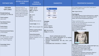
1. PERTINENT DATA
HISTORY OF PRESENT
ILLNESS
PHYSICAL
EXAMINATION
DIAGNOSTICS PREOPERATIVE DIAGNOSIS
DLB 26/M
G2P1 (1001)
Prenatal check-up:
Camiling HC 2x; PMD 10x
Chief complaint:
Labor pains
Past Medical History:
Unremarkable
Family History:
Unremarkable
Personal and Social History:
Unremarkable
OB History:
G1- NSD, live,Term, Health
center, Midwife assisted
G2 - Present Pregnancy
Gynecologic History:
Unremarkable
Few hours prior to
admission, patient had
labor pains increasing in
intensity associated with
watery vaginal discharge
prompting consultation,
hence admission.
Weight: 153 kg
Height: 58 cm
BMI: 24.8 kg/m2
Normal
BP 120/80
HR 92
RR 19
T 36.8
O2 sat: 99% at room air
Fundic Height: 29 cm
FHT: 140s by doppler, RLQ
EFW by cupping method:
2600-2800 grams
Internal Examination:
Cervix fully dilated, fully
effaced, cephalic, St +2, (-)
BOW, clear
Urinalysis
Pus 0-2
RBC 0-3
Protein negative
Glucose negative
RDT: Nonreactive
HBsAg: Nonreactive
PELVIC ULTRASOUND (6/12/23)
• SINGLE LIVE UTERINE PREGNANCY OF 34 WEEKS
GESTATION BY FETAL BIOMETRY
• CEPHALIC PRESENTATION, BPS 8/8, EFW 2,148
GRAMS
• ULTRASONIC EDD 7/24/2023 +/- 2 WEEKS
G2P1 (1001) Pregnancy uterine 40 6/7 weeks Age of
gestation, Cephalic in labor
Plan: Vaginal Delivery
Final Diagnosis
G2P2 (2002) Pregnancy Uterine delivered term, cephalic,
to a live baby boy, AS 8,9 BW 2.7 kg, Ballard Score 38
weeks, AGA via Normal Spontaneous Delivery
*Baby is admitted at NICU
Awake, with alar flaring, grunting, retractions and
cyanosis minutes after delivery
A: Neonatal Pneumonia (Initial Impression)
P: For emergency intubation
Mechanical Ventilation
Start on ampicillin, gentamicin
Chest xray
Wet Reading:
Bowel loops at left chest cavity, Inappreciable Right Lung
mass, with mediastinal shift to the right
* Referred to Dr. Carpio (Surgery Dept)
* Ruled out pneumonia, ruled in Congenital
Diaphragmatic Hernia
* Emergency OR: Exploratory Laparotomy, Repair of
Congenital Diaphragmatic Hernia, Chest Tube
Thoracotomy Insertion, Left
CBC
Hgb 118
Hct 0.356
WBC 14.5
Plt 313,000
2. PERTINENT DATA
HISTORY OF PRESENT
ILLNESS
PHYSICAL
EXAMINATION
DIAGNOSTICS PREOPERATIVE DIAGNOSIS
DLB 26/M
G2P1 (1001)
Prenatal check-up:
Camiling HC 2x; PMD 10x
Chief complaint:
Labor pains
Past Medical History:
Unremarkable
Family History:
Unremarkable
Personal and Social History:
Unremarkable
OB History:
G1- NSD, live,Term, Health
center, Midwife assisted
G2 - Present Pregnancy
Gynecologic History:
Unremarkable
Few hours prior to
admission, patient had
labor pains increasing in
intensity associated with
watery vaginal discharge
prompting consultation,
hence admission.
Weight: 153 kg
Height: 58 cm
BMI: 24.8 kg/m2
Normal
BP 120/80
HR 92
RR 19
T 36.8
O2 sat: 99% at room air
Fundic Height: 29 cm
FHT: 140s by doppler, RLQ
EFW by cupping method:
2600-2800 grams
Internal Examination:
Cervix fully dilated, fully
effaced, cephalic, St +2, (-)
BOW, clear
Urinalysis
Pus 0-2
RBC 0-3
Protein negative
Glucose negative
RDT: Nonreactive
HBsAg: Nonreactive
PELVIC ULTRASOUND (6/12/23)
• SINGLE LIVE UTERINE PREGNANCY OF 34 WEEKS
GESTATION BY FETAL BIOMETRY
• CEPHALIC PRESENTATION, BPS 8/8, EFW 2,148
GRAMS
• ULTRASONIC EDD 7/24/2023 +/- 2 WEEKS
*No intraop pictures*
*Patch Repair of 5cm Diaphragmatic Hole
Surgery:
Left Posterolateral diaphragmatic defect. Herniation of
small and large bowel into the left hemithorax, t/c
Bochdalek Hernia, s/p Exploratory Laparotomy, Repair of
Congenital diaphragmatic hernia under General
Endotracheal Anesthesia, s/p Chest tube thoracostomy,
left
Chest Xray Post Op:
Wet Reading:
* Expanded Right lung, unexpanded left lung
Update Post Op
* Patient still intubated with CTT inserted, left
* 10 hours post op, patient had fever, 1 seizure episode
and reintubation
* Patient had subsequent desaturation despite manual
bagging
*Baby expired after 48 hours of life
Pediatric Final diagnosis:
Acute Respiratory Failure, Congenital Diaphragmatic
Hernia, Left, Neonatal Sepsis, s/p Exploratory
Laparotomy, Repair of Congenital Diaphragmatic Hernia,
s/p Chest Tube Thoracostomy Insertion, Left
CBC
Hgb 118
Hct 0.356
WBC 14.5
Plt 313,000
4. CONGENITAL DIAPHRAGMATIC HERNIA
• Communication between the abdominal and thoracic
cavities with or without abdominal contents in the
thorax
• Etiology is rarely traumatic and usually congenital.
• The defect may be at the esophageal hiatus (hiatal
hernia); paraesophageal, adjacent to the hiatus
(paraesophageal hernia; retrosternal (foramen of
Morgagni hernia); or at the posterolateral portion of
the diaphragm (Bochdalek hernia).
• In congenital diaphragmatic hernia (CDH) the
Bochdalek hernia accounts for up to 90% of the hernias
seen, with 80–90% occurring on the left side. The
Morgagni hernia accounts for 2–6% of CDH.
5. CONGENITAL DIAPHRAGMATIC HERNIA
Pathology and Etiology
• Although CDH is characterized by a structural diaphragmatic defect, a
major limiting factor for survival is the associated pulmonary
hypoplasia.
• Pulmonary hypoplasia is characterized by a reduction in pulmonary
mass and the number of bronchial divisions, respiratory bronchioles,
and alveoli.
6. CONGENITAL DIAPHRAGMATIC HERNIA
Epidemiology
• The incidence of CDH is between 1 in 2,000 and 1 in 5,000 live births
• Females affected twice as often as males.
• Defects are more common on the left (85%) and are occasionally bilateral (<5%).
• Most cases of CDH are sporadic, but familial cases have been reported.
• Associated anomalies have been reported in up to 30% of cases, including CNS lesions,
esophageal atresia, omphalocele, and cardiovascular lesions.
• CDH is recognized as part of several chromosomal syndromes: trisomies 21, 13, and 18
and Fryns, Brachmann–de Lange, Pallister-Killian, and Turner syndromes.
7. CONGENITAL DIAPHRAGMATIC HERNIA
Diagnosis and Clinical Presentation
• In >50% of cases, CDH can be diagnosed
on prenatal ultrasonography (US)
between 16 and 24 wk of gestation.
• US findings may include
polyhydramnios, chest mass,
mediastinal shift, gastric bubble or a
liver in the thoracic cavity, and fetal
hydrops.
• After delivery, a chest radiograph is
needed to confirm the diagnosis
8. CONGENITAL DIAPHRAGMATIC HERNIA
• Respiratory distress is a cardinal sign in babies with CDH
• It may occur immediately after birth, or there may be a “honeymoon” period of up to 48
hr during which the baby is relatively stable.
• Early respiratory distress, within 6 hr after birth, is thought to be a poor prognostic sign.
• Respiratory distress is characterized clinically by tachypnea, grunting, use of accessory muscles,
and cyanosis. Children with CDH may also have a scaphoid abdomen and increased chest wall
diameter.
• Bowel sounds may also be heard in the chest with decreased breath sounds bilaterally.
• The point of maximal cardiac impulse may be displaced away from the side of the hernia
if mediastinal shift has occurred.
• A chest radiograph and passage of a nasal gastric tube are usually sufficient to confirm
the diagnosis.
9. CONGENITAL DIAPHRAGMATIC HERNIA
Treatment
• In the delivery room, infants with respiratory distress should be rapidly stabilized
with endotracheal intubation.
• Arterial (preductal and postductal) and central venous (umbilical) lines are
mandated, as are a urinary catheter and nasogastric tube.
• A preductal arterial oxygen saturation (SpO2) value ≥85% should be the minimum goal
• Gentle ventilation with permissive hypercapnia reduces lung injury
• Routine use of inotropes is indicated in the presence of left ventricular
dysfunction.
• In infants with severe respiratory failure and hypoxemia, sedation and paralysis
may be required.
10. CONGENITAL DIAPHRAGMATIC HERNIA
Prognosis
• Overall survival of liveborn infants with CDH is 71%. Relative predictors of a poor prognosis include an
associated major anomaly, symptoms before 24 hr of age, severe pulmonary hypoplasia, herniation to the
contralateral lung, and the need for ECMO.
• The size of the defect appears to be the strongest predictor of morbidity.
• Gastroesophageal reflux disease (GERD) is reported in >50% of children with CDH.
• Intestinal obstruction
• Recurrent diaphragmatic hernia is reported in 5–20% in most series.
• Children with patch repairs are at highest risk.
• Children with CDH typically have delayed growth in the 1st 2 yr of life.
• Neurocognitive defects are common and may result from the disease or the interventions. The incidence of
neurologic abnormalities is higher in infants who require ECMO (67% vs 24% of those who do not).
• The abnormalities are similar to those seen in neonates treated with ECMO for other diagnoses and include transient
and permanent developmental delay, abnormal hearing or vision, and seizures